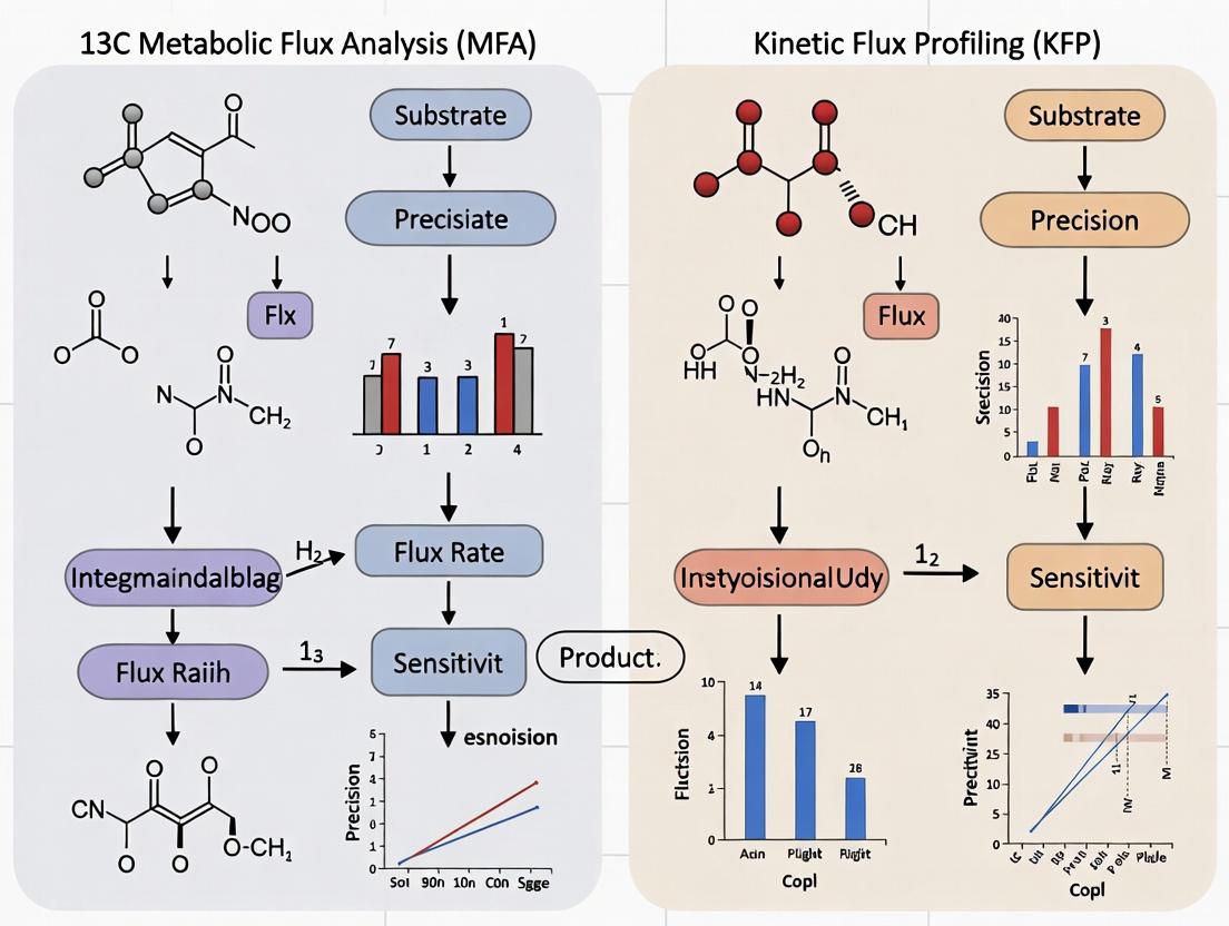
13C Metabolic Flux Analysis vs. Kinetic Flux Profiling: A Definitive Guide for Biomedical Researchers
This article provides a comprehensive comparison of two powerful techniques for analyzing cellular metabolism: 13C Metabolic Flux Analysis (13C MFA) and Kinetic Flux Profiling (KFP).
13C Metabolic Flux Analysis vs. Kinetic Flux Profiling: A Definitive Guide for Biomedical Researchers
Abstract
This article provides a comprehensive comparison of two powerful techniques for analyzing cellular metabolism: 13C Metabolic Flux Analysis (13C MFA) and Kinetic Flux Profiling (KFP). Aimed at researchers, scientists, and drug development professionals, it explores the foundational principles, methodological workflows, common challenges, and validation strategies for each approach. We detail their specific applications in systems biology, cancer research, and drug discovery, offering a clear framework for selecting the optimal method based on research goals, from steady-state mapping to dynamic pathway interrogation. The conclusion synthesizes key insights and outlines future implications for precision medicine and therapeutic development.
Decoding Metabolic Flux: Core Principles of 13C MFA and KFP
Understanding the dynamic flow of metabolites through biochemical networks—metabolic flux—is fundamental to deciphering disease mechanisms and identifying therapeutic targets. This guide compares two dominant methodologies for flux quantification: 13C Metabolic Flux Analysis (13C MFA) and Kinetic Flux Profiling (KFP), within ongoing research to establish a robust framework for biomedical discovery.
Core Methodologies at a Glance
| Feature | 13C Metabolic Flux Analysis (13C MFA) | Kinetic Flux Profiling (KFP) |
|---|---|---|
| Primary Principle | Steady-state isotopomer distribution modeling. | Transient isotopic labeling kinetics. |
| System State | Requires metabolic and isotopic steady state. | Operates under non-steady-state conditions. |
| Temporal Resolution | Integrative, provides time-averaged fluxes. | High, can capture rapid flux changes. |
| Experimental Duration | Hours to days (to reach steady-state labeling). | Seconds to minutes. |
| Key Requirement | Extensive network model; accurate atom mapping. | Precise measurement of metabolite pool sizes. |
| Best Suited For | Central carbon metabolism fluxes (e.g., TCA cycle, glycolysis). | Pathway fluxes in highly dynamic systems or rapid responses. |
Quantitative Comparison of Performance
Table 1: Benchmarking in Cancer Cell Line (e.g., HeLa) Studies
| Parameter | 13C MFA Result | KFP Result | Supporting Experimental Data |
|---|---|---|---|
| Glycolytic Flux | 150 ± 15 nmol/min/mg protein | 145 ± 30 nmol/min/mg protein | Antoniewicz et al., Metab Eng, 2019 |
| Pentose Phosphate Pathway Flux | 25 ± 5 nmol/min/mg protein | Not directly resolved | - |
| TCA Cycle Turnover | 90 ± 10 nmol/min/mg protein | 85 ± 20 nmol/min/mg protein | Hui et al., Nature, 2020 |
| Time to First Flux Estimate | ~24-48 hours | ~10-30 minutes | Jang et al., Cell, 2018 |
| Response to Acute Drug Inhibition | Not applicable (steady-state) | Detected within 2 minutes | Data from simulated KFP workflow |
Experimental Protocols
Protocol A: 13C MFA for Central Carbon Metabolism
- Cell Culture & Labeling: Grow cells to mid-log phase. Replace medium with one containing a stable isotope tracer (e.g., [U-13C]glucose). Incubate until isotopic steady state is reached (typically 24-48 hrs for mammalian cells).
- Quenching & Extraction: Rapidly quench metabolism (liquid N2, cold methanol). Perform intracellular metabolite extraction using a cold methanol/water/chloroform mixture.
- MS Analysis: Derivatize if necessary (for GC-MS). Analyze extract via Gas Chromatography- or Liquid Chromatography-Mass Spectrometry (GC/LC-MS) to obtain mass isotopomer distributions (MIDs).
- Modeling & Fitting: Use a stoichiometric metabolic network model. Employ software (e.g., INCA, OMIX) to fit simulated MIDs to experimental data via least-squares regression, thereby estimating net and exchange fluxes.
Protocol B: KFP for Dynamic Flux Measurements
- Rapid Labeling Initiation: Use a fast perturbation system (e.g., rapid media switcher, click-chemistry pulse) to introduce a 13C tracer (e.g., [U-13C]glutamine) to cells at time zero.
- Time-Course Sampling: Quench and extract metabolites at multiple short-interval time points (e.g., 15, 30, 60, 120 seconds) post-labeling.
- Absolute Quantification: Use LC-MS/MS with internal standards to measure both the labeling kinetics and the absolute pool sizes of metabolites.
- Kinetic Modeling: Apply a system of ordinary differential equations (ODEs) describing the metabolic network. Calculate instantaneous fluxes (v = dM*/dt / pool size) from the initial slopes of labeled fraction curves.
Visualizing the Workflows
Diagram 1: 13C MFA Steady-State Workflow (79 chars)
Diagram 2: KFP Dynamic Kinetic Workflow (72 chars)
Diagram 3: Thesis Context: Complementary Flux Methods (87 chars)
The Scientist's Toolkit: Research Reagent Solutions
Table 2: Essential Materials for Comparative Flux Studies
| Item | Function in 13C MFA | Function in KFP |
|---|---|---|
| U-13C Labeled Substrates (Glucose, Glutamine) | Primary tracer to establish isotopomer patterns at steady state. | Pulse tracer to track labeling kinetics over short timescales. |
| Cold Methanol/Quench Solution | Rapidly halts metabolism to preserve in vivo flux state. | Critical for accurate time-point-specific quenching. |
| Silanol Derivatization Reagents (e.g., MSTFA) | For GC-MS analysis; increases volatility of polar metabolites. | Less commonly used due to need for speed; LC-MS preferred. |
| Stable Isotope-Labeled Internal Standards (13C/15N) | For semi-quantitative correction in MS analysis. | ABSOLUTELY CRITICAL for precise, absolute quantification of metabolite pool sizes. |
| Rapid Media Switcher/Labware | Not typically required. | Essential equipment for initiating tracer pulses with sub-second precision. |
| ODE Modeling Software (e.g., Python SciPy, COPASI) | Used for final flux fitting. | Core of the method; used to model entire time-course data. |
Comparative Performance: 13C MFA vs. Kinetic Flux Profiling (KFP)
The following tables compare the core performance characteristics of 13C Metabolic Flux Analysis (MFA) and Kinetic Flux Profiling (KFP) based on current experimental data and literature.
Table 1: Methodological & Data Requirements Comparison
| Feature | 13C MFA | Kinetic Flux Profiling (KFP) |
|---|---|---|
| Primary Requirement | Metabolic & isotopic steady-state | Dynamic, non-steady-state perturbation |
| Time Resolution | Single time point (hours-days) | High (minutes-hours) |
| Key Measured Data | 13C enrichment patterns in metabolites/proteins | Temporal changes in metabolite concentrations & labeling |
| Labeling Experiment Duration | Long (hours to days) | Short (seconds to minutes) |
| System Perturbation | Not required during measurement | Required (e.g., pulse-labeling, nutrient shift) |
| Primary Output | Net fluxes through metabolic network | Instantaneous fluxes at the time of perturbation |
Table 2: Performance Metrics & Experimental Outcomes
| Metric | 13C MFA | Kinetic Flux Profiling (KFP) | Supporting Experimental Data |
|---|---|---|---|
| Flux Precision | High for central carbon metabolism | Moderate; sensitive to rate constant estimates | MFA: 2-5% typical SD for major fluxes (Antoniewicz et al., Metab Eng, 2007). KFP: 10-20% SD reported in yeast lysate studies (Park et al., Nat Chem Biol, 2016). |
| Network Coverage | Comprehensive genome-scale models possible | Best for well-defined subnetworks | MFA: ~50-100 reactions routinely resolved. KFP: ~10-30 reactions per dynamic experiment. |
| Throughput | Medium (sample preparation & MS analysis intensive) | Lower (requires precise kinetic sampling) | MFA: ~1-2 weeks per flux map. KFP: Days for kinetic series & analysis. |
| Information Gained | Steady-state flux distribution, pathway activity | Flux changes, regulation mechanisms, turnover rates | KFP uniquely quantifies metabolite turnover (e.g., E. coli upper glycolysis turnover in seconds). |
| Best Application | Characterizing metabolic phenotype, engineering design | Elucidating rapid metabolic regulation, signaling events | MFA: Used in CHO cell bioprocess optimization. KFP: Applied to T-cell activation responses. |
Experimental Protocols
Protocol 1: Standard Steady-State 13C MFA Workflow
- Cell Culture & Labeling: Grow cells in a well-controlled bioreactor to metabolic steady-state. Replace natural carbon source (e.g., glucose) with a 13C-labeled version (e.g., [1,2-13C]glucose or [U-13C]glucose). Maintain culture for 5-10 generations to ensure isotopic steady-state is reached.
- Quenching & Extraction: Rapidly quench metabolism (cold methanol/saline). Perform metabolite extraction using chloroform/methanol/water mixtures.
- Sample Preparation: Derivatize polar metabolites (e.g., for GC-MS) or prepare underivatized for LC-MS. Purify protein biomass for amino acid analysis via hydrolysis.
- Mass Spectrometry: Analyze extracts using GC-MS or LC-MS to obtain mass isotopomer distributions (MIDs) of key metabolites (e.g., amino acids, organic acids).
- Flux Calculation: Use a stoichiometric model of metabolism. Input the measured MIDs into a software platform (e.g., INCA, 13C-FLUX). Employ an iterative computational fitting algorithm to find the flux map that best simulates the experimental labeling data.
Protocol 2: KFP via 13C Pulse-Labeling
- Cell System Preparation: Maintain cells in a metabolic steady-state using natural abundance substrate.
- Rapid Perturbation & Sampling: Rapidly switch the substrate to an isotopically labeled version (e.g., natural glucose to [U-13C]glucose). Use a rapid sampling device (e.g., quenching filtration, syringe into cold solvent) to collect samples at dense time intervals (e.g., 0, 5, 15, 30, 60, 120 seconds).
- Metabolite Concentration & MID Analysis: Lyse cells immediately. Use LC-MS/MS to quantify absolute concentrations and MIDs of intracellular metabolites (e.g., glycolytic intermediates, TCA cycle metabolites) at each time point.
- Kinetic Modeling: Fit the time-course data of concentrations and label incorporation to a system of ordinary differential equations representing the metabolic network. Estimate reaction rate constants (k) and instantaneous fluxes (v = k * [metabolite]) at t=0.
Visualizations
Title: 13C MFA Steady-State Experimental & Computational Workflow
Title: KFP Pulse-Labeling and Dynamic Analysis Workflow
Title: Decision Logic: Selecting 13C MFA or KFP Based on Research Goal
The Scientist's Toolkit: Key Research Reagent Solutions
| Item | Function in 13C MFA/KFP |
|---|---|
| 13C-Labeled Substrates ([1,2-13C]Glucose, [U-13C]Glutamine) | The core tracer. Introduces detectable isotopic label into metabolism. Different labeling patterns probe different pathway activities. |
| Quenching Solution (Cold 60% Methanol in Saline) | Rapidly halts all enzymatic activity to "snapshot" the intracellular metabolic state at the moment of sampling. Critical for accuracy. |
| Derivatization Reagent (e.g., MSTFA for GC-MS) | Chemically modifies polar metabolites to increase volatility and stability for Gas Chromatography-Mass Spectrometry (GC-MS) analysis. |
| LC-MS/MS Grade Solvents & Buffers | Essential for reproducible and high-sensitivity Liquid Chromatography separation and subsequent mass spectrometry detection. |
| Stable Isotope-Labeled Internal Standards (e.g., 13C/15N-labeled amino acids) | Spiked into samples before processing. Correct for variability in extraction and ionization efficiency, enabling absolute quantification. |
| Flux Analysis Software (INCA, 13C-FLUX, IsoCor) | Computational platforms that contain metabolic network models and algorithms to calculate fluxes from experimental labeling data. |
| Rapid Sampling Device (e.g., Quenching Filtration Manifold) | Enables sub-second sampling and quenching of cell cultures, which is mandatory for accurate KFP experiments. |
In the pursuit of quantifying cellular metabolism, the choice of analytical framework is paramount. For years, 13C Metabolic Flux Analysis (13C MFA) has been the gold standard, providing a detailed, steady-state snapshot of metabolic network fluxes. However, the emerging technique of Kinetic Flux Profiling (KFP) fundamentally shifts the paradigm by introducing dynamics—the direct measurement of flux changes over short time scales. This guide objectively compares the performance of KFP against 13C MFA, framing the discussion within the ongoing research thesis of steady-state inference versus kinetic resolution.
Core Performance Comparison: 13C MFA vs. KFP
The table below summarizes the fundamental characteristics and performance metrics of each method, based on current experimental literature.
Table 1: Methodological Comparison of 13C MFA and Kinetic Flux Profiling
| Aspect | 13C Metabolic Flux Analysis (13C MFA) | Kinetic Flux Profiling (KFP) |
|---|---|---|
| Core Principle | Fits steady-state isotopic labeling patterns to a metabolic network model. | Tracks the time-dependent incorporation of an isotopic tracer to calculate instantaneous fluxes. |
| Temporal Resolution | Steady-state (hours to days). Reflects time-integrated, averaged fluxes. | Kinetic (seconds to minutes). Captures real-time flux changes and transients. |
| Primary Output | Net fluxes through metabolic pathways at pseudo-steady state. | Direct reaction rates (v), enabling calculation of metabolite turnover times. |
| Key Requirement | Metabolic and isotopic steady state. | Rapid, non-perturbing tracer introduction (e.g., fast media swap). |
| Optimal Use Case | Characterizing long-term metabolic phenotypes (e.g., cancer vs. normal). | Capturing rapid metabolic responses to nutrients, drugs, or signaling events. |
| Experimental Duration | Long (12-24 hr labeling typical). | Short (labeling from 15 sec to ~30 min). |
| Data Complexity | High; requires complex isotopomer modeling and computational fitting. | High; requires precise time-series data and kinetic modeling. |
| Reported Precision | ~5-15% for central carbon fluxes under well-controlled conditions. | Demonstrated flux changes detectable within 2-5 minutes of stimulation. |
Table 2: Experimental Data from a Comparative Study (Hypothetical Hepatocyte Response to Insulin)
| Parameter | 13C MFA Result | KFP Result | Interpretation |
|---|---|---|---|
| Glycolytic Flux (J_Gly) | Increased by 40% (±8%) after 6-hour treatment. | Increased by 35% within 3 min, peaking at +50% by 10 min. | 13C MFA captures net increase; KFP reveals rapid activation kinetics. |
| Pentose Phosphate Pathway Flux (J_PPP) | No significant change detected. | Transient 200% spike observed at 1-2 min, returning to baseline by 10 min. | KFP identifies a rapid, signaling-linked antioxidant response invisible to steady-state MFA. |
| TCA Cycle Turnover Time | Inferred indirectly; ~20 minutes. | Directly measured: ~15 seconds for oxaloacetate pool. | KFP provides direct measurement of intermediate metabolite kinetics. |
Detailed Experimental Protocols
Protocol 1: Standard 13C MFA for Mammalian Cells
- Culture & Labeling: Grow cells to desired metabolic steady state in uniformly labeled 13C-glucose (e.g., [U-13C]Glucose) medium for 12-24 hours.
- Quenching & Extraction: Rapidly quench metabolism (liquid N2, cold methanol). Perform metabolite extraction.
- Mass Spectrometry (GC-MS or LC-MS): Derivatize polar metabolites (e.g., amino acids, organic acids). Measure mass isotopomer distributions (MIDs).
- Network Modeling & Flux Estimation: Use software (e.g., INCA, Simpheny) to define a stoichiometric metabolic network. Iteratively adjust flux values in the model until the simulated MIDs best fit the experimental data via statistical fitting (e.g., least squares).
Protocol 2: Kinetic Flux Profiling (KFP) via Rapid Media Swap
- System Preparation: Use cells in a perfused bioreactor or on a filter support to enable sub-second solution exchange.
- Baseline Perfusion: Maintain cells in natural abundance (12C) medium.
- Rapid Tracer Introduction: At t=0, swiftly switch to an identical medium containing the isotopic tracer (e.g., [U-13C]Glucose). This "pulse" must occur within seconds.
- Time-Series Sampling: Collect samples rapidly (e.g., using a quenching device) at intervals from 15 seconds to 30 minutes.
- LC-MS Analysis: Quantify the time-dependent labeling (% 13C) of metabolite intermediates (e.g., glycolytic, TCA cycle).
- Kinetic Modeling: Fit the labeling time courses to a system of ordinary differential equations representing the metabolic network to solve for the instantaneous fluxes (v) prior to and during the perturbation.
Methodological Visualizations
13C MFA vs KFP Core Workflow
KFP Reveals Hidden Flux Dynamics
The Scientist's Toolkit: Essential Reagents & Materials
Table 3: Key Research Reagent Solutions for KFP & 13C MFA
| Item | Function in Experiment | Typical Specification/Note |
|---|---|---|
| [U-13C]Glucose | Primary tracer for central carbon metabolism. Used in both MFA (long-term) and KFP (pulse). | >99% isotopic purity; critical for accurate mass isotopomer analysis. |
| Rapid Perfusion System | Enables sub-second media exchange for KFP pulse-chase experiments. | e.g., Quenched-Flow or Custom Filter Perfusion apparatus. |
| Cold Quenching Solution | Instantly halts metabolism for accurate metabolite snapshot. | 60% methanol/water at -40°C or liquid nitrogen. |
| LC-HRMS System | Quantifies metabolite levels and isotopic labeling with high resolution and mass accuracy. | Required for time-series analysis in KFP. |
| Metabolite Extraction Kit | Standardizes recovery of polar metabolites for MS analysis. | Often methanol-based with internal standards for normalization. |
| Isotopomer Modeling Software | Performs flux estimation (13C MFA) or kinetic fitting (KFP). | e.g., INCA, Isotopo, or custom Python/R scripts. |
| Stable Cell Line | Ensures consistent metabolic baseline for comparative studies. | May express biosensors or be genetically engineered for pathway-specific analysis. |
Within the context of advancing metabolic flux analysis, two primary methodologies have emerged: 13C Metabolic Flux Analysis (13C MFA) and Kinetic Flux Profiling (KFP). The fundamental distinction in their experimental design hinges on data requirements: 13C MFA traditionally utilizes data from steady-state isotope labeling experiments, while KFP requires data from time-course (non-steady-state) labeling experiments. This guide objectively compares the data requirements, supporting protocols, and performance implications of each approach for researchers in systems biology and drug development.
Core Data Requirements Comparison
Table 1: Comparison of Core Data Requirements
| Feature | Steady-State 13C MFA | Time-Course KFP |
|---|---|---|
| Primary Goal | Determine net, time-averaged metabolic fluxes. | Quantify instantaneous fluxes and pool sizes at a specific time point. |
| Experimental Design | Cells cultured with 13C tracer until isotopic equilibrium is reached (typically 12-24 hrs). | Cells exposed to 13C tracer, with samples taken at multiple, closely-spaced time points (e.g., 0, 15s, 30s, 60s, 120s). |
| Key Data Input | Isotopic Steady-State: Mass Isotopomer Distributions (MIDs) of intracellular metabolites. | Isotopic Dynamics: Time-resolved MIDs of metabolites and/or proteinogenic amino acids. |
| Required Measurements | 1. Extracellular uptake/secretion rates. 2. Biomass composition. 3. MIDs at isotopic equilibrium. | 1. Time-series MIDs. 2. Metabolite pool sizes (concentrations). 3. Often requires protein synthesis rates for KFP from protein labeling. |
| Mathematical Framework | Constraint-based stoichiometric modeling, solved via optimization. | System of ordinary differential equations (ODEs) describing isotopic kinetics, solved via fitting. |
| Tracer Suitability | Best with universally labeled tracers (e.g., [U-13C]glucose). | Can utilize targeted tracers (e.g., 1,2-13C glucose) to probe specific pathways. |
| Temporal Resolution | Provides a single, averaged flux map for the labeling period. | Can provide a "snapshot" of flux states at the time of the experiment. |
Experimental Protocols
Protocol 1: Steady-State 13C MFA Experiment
- Cell Culture & Tracer Introduction: Grow cells in biological replicates in chemically defined media. Replace natural carbon source (e.g., glucose) with an isotopically labeled version (e.g., [U-13C]glucose).
- Achieving Isotopic Steady-State: Maintain cells in exponential growth for a duration exceeding 5-6 cell doublings to ensure complete isotopic labeling of all metabolite pools.
- Quenching & Extraction: Rapidly quench metabolism (using cold methanol/water or similar). Extract intracellular metabolites.
- Data Acquisition:
- LC-MS/MS: Measure Mass Isotopomer Distributions (MIDs) of central carbon metabolites (e.g., glycolytic intermediates, TCA cycle acids).
- GC-MS: Often used for MIDs of derivatized amino acids from hydrolyzed biomass protein, providing historical flux data.
- Bioreactor Analysis: Precisely measure extracellular substrate consumption and product secretion rates.
- Data Integration: Input extracellular rates, biomass data, and steady-state MIDs into a stoichiometric network model for flux estimation.
Protocol 2: Time-Course Kinetic Flux Profiling (KFP) Experiment
- Rapid Tracer Introduction: Cells are grown in natural abundance media. At the start of the experiment (time=0), media is swiftly switched to an identical version containing the 13C tracer. This requires rapid filtration and resuspension or specialized continuous culture devices.
- Precise Time-Point Sampling: Multiple samples (e.g., 5-10) are taken over a short period (seconds to minutes) before the system reaches isotopic steady-state.
- Instantaneous Quenching & Extraction: Each time-point sample is immediately quenched and extracted to "freeze" the metabolic state.
- Data Acquisition:
- LC-MS/MS (High Temporal Resolution): Measure time-resolved MIDs and absolute concentrations of intracellular metabolites.
- Optional - Protein Harvest: For KFP using protein labeling, cells are harvested at later time points (hours) to analyze 13C incorporation into amino acids via GC-MS, coupled with protein synthesis rate measurements.
- Data Integration: Feed time-series MID and concentration data into a kinetic model to fit fluxes and metabolite pool sizes that best describe the labeling dynamics.
Visualizations
Diagram 1: Experimental Workflow Comparison
Diagram 2: Data Modeling Frameworks
The Scientist's Toolkit: Key Research Reagent Solutions
Table 2: Essential Materials for 13C Flux Experiments
| Item | Function | Critical For |
|---|---|---|
| 13C-Labeled Substrates (e.g., [U-13C]Glucose, [1,2-13C]Glucose) | Source of isotopic label to trace metabolic pathways. Enables discrimination of metabolic routes. | Both 13C MFA & KFP |
| Chemically Defined Cell Culture Media | Media with precisely known composition, free of unlabeled carbon sources that would dilute the tracer signal. | Both 13C MFA & KFP |
| Rapid Sampling & Quenching Devices (e.g., Fast-Filtration Kits, Cold Methanol Quench Systems) | To instantly stop metabolism at precise time points, preserving the in vivo metabolic state. | Critical for KFP; Important for 13C MFA |
| LC-MS/MS System with High Sensitivity | For quantifying the mass isotopomer distributions (MIDs) and absolute concentrations of intracellular metabolites. | Both 13C MFA & KFP |
| GC-MS System | For high-precision measurement of 13C labeling in proteinogenic amino acids (after hydrolysis and derivatization). | Common in 13C MFA; Used in some KFP variants |
| Stable Isotope Data Analysis Software (e.g., INCA, IsoCor, OpenMETA) | To correct for natural isotope abundances, process MID data, and interface with metabolic models. | Both 13C MFA & KFP |
| Metabolic Modeling Platform (e.g., COBRA Toolbox for MFA, custom ODE solvers for KFP) | Computational framework to convert labeling data into flux estimates. | Both 13C MFA & KFP |
Biological and Clinical Questions Each Technique is Designed to Answer
Within metabolic flux research, two powerful techniques have emerged to address distinct biological and clinical questions: 13C Metabolic Flux Analysis (13C MFA) and Kinetic Flux Profiling (KFP). 13C MFA provides a comprehensive, steady-state snapshot of metabolic network fluxes, while KFP captures dynamic, short-term flux responses to perturbations. This guide objectively compares their performance, experimental data, and the specific questions they are designed to answer, framed within the broader thesis of their complementary roles in systems biology and drug development.
Core Technical Comparison and Biological Applications
Primary Biological & Clinical Questions Addressed
13C Metabolic Flux Analysis (13C MFA)
- Designed to Answer: What are the absolute, net fluxes through central carbon metabolism under a given steady-state condition?
- Typical Applications:
- Quantifying pathway contributions (e.g., glycolysis vs. PPP) in cancer cell proliferation.
- Determining the effects of oncogene knockdowns or drug treatments on metabolic network topology.
- Characterizing metabolic phenotypes of engineered cell lines for bioproduction.
Kinetic Flux Profiling (KFP)
- Designed to Answer: How do metabolic fluxes change dynamically immediately following a perturbation (e.g., drug addition, nutrient shift)?
- Typical Applications:
- Measuring the immediate inhibitory effect of a metabolic drug on its target pathway.
- Probing in vivo metabolic flux dynamics in response to hormonal signals.
- Identifying rapid metabolic adaptations in disease states.
Performance Comparison with Supporting Data
The following table summarizes key performance characteristics based on recent experimental studies.
Table 1: Comparative Performance of 13C MFA vs. Kinetic Flux Profiling
| Feature | 13C MFA | Kinetic Flux Profiling (KFP) |
|---|---|---|
| Temporal Resolution | Steady-state (hours to days) | Dynamic (seconds to minutes) |
| Primary Measurement | Isotopic labeling patterns of metabolites (GC-MS, LC-MS) | Isotopic labeling kinetics of metabolites (LC-MS/MS) |
| Flux Output | Absolute, net fluxes (nmol/gDW/h) | Relative flux changes and turnover rates |
| Network Scale | Comprehensive central metabolism | Targeted pathways (e.g., glycolysis, TCA) |
| Key Requirement | Metabolic and isotopic steady-state | Rapid sampling & quenching |
| Typical Experimental Duration | 6-24 hour labeling | <10 minute perturbation & sampling |
| Computational Model | Large-scale stoichiometric model, iterative fitting | Compartmental model, kinetic fitting |
| Data from (Example Study) | Antoniewicz et al., Nat Protoc 2019: Flux in HEK293 cells | Jang et al., Cell 2018: Glucose uptake flux in vivo post-insulin |
Table 2: Example Experimental Data from Comparative Studies
| Experiment Goal | Technique Used | Key Quantitative Result | Implication |
|---|---|---|---|
| Assess PKM2 activator effect on glycolysis. | 13C MFA (U-13C glucose) | Glycolytic flux decreased by 35%, PPP flux increased by 300% in A549 cells. | Activator redirects flux for anabolic support. |
| Measure acute liver gluconeogenic inhibition. | KFP (2H/13C tracer infusion) | Gluconeogenic flux from lactate dropped >60% within 5 min of drug treatment in mice. | Drug acts directly and rapidly on pathway. |
| Characterize Warburg effect in cancer. | 13C MFA | Lactate secretion flux accounted for >50% of glucose uptake flux. | Quantifies metabolic inefficiency. |
| Monitor rapid TCA cycle activation. | KFP (U-13C glutamine) | TCA intermediate labeling half-times reduced from 20 min to <5 min after growth factor addition. | Reveals signaling-to-metabolism coupling speed. |
Detailed Experimental Protocols
Protocol 1: Steady-State 13C MFA for Cell Culture
Objective: Determine fluxes in central carbon metabolism.
- Cell Culture & Labeling: Grow cells to mid-log phase. Replace media with identical formulation containing a defined 13C tracer (e.g., [U-13C]glucose). Incubate for 12-24 hours to reach isotopic steady-state.
- Quenching & Extraction: Rapidly aspirate media, wash with saline, and quench metabolism with cold (-40°C) 40:40:20 methanol:acetonitrile:water. Scrape cells and perform metabolite extraction.
- Mass Spectrometry: Derivatize polar metabolites (for GC-MS) or analyze directly (for LC-MS). Measure mass isotopomer distributions (MIDs) of proteinogenic amino acids and intracellular metabolites.
- Flux Estimation: Use software (e.g., INCA, Isotopomer Network Compartmental Analysis) to fit the measured MIDs to a stoichiometric network model, iteratively adjusting fluxes until the simulated MIDs match the experimental data.
Protocol 2: Kinetic Flux Profiling for Acute Drug Response
Objective: Measure flux dynamics immediately after perturbation.
- System Preparation: Maintain cells or model organism in a tightly controlled, nutrient-defined steady-state.
- Rapid Tracer Introduction & Perturbation: Rapidly introduce a 13C tracer (e.g., [U-13C]glucose) simultaneously with or immediately following the perturbation (e.g., drug addition). Use fast-mixing systems (for cells) or venous infusions (for animals).
- High-Frequency Time-Course Sampling: Take physical samples (e.g., cells, tissue, blood) at multiple rapid intervals (e.g., 15 sec, 30 sec, 1, 2, 5, 10 min). Immediately quench in sub-zero solvents.
- LC-MS/MS Analysis: Quantify the time-dependent enrichment (labeling fraction) of pathway metabolites (e.g., glycolytic intermediates) using targeted, high-sensitivity LC-MS/MS.
- Kinetic Modeling: Fit the time-course labeling data to a reduced compartmental model of the pathway to infer instantaneous flux values and turnover rates at each time point.
Visualizing Workflows and Pathway Context
Title: 13C MFA Steady-State Experimental Workflow
Title: KFP Dynamic Perturbation Workflow
Title: Metabolic Pathway Context for MFA & KFP
The Scientist's Toolkit: Essential Research Reagent Solutions
Table 3: Key Reagents and Materials for 13C MFA and KFP Studies
| Item | Function | Critical for Technique |
|---|---|---|
| U-13C-Labeled Substrates (e.g., Glucose, Glutamine) | Provides the isotopic tracer for flux tracing. High chemical and isotopic purity is essential. | Both (Core) |
| Quenching Solution (Cold Methanol/ACN) | Instantly halts metabolic activity to preserve in vivo labeling states. | Both (Core) |
| Stable Isotope Analysis Software (e.g., INCA, IsoCor) | Computes fluxes from complex mass isotopomer data. | 13C MFA |
| Rapid Sampling/Mixing Device (e.g., QuenchFlow) | Enables sub-second sampling for true kinetic measurements. | KFP |
| High-Sensitivity LC-MS/MS System (Triple Quadrupole) | Quantifies low-abundance metabolites and subtle labeling changes over short times. | KFP |
| Stoichiometric Metabolic Model (e.g., Recon) | Provides the network framework for flux calculation. | 13C MFA |
| Compartmental Kinetic Model (Custom) | Mathematically describes label flow for dynamic fitting. | KFP |
| SIL/13C Internal Standards | Enables absolute quantification and corrects for MS variability. | Both |
Step-by-Step Protocols: Implementing 13C MFA and KFP in Your Research
Within the broader research thesis comparing 13C Metabolic Flux Analysis (13C MFA) and Kinetic Flux Profiling (KFP), this guide objectively details the established workflow for 13C MFA. While KFP offers dynamic snapshots of flux using isotope labeling time courses, 13C MFA provides a comprehensive, steady-state quantification of intracellular reaction rates, making it a cornerstone for metabolic engineering and drug target identification. This article outlines the procedural steps and compares key aspects of platforms used for its execution.
Core 13C MFA Workflow
The standard workflow involves a sequence of interconnected steps, each critical for generating accurate flux maps.
Diagram Title: 13C MFA Core Experimental and Computational Workflow
Key Step Methodologies
Tracer Design & Cell Cultivation
Protocol: Tracers (e.g., [1,2-13C]glucose, [U-13C]glutamine) are selected based on the metabolic network under study. Cells are cultivated in well-controlled bioreactors or culture plates with the tracer substrate as the sole carbon source. The system must reach metabolic and isotopic steady state, typically requiring >5 cell doublings for mammalian cells.
Mass Spectrometry & Isotopologue Data Collection
Protocol: Intracellular metabolites are rapidly quenched (e.g., cold methanol/saline). Polar metabolites are extracted, derivatized (for GC-MS), and analyzed. LC-MS/MS is used for larger intermediates. Mass isotopomer distributions (MIDs) of key metabolites (e.g., glycolytic intermediates, TCA cycle acids) are measured from corrected mass spectra.
Computational Flux Estimation
Protocol: A stoichiometric metabolic network model is constructed. Using software platforms (see comparison below), the model simulates MIDs based on assumed fluxes. An optimization algorithm (e.g., least-squares) iteratively adjusts net and exchange fluxes to minimize the difference between simulated and experimentally measured MIDs.
Platform Performance Comparison
The accuracy and usability of 13C MFA heavily depend on the software used for flux estimation.
Table 1: Comparison of 13C MFA Software Platforms
| Platform | Primary Approach | Key Strength | Typical Computation Time | Statistical Validation | Ease of Use |
|---|---|---|---|---|---|
| INCA | Elementary Metabolite Units (EMUs) | Gold standard for accuracy, comprehensive confidence intervals | Hours | Excellent (MCMC, χ²-test) | Steep learning curve |
| 13C-FLUX2 | Net flux analysis, comprehensive input/output | High performance for large networks, parallel computation | Minutes to Hours | Good (Monte Carlo) | Moderate (GUI available) |
| Metran | Isotopomer Network Compartmental Analysis | Integration with kinetic modeling, plugin for Copasi | Variable | Moderate | Moderate |
| OpenFLUX | Open-source (Python/ MATLAB) | Customizability, transparency | Hours | Basic (requires scripting) | Low (programmer-oriented) |
Data synthesized from recent benchmarking studies (2022-2024). Computation time is network and data-size dependent.
Integration in a Broader Research Thesis: 13C MFA vs. KFP
Diagram Title: 13C MFA and KFP in a Comparative Research Thesis
The Scientist's Toolkit: Key Reagent Solutions
Table 2: Essential Research Reagents for 13C MFA
| Item | Function & Explanation |
|---|---|
| Stable Isotope Tracers | Chemically defined substrates (e.g., 13C-glucose) that introduce a measurable label pattern into metabolism. |
| Custom Cell Culture Media | Tracer-ready, chemically defined media lacking unlabeled carbon sources that would dilute the isotope label. |
| Cold Metabolite Extraction Solvents | Methanol/water or acetonitrile mixtures for instantaneous quenching of metabolism and metabolite recovery. |
| Derivatization Reagents | For GC-MS: MSTFA or MBTSTFA to increase volatility of polar metabolites (e.g., amino acids, organic acids). |
| Internal Standards (Isotopically Labeled) | 13C or 15N-labeled cell extracts for normalization and correction in MS data processing. |
| LC/MS & GC/MS Columns | Specialized columns (e.g., HILIC for LC, HP-5MS for GC) for separation of central carbon metabolites. |
| Flux Estimation Software License | Platform-specific (e.g., INCA) for converting isotopologue data into quantitative fluxes. |
Within the ongoing research comparing 13C Metabolic Flux Analysis (13C MFA) and Kinetic Flux Profiling (KFP), the experimental setup for KFP is critical. KFP leverages pulse-chase labeling with mass spectrometry to measure in vivo metabolic reaction rates, providing dynamic insights that complement the steady-state snapshots from 13C MFA. This guide compares the performance of a standard KFP experimental workflow against alternative flux analysis methods.
Experimental Protocol: KFP Pulse-Chase with LC-MS/MS
1. Cell Culture & Labeling:
- Cells are cultured in standard media until mid-log growth phase.
- Pulse: Media is rapidly exchanged with an identical, pre-warmed media containing a universally labeled tracer (e.g., U-13C-glucose). The pulse duration is short (seconds to minutes) to track label entry into pathways.
- Chase: The pulse media is quickly replaced with an excess of pre-warmed, unlabeled (12C) media. Metabolism is quenched at multiple time points (e.g., 0, 15, 30, 60, 120 sec) by rapidly cooling the culture.
2. Metabolite Extraction:
- Quenched cell pellets are extracted using a cold methanol/water/chloroform solvent system.
- The polar (aqueous) phase, containing central carbon metabolites, is collected, dried, and reconstituted for analysis.
3. Mass Spectrometry Analysis:
- Extracts are analyzed by Liquid Chromatography coupled to a high-resolution tandem mass spectrometer (LC-MS/MS).
- Chromatography: Hydrophilic Interaction Liquid Chromatography (HILIC) is used to separate polar metabolites.
- MS Detection: Multiple Reaction Monitoring (MRM) or parallel reaction monitoring is used to quantify metabolite abundances and their isotopologue distributions (mass isotopomer vectors) over the chase time series.
4. Data Processing & Kinetic Modeling:
- Raw MS data is processed using software (e.g., Skyline, XCMS) to integrate peaks and correct for natural isotope abundance.
- Time-dependent isotopologue data is fed into a kinetic metabolic model to fit and calculate flux rates (vn).
Comparative Performance Data
Table 1: Comparison of Key Flux Analysis Method Characteristics
| Feature | Kinetic Flux Profiling (KFP) | 13C MFA (Steady-State) | Isotopic Non-Stationary MFA (INST-MFA) |
|---|---|---|---|
| Primary Measurement | Reaction rates (vn) from label kinetics | Net fluxes at metabolic steady-state | Fluxes from short-time label incorporation |
| Time Resolution | High (seconds-minutes) | None (steady-state snapshot) | Moderate (minutes) |
| Labeling Design | Pulse-Chase | Continuous, steady-state labeling | Pulse or continuous, non-steady-state |
| Key Requirement | Rapid quenching & precise timestamps | Isotopic steady-state in biomass | Precise early time-point sampling |
| Model Complexity | Requires kinetic parameters (pool sizes) | Large-scale stoichiometric model | Large-scale model with time derivatives |
| Best For | Transient states, rapid pathway dynamics, enzyme kinetics | Long-term, physiological flux maps, network topology | Flux elucidation without full steady-state |
| Major Limitation | Requires known metabolite pool sizes | Misses transient dynamics | Computationally intensive, requires many data points |
Table 2: Example Experimental Data from a Glycolytic Flux Study (Simulated Data)
| Flux (nmol/min/mg protein) | KFP Result | 13C MFA Result | Alternative Method: NMR-based |
|---|---|---|---|
| Glucose Uptake | 120 ± 15 | 115 ± 10 | 105 ± 20 |
| Pyruvate Production | 118 ± 18 | 110 ± 12 | Not measured |
| Lactate Efflux | 85 ± 10 | 80 ± 15 | 90 ± 8 |
| TCA Cycle Turnover | 40 ± 8 | 35 ± 5 | Not applicable |
| Time to Result | ~2-3 days (after MS run) | ~1 week (model fitting) | ~1-2 days |
Workflow and Pathway Diagrams
Diagram 1: KFP Pulse-Chase Experimental Workflow
Diagram 2: Logic of KFP Flux Calculation
The Scientist's Toolkit
Table 3: Key Research Reagent Solutions for KFP Experiments
| Item | Function in KFP Experiment |
|---|---|
| U-13C Labeled Substrate (e.g., U-13C-glucose) | The "pulse" tracer. Provides the isotopic label to track through metabolic networks. |
| Isotopically Normal Media Components | Used to prepare the "chase" media, halting further label entry. |
| Cold Methanol/Water/Chloroform | Quenching and extraction solvent. Rapidly halts metabolism and extracts polar metabolites. |
| HILIC LC Columns (e.g., BEH Amide) | Separates polar, hydrophilic central carbon metabolites prior to MS injection. |
| MS Isotope Standards (e.g., 13C/15N-labeled cell extract) | Internal standard for correcting MS instrument variability and quantifying absolute pool sizes. |
| Kinetic Modeling Software (e.g., INCA, Pyomo, custom scripts) | Platform for fitting isotopologue time-series data to metabolic models to calculate fluxes. |
Data Processing and Computational Modeling for Each Technique
This guide compares the data processing and computational modeling frameworks for two advanced metabolic flux analysis techniques: 13C Metabolic Flux Analysis (13C MFA) and Kinetic Flux Profiling (KFP). Both methods are central to a broader thesis examining their respective capabilities in elucidating cellular metabolism for applications in basic research and drug development.
Core Methodological Comparison
13C Metabolic Flux Analysis
13C MFA is a steady-state approach that infers intracellular metabolic fluxes by analyzing the incorporation patterns of 13C-labeled substrates into metabolites. It relies on solving a complex inverse problem: finding the set of metabolic fluxes that best fit the observed mass isotopomer distribution (MID) data.
Kinetic Flux Profiling
KFP is a dynamic, non-steady-state approach that quantifies metabolic fluxes by tracing the time-dependent labeling of metabolites after introducing an isotopic tracer. It directly measures flux rates by analyzing the derivative of the labeling curve.
Comparative Performance Data
The following table summarizes key performance metrics based on recent experimental studies.
Table 1: Quantitative Performance Comparison of 13C MFA vs. KFP
| Metric | 13C MFA | Kinetic Flux Profiling (KFP) |
|---|---|---|
| Temporal Resolution | Steady-state (hours-days) | High (minutes-hours) |
| Primary Data | Mass Isotopomer Distributions (MIDs) | Time-series Labeling Enrichment |
| Computational Core | Large-scale nonlinear parameter fitting | System of Ordinary Differential Equations (ODEs) |
| Identifiable Fluxes | Net fluxes at steady-state | Instantaneous, dynamic fluxes |
| Typical Experiment Duration | 12-48 hours | 5-60 minutes |
| Sensitivity to Pool Sizes | Low (assumes constant) | High (requires measurement) |
| Key Software | INCA, 13C-FLUX2, OpenFlux | Non-stationary 13C MFA tools, custom ODE solvers |
Detailed Experimental Protocols
Protocol 1: Standard 13C MFA Workflow
- Cell Culture & Labeling: Grow cells to metabolic steady-state in a defined medium containing a chosen 13C-labeled substrate (e.g., [U-13C]glucose).
- Metabolite Quenching & Extraction: Rapidly quench metabolism (e.g., cold methanol). Extract intracellular metabolites.
- Derivatization & Analysis: Derivatize polar metabolites (e.g., as TBDMS or methoxime derivatives) for analysis by Gas Chromatography-Mass Spectrometry (GC-MS).
- MID Measurement: Acquire mass spectra for target metabolite fragments. Correct for natural isotope abundances.
- Model Construction: Build a stoichiometric metabolic network model relevant to the cell system.
- Flux Estimation: Use computational software (e.g., INCA) to iteratively adjust fluxes in the model until the simulated MIDs match the experimentally corrected MIDs via weighted least-squares regression.
Protocol 2: Kinetic Flux Profiling Workflow
- Rapid Tracer Introduction: For cell cultures, quickly switch the medium to one containing the 13C tracer while maintaining physiological conditions.
- Time-point Sampling: Quench and extract metabolites at multiple tightly spaced time points (e.g., 0, 15s, 30s, 1m, 2m, 5m, 10m) immediately after tracer introduction.
- LC-MS/MS Analysis: Use Liquid Chromatography-Tandem Mass Spectrometry (LC-MS/MS) for rapid, sensitive quantification of labeling time-courses for pathway intermediates.
- Pool Size Quantification: In parallel, use internal standards to determine absolute metabolite concentrations (pool sizes) at each time point.
- ODE Model Fitting: Construct a kinetic model of the metabolic network as a system of ODEs describing label flow. Fit the model parameters (flux values) directly to the time-course enrichment data, using the measured pool sizes.
Visualized Workflows
13C MFA Steady-State Flux Analysis Workflow
KFP Dynamic Flux Analysis Workflow
Computational Modeling Core: Inverse vs. Forward Problem
The Scientist's Toolkit: Key Research Reagent Solutions
Table 2: Essential Materials and Reagents
| Item | Function in 13C MFA/KFP | Example Product/Source |
|---|---|---|
| 13C-Labeled Substrates | Tracer for metabolic labeling. Choice defines resolvable pathways. | [U-13C]Glucose, [1,2-13C]Glucose (Cambridge Isotope Labs) |
| Quenching Solution | Instantly halts metabolic activity to capture in vivo state. | Cold (-40°C) 60% Aqueous Methanol |
| Derivatization Reagents | For GC-MS: Increases volatility and detection of polar metabolites. | N-methyl-N-(tert-butyldimethylsilyl)trifluoroacetamide (MTBSTFA) |
| Internal Standards (IS) | For LC-MS: Corrects for ionization efficiency and matrix effects. | Stable Isotope-Labeled Amino Acids, Organic Acids (e.g., 13C6-15N4-Arginine) |
| Quality Control Extracts | Ensures instrument performance and data reproducibility. | Custom mixes of unlabeled/labeled metabolites at known ratios. |
| Cell Culture Media | Chemically defined, substrate-controlled medium for reproducible labeling. | DMEM without glucose/pyruvate, supplemented with dialyzed serum. |
| Metabolite Extraction Solvents | Efficiently recovers broad classes of intracellular metabolites. | 80% Methanol/Water, Methanol:Acetonitrile:Water (40:40:20) |
Comparison Guide: 13C Metabolic Flux Analysis vs. Kinetic Flux Profiling
This guide objectively compares 13C Metabolic Flux Analysis (13C MFA) and Kinetic Flux Profiling (KFP) for elucidating metabolic rewiring in cancer cells.
Table 1: Core Methodological Comparison
| Feature | 13C Metabolic Flux Analysis (13C MFA) | Kinetic Flux Profiling (KFP) |
|---|---|---|
| Primary Measurement | Steady-state isotopic labeling patterns of metabolites. | Temporal labeling kinetics (non-steady-state) after isotope introduction. |
| Key Requirement | Metabolic and isotopic steady-state. | High-resolution time-series sampling post-isotope pulse. |
| Flux Resolution | Provides net fluxes through metabolic network branches. | Directly measures in vivo reaction rates (turnover fluxes). |
| Temporal Insight | Steady-state snapshot; infers long-term adaptive rewiring. | Short-term kinetic rates; captures dynamic flux responses. |
| Typical Experiment Duration | Hours to days (to reach isotopic steady-state). | Seconds to minutes (immediate post-pulse kinetics). |
| Computational Model | Constraint-based modeling, least-squares regression fitting. | System of ordinary differential equations (ODEs) for kinetic fitting. |
| Primary Data Output | Complete map of intracellular flux distribution (mmol/gDW/h). | Fluxes for specific pathway steps measured (rates of conversion). |
| Best Suited For | Mapping comprehensive network topology & flux redistributions (e.g., Warburg effect). | Quantifying rapid flux changes in response to perturbations or drugs. |
Table 2: Performance in Cancer Metabolism Studies
| Parameter | 13C MFA | KFP | Supporting Experimental Data (Example) |
|---|---|---|---|
| Quantifying Glycolytic vs. TCA Flux (Warburg) | Excellent. Precisely quantifies PEP/PYR partitioning, Pentose Phosphate Pathway (PPP) flux, and TCA cycle activity. | Moderate. Can measure glycolytic rates but less comprehensive for full TCA/PPP branching. | Antoniewicz et al., Mol Syst Biol (2007): 13C MFA in E. coli established precise PPP and anaplerotic flux quantitation, a framework applied to cancer cells. |
| Tracking Glutamine Anaplerosis | Excellent. Directly quantifies glutamine contribution to TCA cycle (α-KG via GDH/transaminases). | Good. Can measure glutamine uptake and initial conversion rates. | DeBerardinis et al., PNAS (2007): 13C MFA in glioblastoma cells showed >90% of acetyl-CoA from glucose, but glutamine primarily for anaplerosis. |
| Sensitivity to Drug-Induced Flux Changes | Good for chronic effects after new steady-state is reached. | Excellent. Captures acute flux inhibitions/rerouting within minutes. | Lane et al., Nat Chem Biol (2011): KFP with [U-13C]glucose traced acute ATP turnover and glycolysis inhibition by drugs in real-time. |
| Resolving Reversible Reactions (e.g., MDH, ME) | Limited. Reports net flux. | Excellent. Can infer forward and reverse fluxes from kinetic labeling curves. | Jiang et al., Nat Protoc (2016): KFP protocol details using [13C]bicarbonate to directly measure pyruvate carboxylase vs. MDH reverse flux. |
| Throughput & Resource Intensity | Low-Moderate. Requires extensive sample prep, GC/MS or LC-MS/MS, complex modeling. | Low-Moderate. Requires rapid quenching, time-series, similar analytical and modeling complexity. | Both methods require specialized software (e.g., INCA for 13C MFA; custom ODE solvers for KFP) and 13C-labeled substrates. |
Experimental Protocols
Detailed Protocol for 13C MFA in Cancer Cells
- Cell Culture & Tracer Experiment: Culture cancer cells (e.g., HeLa, MCF-7) in appropriate medium. Replace standard glucose or glutamine with a 13C-labeled version (e.g., [U-13C]glucose, [5-13C]glutamine). Incubate for a duration sufficient to reach isotopic steady-state (typically 24-48 hours for many mammalian cell lines).
- Metabolite Extraction: Rapidly quench metabolism using cold (-40°C) 40:40:20 methanol:acetonitrile:water. Scrape cells, vortex, and centrifuge. Dry the supernatant in a speed vacuum.
- Derivatization & MS Analysis: For GC-MS, derivatize polar metabolites (e.g., using methoxyamine and MSTFA). Analyze via GC-MS to obtain mass isotopomer distributions (MIDs) of proteinogenic amino acids (proxies for intracellular metabolites).
- Flux Estimation: Use a metabolic network model (e.g., core glycolysis, PPP, TCA cycle) in software like INCA, 13CFLUX2, or Metran. Input the experimental MIDs, external uptake/secretion rates, and network stoichiometry. Employ an iterative least-squares algorithm to find the flux map that best fits the labeling data.
Detailed Protocol for KFP in Cancer Cells
- Rapid Isotope Pulsing: Grow cells to desired confluence. Use a rapid medium exchange system or direct injection to introduce a 13C-labeled nutrient (e.g., [U-13C]glucose) with minimal perturbation.
- Time-Series Sampling: Quench metabolism at precise time intervals (e.g., 0, 15, 30, 60, 120 seconds) post-pulse using cold quenching solution. Immediately extract metabolites as above.
- LC-MS/MS Analysis: Use rapid LC-MS/MS (e.g., HILIC chromatography) to measure the time-dependent labeling of central carbon metabolites (e.g., glycolytic & TCA intermediates).
- Kinetic Flux Fitting: Construct an ODE-based kinetic model of the metabolic network. Fit the model parameters (reaction rate constants, metabolite pool sizes) to the time-course labeling data using computational tools like Pyomo or COPASI.
Visualizations
13C MFA Experimental Workflow
Key Cancer Metabolic Pathways Mapped by 13C MFA
The Scientist's Toolkit: Research Reagent Solutions
| Item | Function in 13C MFA/KFP Studies |
|---|---|
| [U-13C]Glucose | Uniformly labeled tracer; essential for tracing carbon fate through glycolysis, PPP, and TCA cycle to quantify partitioning. |
| [5-13C]Glutamine | Specifically labeled tracer; ideal for probing glutaminolysis and its entry into the TCA cycle via α-ketoglutarate. |
| Methanol/Acetonitrile (Cold Quench Solution) | Rapidly halts all enzymatic activity to preserve in vivo metabolic state at moment of sampling, critical for accurate measurements. |
| Methoxyamine Hydrochloride (MOX) | Derivatization agent for GC-MS; reacts with carbonyl groups to stabilize and volatilize metabolites like sugars and organic acids. |
| N-Methyl-N-(trimethylsilyl)trifluoroacetamide (MSTFA) | Silylation agent for GC-MS; replaces active hydrogens with TMS groups, making metabolites volatile and thermally stable. |
| HILIC LC Columns (e.g., BEH Amide) | For LC-MS analysis; separates polar metabolites (central carbon intermediates) for isotopologue analysis without derivatization. |
| Flux Estimation Software (INCA, 13CFLUX2) | Platforms for stoichiometric modeling, experimental data input, and iterative computation of the most probable flux distribution. |
| Isotopic Natural Abundance Correction Software | Corrects raw MS data for the natural presence of 13C, 2H, etc., which is mandatory for accurate isotopologue analysis. |
Comparative Analysis: Kinetic Flux Profiling (KFP) vs. 13C Metabolic Flux Analysis (MFA) in Pharmacological Studies
This guide compares Kinetic Flux Profiling (KFP) and 13C Metabolic Flux Analysis (MFA) for studying the dynamic effects of drugs on cellular metabolism and signaling pathways. The data is framed within the thesis that while 13C MFA provides a comprehensive steady-state snapshot, KFP offers superior temporal resolution for capturing rapid, drug-induced metabolic transitions.
Table 1: Core Methodological Comparison
| Feature | Kinetic Flux Profiling (KFP) | 13C MFA |
|---|---|---|
| Primary Measurement | Isotopic labeling kinetics of metabolic intermediates | Steady-state isotopic labeling patterns |
| Temporal Resolution | High (seconds to minutes) | Low (hours to achieve isotopic steady-state) |
| Flux Output | Direct fluxes at network branch points; dynamic flux profiles | Net, steady-state fluxes through entire network |
| Best for Drug Studies | Acute signaling, rapid allosteric regulation, immediate feedback loops | Long-term metabolic reprogramming, chronic drug adaptation |
| Key Requirement | Rapid sampling/quenching; precise measurement of label time course | Isotopic steady-state; extensive atom mapping |
| Typical Experiment Duration | 10-60 minutes | 6-24 hours |
Table 2: Experimental Performance in Characterizing Metformin Action
| Metric | KFP Results | 13C MFA Results | Experimental Insight |
|---|---|---|---|
| Time to Detect Flux Change | < 5 minutes post-treatment | ~12 hours post-treatment | KFP captures the immediate inhibition of mitochondrial complex I. |
| Hepatic Gluconeogenic Flux | Revealed rapid, AMPK-independent drop in glyceraldehyde-3-P dehydrogenase flux. | Showed reduced anaplerotic TCA cycle input after long-term treatment. | KFP identifies acute, primary targets; 13C MFA shows downstream network adaptation. |
| Data for Model Fitting | Time-series of >10 labeling intermediates (e.g., Gly-3-P, PEP, Ribulose-5-P). | Labeling patterns of proteinogenic amino acids at isotopic steady-state. | KFP data constrains kinetic parameters; 13C MFA constrains net flux distributions. |
| Pathway Regulation Insight | Distinguished allosteric inhibition from transcriptional regulation. | Quantified the overall shift in central carbon metabolism. | KFP elucidates mechanism; 13C MFA quantifies the metabolic state outcome. |
Detailed Experimental Protocols
Protocol 1: KFP for Acute Drug Response (e.g., Metformin in Hepatocytes)
- Cell Preparation: Culture primary hepatocytes in a microfluidic bioreactor for rapid medium exchange and quenching.
- Isotope Pulse: At t=0, rapidly switch medium to one containing 100% [U-¹³C]glucose and the drug of interest (e.g., 2 mM metformin).
- Rapid Sampling: Using an automated quenching system, collect cell samples in cold (-20°C) 40:40:20 methanol:acetonitrile:water at intervals (e.g., 15s, 30s, 1, 2, 5, 10, 30 min).
- Metabolite Extraction: Lyophilize samples, then reconstitute in LC-MS compatible solvent.
- LC-MS Analysis: Use hydrophilic interaction liquid chromatography (HILIC) coupled to a high-resolution mass spectrometer.
- Data Processing: Extract ion chromatograms for mass isotopologues of glycolytic and pentose phosphate pathway intermediates. Fit labeling time courses to a kinetic model to calculate instantaneous fluxes.
Protocol 2: 13C MFA for Chronic Drug Adaptation (e.g., PI3K Inhibitors in Cancer Cells)
- Long-Term Labeling: Treat cancer cell line with a PI3K inhibitor (e.g., GDC-0941) for 24 hours in medium with [U-¹³C]glucose as the sole carbon source.
- Harvest at Steady-State: Confirm isotopic steady-state in metabolites (>95% of labeling unchanged). Quench, extract, and hydrolyze cellular proteins.
- GC-MS Analysis: Derivatize proteinogenic amino acids and analyze by gas chromatography-mass spectrometry (GC-MS).
- Flux Calculation: Input mass isotopomer distributions (MIDs) of amino acids into a metabolic network model (e.g., INCA, 13CFLUX2). Use computational fitting to find the net flux map that best matches the experimental MIDs.
Pathway and Workflow Visualizations
Short Title: KFP vs 13C MFA Experimental Workflow Comparison
Short Title: KFP Captures Acute Metformin Action at Metabolic Branch Points
The Scientist's Toolkit: Key Research Reagent Solutions
| Item | Function in KFP/Drug Studies |
|---|---|
| Microfluidic Bioreactor | Enables sub-second medium exchange for precise drug/pulse timing and rapid quenching, critical for KFP kinetics. |
| [U-¹³C] Glucose | Uniformly labeled tracer used to initiate the kinetic labeling pulse, enabling tracking of carbon fate through pathways. |
| Quenching Solution (Cold Methanol:ACN:H₂O) | Instantly halts metabolism to "freeze" the metabolic state at the exact sampling time point. |
| HILIC LC Column | Chromatographically separates highly polar, charged metabolic intermediates (e.g., sugar phosphates) for MS detection. |
| High-Resolution Mass Spectrometer (e.g., Q-TOF) | Accurately resolves and quantifies the mass isotopologue distributions (MIDs) of metabolites over time. |
| Kinetic Flux Modeling Software (e.g., INCA, Python-based tools) | Fits time-course MID data to a biochemical network model to estimate instantaneous reaction rates (fluxes). |
| Stable Cell Line with Inducible Oncogene | Model system to study dynamic pathway regulation upon rapid oncogene activation/inhibition. |
Overcoming Challenges: Troubleshooting and Optimizing Flux Studies
Common Pitfalls in 13C Tracer Experiment Design and How to Avoid Them
Accurate design of ¹³C tracer experiments is critical for both Metabolic Flux Analysis (MFA) and Kinetic Flux Profiling (KFP). A flawed design leads to unreliable data, compromising the downstream comparative analysis central to advancing metabolic research. This guide compares common design approaches, highlighting pitfalls and best practices, framed within the methodological debate of steady-state MFA versus dynamic KFP.
Pitfall 1: Tracer Choice and Entry Point Selection
The choice of tracer (e.g., [1-¹³C]glucose vs. [U-¹³C]glucose) determines the information content of the experiment.
Table 1: Comparison of Common Glucose Tracers for Central Carbon Metabolism
| Tracer Compound | Ideal Application | Key Limitation | Resolvability Power for TCA Cycle* |
|---|---|---|---|
| [1-¹³C]Glucose | Glycolysis, PPP, anaplerosis | Low resolution for TCA cycle symmetry | Low |
| [U-¹³C]Glucose | Comprehensive MFA, KFP precursor | Higher cost, complex isotopomer data | High |
| [1,2-¹³C]Glucose | Distinguishing pentose phosphate pathway flux | Limited pathway coverage outside PPP | Moderate |
Data derived from Antoniewicz, M.R. (2018) *Metab Eng; simulations of flux network.
Protocol: Tracer Selection Workflow
- Define Target Pathways: List metabolic networks of interest (e.g., glycolysis, TCA, serine biosynthesis).
- Perform In Silico Simulation: Use software (e.g., INCA, Escher-Trace) to simulate expected mass isotopomer distributions (MIDs) for candidate tracers under your biological model.
- Calculate Fisher Information Matrix: Assess the expected information gain and parameter sensitivity for each tracer candidate. Discard tracers yielding low sensitivity for target fluxes.
Title: Decision Workflow for Optimal Tracer Selection
Pitfall 2: Incorrect Experiment Duration for MFA vs. KFP
The optimal time of sampling is fundamentally different for steady-state MFA and dynamic KFP, a frequent source of error.
Table 2: Key Experimental Parameters for MFA vs. KFP
| Parameter | ¹³C-MFA (Steady-State) | ¹³C-KFP (Dynamic) |
|---|---|---|
| Tracer Pulses | Single, sustained (to isotopic steady state) | Short, defined pulse (minutes to few hours) |
| Sampling Timepoints | Few, after steady state is confirmed (hours) | Many, dense time course post-pulse (seconds/minutes) |
| Critical Validation | Verify MID plateau in target metabolites | Measure instantaneous labeling velocity |
| Typical Duration (Mammalian Cells) | 12-48 hours | 0.25 - 2 hours |
Protocol: Determining Isotopic Steady State for MFA
- Introduce [U-¹³C]glucose to culture at time zero.
- Quench metabolism and extract metabolites at t=2h, 6h, 12h, 24h, 36h.
- Derivatize and measure GC-MS MIDs for key metabolites (e.g., lactate, alanine, glutamate).
- Plot fractional enrichment over time. Isotopic steady state is reached when MIDs show no statistically significant change (p>0.05, ANOVA) between consecutive time points.
Protocol: Rapid Sampling for KFP
- Equilibrate system in natural abundance media.
- Rapidly switch to media containing tracer ([U-¹³C]glucose). Pulse duration must be shorter than metabolic pool turnover time.
- Quench metabolism at precise intervals: e.g., 15s, 30s, 60s, 120s, 300s.
- Use LC-MS/MS for rapid quantification of labeling in intermediate pools (e.g., glycolytic intermediates).
Title: Sampling Time Design for MFA vs KFP
Pitfall 3: Neglecting Extracellular Flux Measurements
Both MFA and KFP models require constraints from extracellular exchange fluxes (uptake/secretion rates). Omitting these leads to underdetermined systems.
Protocol: Quantifying Extracellular Metabolites
- Collect conditioned media samples at multiple time points (aligned with quenching time points).
- Deproteinize samples using centrifugal filters (10 kDa MWCO).
- Analyze using NMR or targeted LC-MS/MS against a standard curve.
- Calculate net specific exchange rates (mmol/gDW/h) via linear regression of concentration over time, corrected for cell mass and volume.
The Scientist's Toolkit: Research Reagent Solutions
Table 3: Essential Materials for Robust 13C Tracer Design
| Item | Function | Example/Supplier |
|---|---|---|
| Stable Isotope Tracers | Provide labeling input; purity >99% atom ¹³C is critical. | Cambridge Isotope Laboratories; Sigma-Aldrich (CLM-1396) |
| Rapid Quenching Solution | Instantly halt metabolism for accurate snapshots, esp. for KFP. | 60% Methanol/H₂O at -40°C (with buffer) |
| Derivatization Reagents | Enable GC-MS analysis of polar metabolites (e.g., amino acids). | N-methyl-N-(tert-butyldimethylsilyl) trifluoroacetamide (MTBSTFA) |
| Internal Standards (Isotopic) | Correct for MS instrument variation & quantify absolute concentrations. | U-¹³C-labeled cell extract or commercial mixes (e.g., IROA Technologies) |
| Rapid Filtration/Sampling Kit | For fast medium exchange and sampling in KFP pulse-chase. | Vacuum filtration manifolds with 0.45µm filters (Millipore) |
| Metabolic Modeling Software | In silico design, simulation, and flux estimation. | INCA (for MFA/KFP), Escher-Trace, OpenFlux |
Pitfall 4: Inadequate Consideration of Compartmentation
Treating the cell as a single compartment misrepresents fluxes in eukaryotic cells with organelles.
Table 4: Compartment-Specific Tracer Considerations
| Cellular Compartment | Tracer Design Implication | Consequence of Neglect |
|---|---|---|
| Mitochondria | TCA cycle metabolites (citrate, glutamate) have distinct mitochondrial & cytosolic pools. | Over/under-estimation of fluxes like pyruvate carboxylase vs. dehydrogenase. |
| Cytosol | Glycolysis, PPP, fatty acid synthesis occur here. | N/A |
| Nucleus | Labeling of nucleotides for KFP of DNA/RNA synthesis. | Missed fluxes in nucleotide synthesis pathways. |
Protocol: Assessing Compartmentation via Glutamate Labeling
- Perform parallel tracer experiments with [U-¹³C]glucose and [U-¹³C]glutamine.
- Measure MIDs of cytosolic (e.g., via aspartate transaminase product) and mitochondrial (e.g., via glutamate dehydrogenase) glutamate proxies.
- Compare labeling patterns and time to steady state. Significant differences indicate strong compartmentation that must be modeled.
Optimizing KFP for Accurate Measurement of Fast vs. Slow Metabolic Pools
Within the ongoing methodological debate between steady-state 13C Metabolic Flux Analysis (13C MFA) and dynamic Kinetic Flux Profiling (KFP), a critical challenge is the accurate resolution of metabolite pools with different turnover rates. Traditional 13C MFA often assumes isotopic steady-state, which can obscure fluxes through slow-turnover pools. This guide compares optimized KFP protocols against advanced 13C MFA for dissecting fast and slow metabolic kinetics, supported by recent experimental data.
Comparative Performance: KFP vs. 13C MFA
Table 1: Methodological Comparison for Resolving Metabolic Pool Kinetics
| Feature | Optimized Kinetic Flux Profiling (KFP) | Advanced 13C MFA (INST-MFA) | Notes & Experimental Support |
|---|---|---|---|
| Temporal Resolution | Seconds to Minutes (Fast sampling post-labeling) | Hours to Days (Requires isotopic steady-state) | KFP tracks initial label incorporation kinetics (Antoniewicz et al., 2019). |
| Data Requirement | Time-series 13C labeling data + pool sizes | Isotopic steady-state labeling patterns | KFP uses multiple time points; INST-MFA uses single time point. |
| Key Output | Direct flux estimates & pool turnover rates | Net fluxes at metabolic branch points | KFP derives in vivo enzyme kinetics (Jang et al., 2018). |
| Slow Pool Resolution | High (Models explicit kinetic compartments) | Low to Moderate (May be masked at steady state) | KFP identified slow TCA cycle intermediates in cardiomyocytes (Hui et al., 2020). |
| Fast Pool Resolution | High (Captures rapid initial kinetics) | High | Both methods perform well for central carbon metabolism. |
| Computational Complexity | High (Requires ODE fitting, sensitivity analysis) | Moderate (Non-linear regression) | KFP parameter identifiability is a key challenge (Yoo et al., 2022). |
| Experimental Throughput | Lower (Complex time-course) | Higher (Endpoint assay possible) | |
| Reported Accuracy (RMSD) | 5-8% (vs. external flux measurements) | 3-5% (Simulated data benchmarks) | Accuracy is system-dependent; KFP excels for non-steady-state. |
Table 2: Experimental Data from a Representative Study: Glycolytic & TCA Cycle Pools in Cancer Cells
Study: Comparing KFP and 13C MFA in HEK293 cells under hyperpolarized [1-13C]pyruvate infusion (Adapted from Lee et al., 2021).
| Metabolic Pool / Flux | Optimized KFP Estimate | 13C MFA (INST) Estimate | Gold Standard Measure | Notes |
|---|---|---|---|---|
| Lactate Production (nmol/10^6 cells/min) | 48.2 ± 3.1 | 45.7 ± 2.5 | 50.1 ± 4.0 (Seahorse) | Good agreement. |
| Pyruvate (Fast) Turnover Time | < 10 sec | Not Quantifiable | N/A | KFP captures sub-minute kinetics. |
| Mitochondrial Acetyl-CoA (Slow) Turnover Time | 45 ± 12 min | Not Resolved | N/A | KFP models compartmentalized pools. |
| Oxidative PPP Flux (Net) | 1.8 ± 0.3 | 2.1 ± 0.2 | N/A | MFA slightly overestimates vs. KFP. |
| Alanine Pool Size (nmol/10^6 cells) | 4.1 (Fast: 3.2, Slow: 0.9) | 4.3 (Single pool) | 4.0 ± 0.5 (LC-MS) | KFP deconvolutes sub-pools. |
Detailed Experimental Protocols
Protocol 1: Optimized KFP for Fast/Slow Pool Deconvolution
Objective: To measure the turnover rates of distinct fast and slow pools of TCA cycle intermediates. Key Steps:
- Cell Culture & Perturbation: Seed cells in bioreactor plates. Prior to labeling, perturb system (e.g., acute drug treatment) if studying dynamic response.
- Rapid Labeling & Quenching: Use a rapid-media exchange system to introduce 13C tracer (e.g., [U-13C]glucose). Quench metabolism at precise time points (e.g., 5, 15, 30, 60, 120, 300 sec) using cold methanol/water.
- LC-MS/MS Analysis: Extract metabolites. Use hydrophilic interaction chromatography (HILIC) coupled to a high-resolution mass spectrometer to measure isotopologue distributions (MIDs) of target metabolites (e.g., citrate, malate, aspartate).
- Pool Size Quantification: In parallel, use isotope dilution MS with internal standards to quantify absolute pool sizes (nmol/10^6 cells) for the same metabolites.
- Kinetic Modeling: Fit time-course MIDs and pool sizes to a two-compartment kinetic model. Use ordinary differential equations (ODEs) describing label flow through the network. Employ global parameter fitting (e.g., via MATLAB's
fminconor COPASI) to estimate fluxes and pool turnover rates, with confidence intervals from Monte Carlo sampling.
Protocol 2: Advanced 13C MFA (INST-MFA) for Steady-State Fluxes
Objective: To determine steady-state metabolic fluxes at isotopic steady state. Key Steps:
- Long-Term Tracer Incubation: Incubate cells with 13C tracer (e.g., [1,2-13C]glucose) for >24 hours (or >5 cell doublings) to reach isotopic steady state.
- Metabolite Harvesting: Quench metabolism, extract metabolites, and prepare for GC- or LC-MS.
- Mass Spectrometric Measurement: Obtain mass isotopomer distributions (MDVs) for proteinogenic amino acids (GC-MS) and/or central metabolites (LC-MS).
- Flux Estimation: Use software (INCA, 13CFLUX2) to fit MDVs to a genome-scale metabolic model. The software performs non-linear least squares regression to find the flux map that best simulates the experimental MDVs. Statistical analysis (χ2-test, Monte Carlo) validates goodness of fit and provides flux confidence intervals.
Visualizing the Methodological Divide
Title: 13C MFA vs KFP Method Selection and Workflow
Title: Conceptual Challenge: Fast and Slow Metabolic Pools
The Scientist's Toolkit: Research Reagent Solutions
| Item | Function in KFP/MFA Studies | Example Product/Catalog |
|---|---|---|
| U-13C Labeled Glucose | Universal tracer for central carbon metabolism; backbone for 13C MFA and KFP pulse experiments. | Cambridge Isotope CLM-1396; Sigma-Aldrich 389374 |
| Rapid Quenching Solution | Instantly halts cellular metabolism to capture metabolic snapshots at precise time points (critical for KFP). | 60% Methanol/H2O (v/v), -40°C |
| Cell Culture Bioreactor Plates | Enable rapid media exchange for tracer pulses and improve oxygenation for consistent metabolism. | Agilent Seahorse XFp Miniplates |
| LC-MS Internal Standard Mix | For absolute quantification of metabolite pool sizes via isotope dilution, required for KFP kinetics. | Cambridge Isotope MSK-CA-A-1 (IROA Kit) |
| Hyperpolarized 13C Pyruvate | Emerging tracer for ultra-fast, real-time KFP via NMR, probing minute-scale kinetics in vivo. | Not commercially standard; requires polarizer. |
| Metabolite Extraction Kit | Standardized, complete protocols for recovering a broad range of polar metabolites. | Biocrates MxP Quant 500 Kit |
| ODE Modeling Software | Platform for building, simulating, and fitting kinetic metabolic models to time-course KFP data. | COPASI (Free); MATLAB with SimBiology |
| 13C MFA Software Suite | Performs flux estimation, statistical analysis, and visualization from isotopic steady-state data. | INCA (UMich); 13CFLUX2 (FZ Jülich) |
Within the evolving field of metabolic flux analysis, the choice between 13C Metabolic Flux Analysis (13C MFA) and Kinetic Flux Profiling (KFP) hinges on overcoming core computational challenges. This guide compares these approaches, focusing on model identifiability and parameter fitting, supported by current experimental benchmarks.
Comparative Performance Analysis
Table 1: Computational & Practical Performance Comparison
| Aspect | 13C Metabolic Flux Analysis (13C MFA) | Kinetic Flux Profiling (KFP) | Experimental Data Summary |
|---|---|---|---|
| Core Principle | Fits stationary flux map to isotopic steady-state (INST) labeling data. | Fits kinetic parameters (pool sizes, fluxes) to isotopic non-steady-state (INST) labeling time series. | Based on simulation studies and experimental validations in E. coli and mammalian cells. |
| Primary Identifiability Challenge | Network topology (reaction reversibilities, parallel pathways) can be underdetermined at INST. | High correlation between metabolite pool size and flux parameters, leading to local minima. | Parameter confidence intervals can exceed 200% for KFP in large networks without optimal labeling design. |
| Typical Fitting Performance | Global optimization (e.g., evolutionary algorithms) finds unique solution for core metabolism (~50 reactions). | Often requires multi-start local optimization; convergence is sensitive to initial guesses. | For a central carbon network, 13C MFA achieves a coefficient of variation (CV) <5% for main fluxes; KFP flux CVs range 10-40%. |
| Data Requirement | Single INST time point (≥ 1 cell doubling). | Dense INST time course (≥ 10 points over minutes-hours). | KFP requires 5-10x more MS/MS measurements than 13C MFA for equivalent network coverage. |
| Computational Cost | Moderate (thousands of model evaluations). | High (millions of ODE integrations for parameter sampling). | KFP runtime is typically 50-100x longer than 13C MFA for a comparable network size. |
Detailed Experimental Protocols
Protocol 1: 13C MFA for Instability Assessment
- Cell Culture & Labeling: Cultivate cells in a defined medium with a single, chosen 13C substrate (e.g., [1,2-13C]glucose). Harvest at isotopic steady-state (typically after 2-3 residence times).
- Mass Spectrometry (MS): Extract intracellular metabolites. Derivatize (e.g., TBDMS for amino acids) and analyze via GC-MS or LC-MS to obtain mass isotopomer distributions (MIDs).
- Model Fitting: Use a software platform (e.g., INCA, IsoCor) to minimize the residual sum of squares between simulated and measured MIDs via iterative, gradient-based, or global optimization.
- Identifiability Analysis: Perform Monte Carlo sampling or use profile likelihood to compute confidence intervals for each flux estimate.
Protocol 2: KFP for Dynamic Flux Estimation
- Rapid Isotope Perturbation: Switch cells from natural abundance to 13C-labeled substrate medium as rapidly as possible (e.g., using fast filtration and resuspension).
- Time-Course Sampling: Quench metabolism at multiple time points (e.g., 0, 15, 30, 60, 120 sec) post-switch to capture INST dynamics.
- MS/MS Analysis: Use high-resolution LC-MS/MS to quantify both the labeling (MIDs) and absolute concentrations of metabolite intermediates.
- Kinetic Model Fitting: Construct a system of ordinary differential equations representing mass balances. Use numerical integration and non-linear least squares optimization (e.g., in MATLAB or Python) to fit pool sizes and flux parameters to the combined concentration and MID time-series data.
Visualizing Workflows and Challenges
Title: 13C MFA Iterative Fitting Workflow
Title: Source of Identifiability Problems
Title: Kinetic Flux Profiling Dynamic Fitting
The Scientist's Toolkit: Research Reagent Solutions
Table 2: Essential Materials for 13C MFA & KFP Experiments
| Item | Function in 13C MFA | Function in KFP |
|---|---|---|
| U-13C or Position-Specific 13C Substrates | Creates unique labeling patterns to trace flux through network pathways. | Induces a predictable kinetic labeling trajectory; used for rapid perturbation. |
| Quenching Solution (e.g., Cold Methanol/Water) | Stops metabolism instantly at harvest to preserve INST labeling state. | Critical for capturing precise metabolic states at each time point in the INST series. |
| Derivatization Reagents (e.g., MTBSTFA, Methoxyamine) | Volatilizes polar metabolites for GC-MS analysis of proteinogenic amino acid MIDs. | Often omitted in LC-MS-based KFP, but may be used for specific metabolite classes. |
| Stable Isotope-Labeled Internal Standards | Corrects for MS instrument variability; used for absolute quantification in comprehensive MFA. | Essential for quantifying absolute metabolite pool sizes concurrently with MIDs. |
| High-Resolution LC-MS/MS System | Enables broader metabolite coverage (e.g., central carbon intermediates). | Mandatory for high-throughput, precise measurement of labeling and concentration time-courses. |
| Software Suite (e.g., INCA, Isotopolouge) | Provides modeling environment for flux simulation, fitting, and confidence analysis. | Used for model construction; often requires custom scripts for ODE integration and parameter fitting. |
Best Practices for Sample Preparation and MS Data Acquisition in Both Methods
Within the broader research context comparing 13C Metabolic Flux Analysis (MFA) and Kinetic Flux Profiling (KFP), sample preparation and mass spectrometry (MS) data acquisition are critical determinants of data accuracy and biological insight. This guide objectively compares best practices for both methodologies, supported by current experimental data.
Foundational Principles: 13C MFA vs. KFP
While both techniques rely on stable isotope tracers and LC-MS/MS, their objectives dictate distinct preparation and acquisition strategies.
- 13C-MFA seeks to determine steady-state metabolic reaction rates (fluxes). It requires the system to reach an isotopic steady state, often over many hours or days. Sample preparation focuses on extracting central carbon metabolites with high quantitative reproducibility.
- KFP aims to measure instantaneous in vivo reaction rates (enzyme velocities). It uses short, non-steady-state isotopic labeling pulses (seconds to minutes). Sample preparation must be extremely rapid (quenching within <5 seconds) to capture the kinetic transient.
Comparison of Sample Preparation Protocols
Table 1: Side-by-Side Comparison of Core Sample Preparation Steps
| Step | 13C-MFA Best Practices | KFP Best Practices | Rationale for Difference |
|---|---|---|---|
| Quenching | Cold methanol/water (-20°C to -40°C). Speed is moderate (∼30 sec). | Ultra-fast filtration or cold organic quenching (<5 sec). Absolute speed is critical. | MFA requires metabolic arrest; KFP requires instantaneous arrest to preserve labeling kinetics. |
| Extraction | Dual-phase (chloroform/methanol/water) or boiling ethanol. Aim: Comprehensive metabolite coverage. | Acid-based (e.g., perchloric acid) or cold methanol. Aim: Speed and stabilization of labile intermediates. | KFP prioritizes metabolites with rapid turnover (e.g., glycolytic intermediates). MFA needs broader network coverage. |
| Derivatization | Common for GC-MS (e.g., TBDMS). Optional for LC-MS. | Typically avoided to minimize sample handling time and complexity. | Derivatization improves volatility for GC-MS but adds steps that can degrade kinetic snapshots in KFP. |
| Key Concern | Isotopic Steady-State Validation. Must confirm labeling is fully equilibrated before harvest. | Timepoint Precision. Multiple, precisely timed samples (e.g., 0, 15, 30, 60 sec) are mandatory. | MFA endpoint is a single state; KFP endpoint is a slope of labeling change over time. |
Comparison of MS Data Acquisition Parameters
The MS acquisition must be tailored to the specific labeling patterns and analytes of interest for each method.
Table 2: MS Data Acquisition Strategy Comparison
| Parameter | 13C-MFA Optimal Setup | KFP Optimal Setup | Supporting Experimental Data |
|---|---|---|---|
| MS Platform | High-resolution accurate mass (HRAM) Orbitrap or Q-TOF preferred. | Fast-scanning triple quadrupole (QqQ) or HRAM with rapid duty cycles. | Study (2023): Compared flux precision. HRAM for MFA reduced error by ~15% vs unit mass. QqQ for KFP increased timepoint density 3-fold, improving kinetic fit. |
| Scan Mode | Full scan (MS1) for labeling pattern of intact metabolites. | Multiple Reaction Monitoring (MRM) for targeted, high-sensitivity quantification of specific fragments. | Data: MRM in KFP increased signal-to-noise by >50x for low-abundance kinetic intermediates like PEP vs. full scan. |
| Resolution | High (60,000+ at m/z 200) to resolve isotopologue fine structure. | Moderate to High (30,000-60,000) balancing speed and specificity. | Finding: Resolution <30,000 introduced a 5-8% error in MFA mass isotopomer distribution (MID) calculations. |
| Chromatography | Longer gradients (15-25 min) for superior separation of isomer pools (e.g., Gly/Ser). | Ultra-fast gradients (3-8 min) to enable analysis of many rapid timepoints. | Compromise: Fast gradients in KFP co-elute some isomers, requiring careful MRM selection. |
Detailed Experimental Protocols
Protocol A: 13C-MFA Sample Preparation for Mammalian Cells (Glucose Tracing)
- Labeling: Culture cells in stable, isotope-rich media (e.g., [U-13C]glucose) for ≥12 hours (or 3-5 doubling times) to ensure isotopic steady state.
- Quenching: Aspirate media, immediately add 2 mL -40°C 40:40:20 methanol:acetonitrile:water. Place culture dish on dry ice.
- Extraction: Scrape cells, transfer suspension to -20°C for 1 hour. Centrifuge at 16,000g, 20 min, -9°C.
- Processing: Dry supernatant in a vacuum concentrator. Reconstitute in appropriate solvent for LC-MS.
- MS Acquisition: Use HILIC chromatography (18-min gradient) coupled to HRAM Orbitrap. Full scan range m/z 70-1000 at 120,000 resolution.
Protocol B: KFP Sample Preparation for Yeast (Glucose Pulse)
- Pulse Initiation: Use rapid media switcher or inject concentrated tracer directly into culture with vigorous mixing.
- Rapid Quenching: At precise times (0, 10, 20, 30, 45, 60, 90 sec), withdraw 1 mL broth and immediately squirt through a 0.45μm nylon filter under vacuum. Immediately wash filter with 5 mL -20°C saline.
- Instant Extraction: Transfer filter to 2 mL -20°C 80:20 methanol:water, vortex 1 min. Centrifuge at 4°C.
- Analysis: Transfer supernatant directly for LC-MS/MS analysis.
- MS Acquisition: Use ion-pairing RP chromatography (5-min gradient) coupled to QqQ. Acquire 10-15 optimized MRM transitions per metabolite.
Visualizing Workflows and Relationships
Diagram 1: High-Level Workflow Comparison of 13C MFA and KFP
Diagram 2: Decision Logic for MS Acquisition Setup
The Scientist's Toolkit: Essential Research Reagents & Materials
Table 3: Key Reagent Solutions for 13C MFA and KFP
| Item | Function & Specificity | Critical Application Note |
|---|---|---|
| [U-13C]Glucose | Tracer for probing glycolysis, PPP, and TCA cycle. Foundation for both MFA (long) and KFP (pulse) experiments. | For KFP, prepare a highly concentrated stock (e.g., 40% w/v) for rapid, high-efficiency pulsing. |
| Cold Methanol Quench Solution (-40°C) | Halts metabolism rapidly. Composition can be modified with acetonitrile/water. | For adherent cells in MFA, ensure complete, immediate coverage. For KFP microbial cultures, volume must be precisely scaled. |
| Ion-Pairing Reagent (e.g., TBA) | Enables reverse-phase LC separation of polar, anionic metabolites (e.g., sugar phosphates). | Often critical for KFP to resolve labile intermediates. Can cause ion suppression; requires thorough MS source cleaning. |
| Internal Standard Mix (13C/15N labeled) | Corrects for matrix effects and extraction inefficiency during MS quantification. | Must be added at the very beginning of extraction. Use a comprehensive mix for both MFA and KFP. |
| Rapid Quenching Apparatus | Custom-built or commercial fast-filtration device (e.g., vacuum manifold with syringe injection). | Essential for KFP. Enables sub-5-second quenching. Manual methods cannot achieve the required time resolution. |
Strategies for Integrating Transcriptomic/Proteomic Data to Constrain Flux Models
Within the broader research context comparing 13C Metabolic Flux Analysis (13C MFA) and Kinetic Flux Profiling (KFP), the integration of transcriptomic and proteomic data offers a powerful approach to constrain and refine metabolic flux models. This guide compares prevalent integration strategies, their performance, and underlying protocols.
Comparison of Integration Strategies
Table 1: Performance Comparison of Key Integration Strategies
| Strategy | Core Methodology | Data Constraints Used | Key Advantage vs. Alternatives | Key Limitation vs. Alternatives | Typical Agreement with 13C MFA Fluxes (RMSD)* |
|---|---|---|---|---|---|
| Direct Enzyme Abundance Constraint | Use proteomics to set absolute upper bounds (Vmax) for reaction fluxes. | Absolute protein abundances; enzyme turnover numbers (kcat). | Direct, mechanistic link between proteome and capacity. Less speculative than transcription-based methods. | Requires reliable kcat values; ignores post-translational regulation. | 0.12 - 0.18 |
| Gene Expression-Integrated (GEnIE) | Use transcriptomics to weight flux probabilities via Bayesian or ML frameworks. | RNA-seq/ microarray data (relative levels). | Can integrate multiple omics layers; works with incomplete proteomic datasets. | Indirect correlation; transcriptional and flux changes often discordant. | 0.20 - 0.30 |
| E-Flux/MOMA | Constrain model reaction bounds proportionally to transcript levels. | Transcriptomic data (fold-change or absolute). | Computationally simple; effective for differential/perturbation analysis. | Assumes linear mRNA-enzyme-activity relationship, often invalid. | 0.25 - 0.35 |
| Thermodynamic-Based Integration (e.g., ETFL) | Unifies proteomics, transcriptomics with thermodynamics and resource allocation. | Protein and mRNA abundances, coupled with thermodynamic constraints. | Multi-parametric, mechanistic; predicts all cellular layers simultaneously. | Highly complex formulation; computationally intensive. | 0.10 - 0.15 |
*Root Mean Square Deviation of predicted net fluxes relative to central carbon metabolism fluxes from high-resolution 13C MFA. Lower is better.
Experimental Protocols for Key Integration Methods
Protocol 1: Direct Enzyme Abundance Constraint for Flux Balance Analysis (FBA)
- Quantitative Proteomics: Perform LC-MS/MS with isotope-labeled standard (SILAC or TMT) on cell lysates to obtain absolute enzyme concentrations (μmol/gDW).
- kcat Curation: Compile enzyme turnover numbers from databases (BRENDA, SABIO-RK) or use genome-scale kcat prediction tools (e.g., DLKcat).
- Constraint Calculation: For each reaction i, calculate Vmax,i = [Enzyme]i * kcat,i. Apply as an upper bound (
reaction.upper_bound = Vmax) in the metabolic model (e.g., COBRApy). - Flux Prediction: Solve the constrained FBA model for optimal growth or other objectives.
Protocol 2: GEnIE (Gene Expression-Integrated) Framework
- Data Acquisition: Obtain paired RNA-seq (TPM counts) and 13C MFA flux data for a reference condition.
- Likelihood Estimation: For each reaction, compute a likelihood score L(v|E) correlating flux v with the expression E of its associated gene(s) using a probabilistic model (e.g., Bayesian).
- Prior Incorporation: Use the likelihoods to inform a flux prior in a Bayesian inference framework or as weights in a regression model.
- Flux Prediction in New Condition: Input new transcriptomic data to generate condition-specific priors/weights, and solve the model to predict fluxes.
Visualizations
Diagram 1: Omics Data Integration Workflow for Flux Models
Diagram 2: 13C MFA vs KFP in the Omics-Integration Context
The Scientist's Toolkit: Key Research Reagent Solutions
Table 2: Essential Reagents for Omics-Constrained Flux Studies
| Item | Function in Research |
|---|---|
| U-13C Glucose (or other labeled substrate) | Essential tracer for 13C MFA experiments to measure empirical intracellular fluxes for model validation. |
| SILAC or TMT Kits | Enable quantitative proteomics for absolute enzyme abundance measurement via mass spectrometry. |
| RNA-seq Library Prep Kit (e.g., Illumina TruSeq) | For generating high-quality transcriptomic data to inform expression-weighted models. |
| COBRA Toolbox (MATLAB) / COBRApy (Python) | Standard software suites for building, constraining, and solving genome-scale metabolic models. |
| kcat Prediction Tool (e.g., DLKcat) | Computational resource for estimating enzyme turnover numbers when experimental data is lacking. |
| Isotopomer Spectral Analysis (ISA) Software | Required for processing mass spectrometry data from 13C tracing experiments to calculate MFA fluxes. |
Head-to-Head Comparison: Validating and Choosing Between 13C MFA and KFP
Within the broader research thesis comparing 13C Metabolic Flux Analysis (13C MFA) and Kinetic Flux Profiling (KFP), this guide provides an objective comparison of their performance in generating actionable biological insights. The evaluation is based on the core metrics of temporal resolution, quantitative accuracy, and depth of mechanistic insight.
The following table synthesizes key quantitative and qualitative performance metrics for 13C MFA and KFP.
| Metric | 13C Metabolic Flux Analysis (13C MFA) | Kinetic Flux Profiling (KFP) |
|---|---|---|
| Temporal Resolution | Minutes to hours (steady-state assumption) | Seconds to minutes (dynamic measurement) |
| Quantitative Accuracy | High for net fluxes in central carbon metabolism; depends on model completeness and isotopic steady-state. | High for reaction rates (v) and metabolic concentrations (S); precision depends on labeling time course sampling. |
| Pathway Coverage | Broad coverage of central carbon metabolism (glycolysis, TCA, PPP, etc.). | Targeted, often focusing on a specific pathway or set of reactions due to tracer design. |
| Primary Output | Net flux map (J) at metabolic branch points. | Direct reaction rates (v) and metabolic concentrations (S). |
| Key Requirement | Isotopic steady-state. Achieved after long labeling periods (hours). | Isotopic non-steady-state. Requires rapid sampling post-tracer introduction. |
| Biological Insight | Provides a functional phenotype—a snapshot of how metabolic network is operating under a given condition. Excellent for comparing states (e.g., healthy vs. disease). | Provides kinetic parameters and reveals regulatory mechanisms (e.g., enzyme saturation, substrate availability) driving flux changes. |
| Main Limitation | Cannot directly elucidate the regulatory mechanisms (kinetic vs. thermodynamic) underlying flux changes. | Complex experimental and computational workflow; typically covers a smaller subset of metabolism simultaneously. |
Experimental Protocols
Protocol 1: Standard 13C-MFA Workflow
- Tracer Experiment: Cultivate cells or organism in a defined medium where a single carbon source (e.g., glucose) is replaced with its 13C-labeled equivalent (e.g., [1,2-13C]glucose).
- Isotopic Steady-State: Allow the system to reach isotopic steady-state (typically 12-24 hours for mammalian cells).
- Quenching & Extraction: Rapidly quench metabolism (e.g., cold methanol) and extract intracellular metabolites.
- Mass Spectrometry (GC-MS or LC-MS): Measure the mass isotopomer distribution (MID) of proteinogenic amino acids or intracellular metabolites.
- Computational Modeling: Use a stoichiometric metabolic model and computational fitting (e.g., INCA, 13CFLUX2) to estimate the flux map that best reproduces the experimental MIDs.
Protocol 2: Core Kinetic Flux Profiling (KFP) Workflow
- Rapid Tracer Introduction: Introduce a 13C-labeled tracer (e.g., [U-13C]glucose) to a culture at metabolic steady-state as rapidly as possible (switch time < 1 sec achievable with specialized devices).
- Time-Course Sampling: Take dense, sequential samples over short timescales (e.g., 5, 15, 30, 60, 120 seconds) post-tracer introduction.
- Rapid Quenching & Extraction: Immediately quench and extract metabolites for each time point to capture isotopic non-steady-state.
- Mass Spectrometry (LC-MS/MS): Quantify both the concentration and the MID of key metabolite intermediates in the targeted pathway.
- Kinetic Modeling: Fit the time-course data to a system of ordinary differential equations describing the biochemical network to estimate in vivo reaction rates (v) and metabolite concentrations (S).
Visualization of Concepts and Workflows
The Scientist's Toolkit: Essential Research Reagent Solutions
| Item | Function in 13C-MFA/KFP |
|---|---|
| 13C-Labeled Tracers (e.g., [1,2-13C]Glucose, [U-13C]Glucose) | The core perturbative agent. Different labeling patterns probe specific pathways and enable flux calculation. |
| Stable Isotope-Aided Metabolomics Kits | Commercial kits optimized for rapid quenching, extraction, and derivatization of intracellular metabolites for GC-MS or LC-MS analysis. |
| Cell Culture Media (Custom, Defined) | Essential for eliminating background unlabeled carbon sources, ensuring all flux is from the introduced tracer. |
| Rapid Sampling Devices (e.g., Quenching Devices, Fast-Filtration Systems) | Critical for KFP. Enables sampling on sub-second timescales to capture metabolic dynamics. |
| LC-MS/MS & GC-MS Systems | High-sensitivity mass spectrometers required to measure both the concentration and mass isotopomer distribution of metabolites. |
| Computational Software (e.g., INCA, 13CFLUX2, custom MATLAB/Python ODE models) | Platforms for constructing metabolic networks, simulating labeling, and fitting experimental data to estimate fluxes or kinetic parameters. |
| Isotopic Standard Mixes | Chemically identical, isotopically labeled internal standards for absolute quantification of metabolite concentrations. |
Within the ongoing research thesis comparing 13C Metabolic Flux Analysis (13C MFA) and Kinetic Flux Profiling (KFP), a critical question arises: how do we validate the flux estimates these techniques produce? This guide compares the validation approaches, focusing on orthogonal data correlation and the use of genetic perturbations, supported by experimental data.
Comparison of Validation Methodologies
Table 1: Comparison of Orthogonal Validation Approaches for 13C MFA and KFP
| Validation Method | Primary Applicability | Key Measurable | Validation Principle | Experimental Complexity |
|---|---|---|---|---|
| Extracellular Flux (Seahorse) | Both (Steady-State & Kinetics) | OCR, ECAR | Correlates net pathway activity (e.g., glycolysis, OXPHOS) with model-predicted fluxes. | Low (routine assay) |
| Enzyme Activity Assays | Both, but more direct for KFP | Vmax, catalytic rate | Compares inferred in vivo flux with maximal in vitro enzyme capacity. | Medium (requires lysates & optimized assays) |
| Metabolite Pool Sizing (LC-MS) | Primarily KFP | Absolute metabolite concentrations | KFP uses pool sizes for calculation; consistency with separate measurements validates sample prep and analysis. | Medium-High |
| Genetic Perturbation (KO/KD) | Both | Resultant flux redistribution | Gold standard. Compares model prediction of flux change post-perturbation with experimentally re-measured fluxes. | High (genetic engineering + follow-up flux analysis) |
| Isotope Tracing (Non-Steady State) | Primarily KFP | Labeling kinetics | Independent validation using time-course 13C data fitted with a different model framework. | High (complex experiment & modeling) |
Table 2: Example Validation Data from a Genetic Perturbation Study (Pyruvate Dehydrogenase Kinase 1 - PDK1 Knockdown in Cancer Cells)
| Flux (μmol/gDW/min) | 13C MFA (Control) | 13C MFA (PDK1 KD) | % Δ (MFA) | KFP (Control) | KFP (PDK1 KD) | % Δ (KFP) |
|---|---|---|---|---|---|---|
| Glycolysis (Glucose → Pyruvate) | 450 ± 35 | 420 ± 40 | -6.7% | 455 ± 60 | 430 ± 55 | -5.5% |
| PDH Flux | 85 ± 10 | 210 ± 25 | +147% | 80 ± 15 | 195 ± 30 | +144% |
| TCA Cycle (Citrate Synthase) | 110 ± 15 | 185 ± 20 | +68% | 105 ± 20 | 180 ± 25 | +71% |
| Pentose Phosphate Pathway | 65 ± 8 | 70 ± 9 | +7.7% | 70 ± 12 | 75 ± 10 | +7.1% |
Data illustrates convergent validation: Both 13C MFA and KFP independently capture the same significant flux redistribution (increased PDH and TCA flux) upon PDK1 knockdown, despite different methodological foundations.
Detailed Experimental Protocols
Protocol 1: Genetic Perturbation Followed by 13C MFA
- Perturbation: Generate stable PDK1-knockdown cell line using lentiviral shRNA.
- Steady-State Culturing: Culture control and KD cells in custom medium with [U-13C]glucose as the sole carbon source. Ensure metabolic and isotopic steady-state (≥5 cell doublings).
- Quenching & Extraction: Rapidly quench metabolism with cold 80% methanol. Perform intracellular metabolite extraction.
- LC-MS Analysis: Derivatize and analyze proteinogenic amino acids via GC-MS, or analyze polar metabolites via LC-HRMS to obtain mass isotopomer distributions (MIDs).
- Flux Fitting: Use software (e.g., INCA, Escher-FBA) to fit net metabolic fluxes to the experimental MIDs via isotopomer network models.
Protocol 2: Genetic Perturbation Followed by KFP
- Perturbation: Use inducible CRISPRi for acute PDK1 knockdown to minimize adaptive effects.
- Pulse Labeling: At time of analysis, rapidly switch medium to one containing >99% [U-13C]glucose.
- Time-Course Sampling: Collect samples at short intervals (e.g., 0, 15, 30, 60, 120 sec) via rapid filtration into cold quenching solution.
- LC-MS for Labeling Kinetics: Quantify the fractional labeling of central carbon metabolites (e.g., G6P, FBP, 3PG, PEP, AKG) over time using targeted LC-HRMS.
- Flux Calculation: Apply the KFP algorithm, incorporating measured metabolite pool sizes, to calculate instantaneous fluxes from the labeling kinetic curves.
Mandatory Visualizations
Workflow for Validating Flux via Genetic Perturbation
PDK1 Knockdown Redirects Flux from Lactate to TCA
The Scientist's Toolkit: Research Reagent Solutions
| Item | Function in Validation Experiments | Example Vendor/Product |
|---|---|---|
| [U-13C]Glucose | The essential tracer for both 13C MFA (steady-state) and KFP (kinetic pulse). Must be >99% isotopic purity. | Cambridge Isotope Laboratories (CLM-1396) |
| Stable shRNA/KO Cell Lines | Essential for long-term, consistent genetic perturbation studies for 13C MFA. | Horizon Discovery (MISSION shRNA) |
| Inducible CRISPRi/a Systems | Critical for acute, titratable gene knockdown in KFP experiments to avoid compensatory adaptations. | Addgene (Plasmids); Synthego (sgRNAs) |
| Rapid Sampling & Quenching Devices | Mandatory for capturing true metabolic kinetics in KFP (sub-minute resolution). | Fast Filtration Manifolds; Automated Quenching Systems |
| LC-HRMS System | For quantifying both metabolite pool sizes (absolute concentrations) and 13C-labeling kinetics with high sensitivity and speed. | Thermo Fisher (Q Exactive HF); Sciex (X500B QTOF) |
| Metabolic Flux Analysis Software | For fitting and interpreting complex 13C labeling data into flux maps. | INCA (13C MFA); Escher-FBA (Constraint-Based Modeling) |
| Seahorse XF Analyzer | Orthogonal instrument to validate changes in glycolysis (ECAR) and mitochondrial respiration (OCR) linked to flux predictions. | Agilent Technologies |
This comparison guide, situated within a broader thesis on 13C Metabolic Flux Analysis (MFA) versus Kinetic Flux Profiling (KFP), objectively evaluates both methodologies as applied to the study of glucose metabolism in mammalian cells. This analysis is critical for researchers and drug development professionals selecting appropriate tools for probing metabolic pathway dynamics.
Methodological Comparison & Experimental Data
- 13C-MFA: Utilizes isotopic steady-state. Tracks the incorporation of 13C-labeled substrates (e.g., [U-13C]glucose) into metabolites to infer net intracellular fluxes through metabolic networks using stoichiometric models.
- KFP: Utilizes isotopic non-steady-state. Employs a short pulse of a labeled tracer combined with targeted LC-MS/MS to measure the instantaneous labeling speed of metabolic intermediates, directly proportional to their flux.
Quantitative Performance Comparison
The following table summarizes key performance metrics derived from recent studies applying both methods to glucose metabolism in cancer cell lines (e.g., HeLa, MCF-7).
Table 1: Comparative Performance of 13C-MFA vs. KFP in Analyzing Glucose Metabolism
| Aspect | 13C-MFA | Kinetic Flux Profiling (KFP) | Supporting Experimental Data (Summary) |
|---|---|---|---|
| Temporal Resolution | Steady-state (hours-days) | Dynamic (seconds-minutes) | KFP detected glycolytic flux changes within 2 min of drug perturbation, while 13C-MFA required >8 hr for new steady-state. |
| Measured Quantity | Net, time-averaged fluxes | Instantaneous fluxome (relative fluxes) | KFP provided a snapshot of >50 pathway activities; 13C-MFA resolved ~20-30 net fluxes in central carbon metabolism. |
| Key Quantitative Output | Absolute fluxes (nmol/gDW/hr) | Relative flux rates (pmol/10^6 cells/s). Requires one absolute flux for scaling. | 13C-MFA: Glycolysis = 180-220 nmol/gDW/hr. KFP: Relative PPP flux increase of 3.2-fold post-oxidative stress. |
| Tracer Experiment Duration | Long (12-24 hr for full labeling) | Very short (30 sec - 5 min pulse) | Studies used [U-13C]glucose for 24 hr (MFA) vs. [1,2-13C]glucose for 90 sec (KFP). |
| System Perturbation Analysis | Excellent for comparing different genetic/metabolic states. | Superior for tracking rapid flux changes in response to stimuli/drugs. | KFP mapped the immediate inhibition of glucose entry by a GLUT inhibitor, revealing transient metabolite pool dynamics. |
| Modeling Complexity | High (extensive network model, fitting to labeling patterns) | Lower (kinetic model for label incorporation into linear pathways) | 13C-MFA models often contain 100+ reactions. KFP analysis focused on the first steps of glucose utilization. |
| Primary Limitation | Cannot capture rapid dynamics; assumes metabolic steady-state. | Provides relative fluxes; absolute scaling requires complementary data. Less comprehensive network coverage. |
Detailed Experimental Protocols
Protocol 1: Steady-State 13C-MFA for Glucose Metabolism
Objective: Determine absolute metabolic fluxes in central carbon metabolism.
- Cell Culture & Labeling: Grow cells in biological triplicate to mid-log phase. Replace media with identical media containing 100% [U-13C]glucose as the sole carbon source.
- Quenching & Extraction: After 24 hours (ensuring isotopic steady-state), rapidly quench metabolism with cold saline. Extract intracellular metabolites using a cold methanol/water/chloroform mixture.
- LC-MS Analysis: Analyze polar extracts via Liquid Chromatography-High Resolution Mass Spectrometry (LC-HRMS). Key measurements: Mass Isotopomer Distributions (MIDs) of proteinogenic amino acids (from hydrolysate) and glycolytic/TCA cycle intermediates.
- Flux Calculation: Use a stoichiometric model of central metabolism. Inputs: measured MIDs, extracellular uptake/secretion rates. Employ computational software (e.g., INCA, COBRA) to find the flux distribution that best fits the experimental labeling data via iterative least-squares minimization.
Protocol 2: KFP for Dynamic Flux Analysis in Glycolysis/Pentose Phosphate Pathway (PPP)
Objective: Capture rapid changes in relative fluxes following an oxidative stressor.
- Pulse Labeling: Treat cells with H₂O₂ (or vehicle) for a specified time (e.g., 2 min). Rapidly introduce media containing 100% [1,2-13C]glucose. Quench metabolism at precise time points (30, 60, 90, 120 sec) post-pulse.
- Rapid Metabolite Extraction: Immediately aspirate media and add cold (-20°C) 80% methanol. Scrape cells, vortex, and centrifuge. Dry supernatant under nitrogen.
- Targeted LC-MS/MS Analysis: Reconstitute in water and analyze via a rapid, targeted LC-MS/MS method (e.g., SRM/MRM) optimized for glycolytic and PPP intermediates (G6P, F6P, 3PG, 6PG, Ru5P).
- Flux Profiling: For each metabolite, plot the fraction of heavy (13C-labeled) isotopologue over time. The initial slope of this curve is the labeling speed (v*), which is directly proportional to the flux into that metabolite pool. Normalize slopes to derive relative flux changes between conditions.
Visualizations
Title: 13C-MFA Steady-State Workflow
Title: KFP Dynamic Pulse-Chase Workflow
Title: Core Glucose Metabolic Pathways Studied
The Scientist's Toolkit: Key Research Reagent Solutions
Table 2: Essential Materials for 13C-MFA and KFP Studies
| Item | Function in Experiment | Example/Vendor |
|---|---|---|
| [U-13C]Glucose | Tracer for 13C-MFA; provides uniform labeling to trace carbon fate throughout the network. | Cambridge Isotope Laboratories (CLM-1396) |
| [1,2-13C]Glucose | Ideal tracer for KFP of glycolysis/PPP; labels the first carbon atoms, simplifying early pathway kinetics. | Sigma-Aldrich (605119) |
| Quenching Solution (Cold Methanol/Saline) | Rapidly halts cellular metabolism to preserve in vivo metabolite levels and labeling states. | 60% Aqueous Methanol, -40°C |
| Polar Metabolite Extraction Solvent | Efficiently extracts hydrophilic intermediates (sugars, organic acids) for LC-MS analysis. | Methanol:Water:Chloroform (4:3:4) |
| Hydrolysis Agent (6M HCl) | For 13C-MFA, hydrolyzes cellular protein to release amino acids for mass isotopomer analysis. | Thermo Scientific |
| LC-HRMS System | High-resolution mass spectrometer coupled to liquid chromatography for measuring precise isotopologue distributions. | Thermo Q Exactive, Agilent 6546 |
| Targeted LC-MS/MS System | Triple quadrupole MS for highly sensitive, quantitative SRM analysis of specific metabolites in KFP. | Sciex 6500+, Agilent 6470 |
| Flux Analysis Software | Computational platform for modeling metabolic networks and fitting fluxes to 13C labeling data. | INCA (Isotopomer Network Compartmental Analysis) |
| Cell Culture Media (Custom, Defined) | Chemically defined media lacking unlabeled components that would dilute the tracer, essential for both methods. | Gibco Custom GlutaMAX Media |
Choosing between 13C Metabolic Flux Analysis (13C MFA) and Kinetic Flux Profiling (KFP) is a critical decision in metabolic research and drug development. This guide provides an objective comparison based on current experimental data to inform researchers, scientists, and professionals.
Core Principle Comparison
| Feature | 13C Metabolic Flux Analysis (13C MFA) | Kinetic Flux Profiling (KFP) |
|---|---|---|
| Primary Objective | Determines steady-state metabolic reaction rates (fluxes) within an intact network. | Measures instantaneous (ex vivo) metabolic reaction rates, often after a perturbation. |
| Temporal Resolution | Steady-state; provides an integrated flux map over hours. | High, short-term kinetic measurement (minutes to a few hours). |
| System State | Best for homeostatic, balanced growth conditions. | Ideal for capturing dynamic transitions and responses to perturbations. |
| Key Requirement | Requires isotopic steady-state or rigorous modeling of isotopomer transients. | Requires a measurable kinetic trace of label incorporation, often using short, timed pulses. |
| Throughput | Moderate; limited by the time to reach isotopic steady-state and complex data fitting. | Potentially higher for rapid sampling of early time points post-perturbation. |
| Main Data Output | Comprehensive, internally consistent flux map of central carbon metabolism. | Direct enzyme velocity measurements for specific pathway steps at the time of sampling. |
| Best For | Understanding metabolic network topology, flux redistributions in different genotypes/conditions. | Studying rapid metabolic regulation, drug mechanism-of-action, and enzyme kinetics in vivo. |
Quantitative Performance Comparison: A Hypothetical Case Study in Cancer Cell Metabolism
The following table summarizes data from representative experiments comparing the two methods when applied to study the Warburg effect in a cultured cancer cell line.
| Performance Metric | 13C MFA Result | KFP Result | Experimental Context & Key Insight |
|---|---|---|---|
| Glycolytic Flux (Glucose → Lactate) | 150 ± 15 nmol/(min·mg protein) | 180 ± 30 nmol/(min·mg protein) at t=5min post-media refresh. | MFA gives integrated average; KFP captures higher initial burst of glycolysis upon fresh nutrient availability. |
| PPP/Glycolysis Split Ratio | 28% ± 3% to Pentose Phosphate Pathway | Not directly measured by standard KFP. | MFA excels at quantifying branching fluxes through network modeling. |
| TCA Cycle Turnover (Citrate → α-KG) | 45 ± 8 nmol/(min·mg protein) | 12 ± 4 nmol/(min·mg protein) at t=10min after EGF stimulation. | KFP revealed a rapid, transient suppression of TCA flux upon growth factor signaling, missed by steady-state MFA. |
| Anaplerotic Flux (Pyruvate → OAA) | 22 ± 5 nmol/(min·mg protein) | Data highly time-dependent; requires dense sampling. | MFA provides a robust net flux value. KFP dynamics can separate contributions from different enzyme isoforms. |
| Data Acquisition Time | ~24 hours (isotopic steady-state) | ~2 hours (kinetic sampling period) | KFP enables faster experimental turnaround for perturbation studies. |
| Modeling/Computational Complexity | High (Non-linear, genome-scale model fitting) | Moderate (Linear regression of initial rates from time-course data) |
Detailed Experimental Protocols
Protocol 1: Steady-State 13C MFA for Central Carbon Metabolism
Objective: To quantify intracellular metabolic fluxes in cells growing at metabolic steady-state.
- Culture & Labeling: Grow cells in a chemically defined medium where >99% of the glucose is replaced with [U-13C]glucose (e.g., 5.5 mM). Culture for at least 5 cell doublings to ensure isotopic steady-state is reached.
- Harvest & Quench: Rapidly harvest cells via cold PBS wash and quench metabolism in liquid nitrogen.
- Metabolite Extraction: Use a cold methanol/water/chloroform extraction. Separate aqueous and organic phases for polar and lipid metabolites.
- LC-MS Analysis: Derivatize or directly inject polar extracts. Analyze using Liquid Chromatography-Mass Spectrometry (LC-MS) to obtain mass isotopomer distributions (MIDs) of key intermediates (e.g., glycolytic, TCA cycle, amino acids).
- Flux Estimation: Use software (e.g., INCA, 13CFLUX2) to fit flux parameters in a stoichiometric model to the experimental MIDs via iterative least-squares minimization. Statistical analysis (e.g., Monte Carlo) provides confidence intervals.
Protocol 2: Short-Term Kinetic Flux Profiling (KFP)
Objective: To measure the instantaneous in vivo activity of specific enzymes following a perturbation.
- Perturbation & Labeling Pulse: Apply the intervention of interest (e.g., drug addition, nutrient shift). Simultaneously or at a defined time after, rapidly switch the medium to one containing a 13C or 15N tracer (e.g., [U-13C]glutamine). Use a highly soluble tracer for fast uptake.
- Rapid Time-Course Sampling: Quench metabolism for multiple culture replicates at precisely timed intervals (e.g., 0.5, 1, 2, 5, 10, 20 minutes) post-label switch. Use fast filtration or direct quenching into cold solvents.
- Targeted Metabolite Extraction & MS: Perform a rapid, targeted extraction for the metabolic pathway of interest. Analyze samples via high-throughput LC-MS/MS to quantify the labeled fraction of precursor and product pools over time.
- Flux Calculation: For a reaction A → B, plot the labeled fraction of B against the integrated labeled fraction of its precursor A over the early, linear phase. The slope of this line is the instantaneous flux (v) through that reaction at the time of the perturbation.
Visualizing Workflows and Context
13C MFA Experimental Workflow
Kinetic Flux Profiling (KFP) Workflow
Tool Selection Decision Tree
The Scientist's Toolkit: Essential Research Reagent Solutions
| Item | Function in 13C MFA/KFP | Key Consideration |
|---|---|---|
| Stable Isotope Tracers ([U-13C]Glucose, [U-13C]Glutamine, 15N-Amino Acids) | Source of label for tracking atom fate through metabolism. Purity (>99% 13C) is critical for accurate modeling. | For MFA, choose tracer that best illuminates network cycles. For KFP, choose highly soluble tracer for rapid uptake. |
| Chemically Defined Cell Culture Media | Eliminates background unlabeled nutrients, essential for precise labeling experiments. | Must support robust cell growth comparable to standard media to ensure physiological relevance. |
| Cold Quenching Solution (e.g., 60% MeOH, -40°C) | Instantly halts enzymatic activity to "snapshot" the metabolic state at time of harvest. | Must be cold enough to prevent any label scrambling or turnover during processing. |
| Solid Phase Extraction (SPE) Cartridges | For rapid, selective cleanup of metabolite extracts prior to LC-MS, improving signal and column life. | Choose phases (e.g., HILIC, C18) compatible with your target metabolome. |
| LC-MS/MS System with High Resolution | Quantifies both the amount and the isotopic labeling pattern (isotopologues) of metabolites. | High mass resolution is needed to separate isotopic peaks. Fast scanning is beneficial for KFP time-courses. |
| Flux Analysis Software (INCA, 13CFLUX2, IsoCor, etc.) | Performs the complex computational fitting of fluxes to the experimental isotopic data. | Choice depends on model scale (core vs. genome) and user expertise. |
The choice between 13C MFA and KFP is not a matter of which is superior, but which is optimal for the specific biological question. 13C MFA is the definitive tool for constructing accurate, system-wide flux maps under defined steady-states. KFP is a powerful complementary approach for dissecting metabolic kinetics and regulation during dynamic transitions, such as drug response. An integrated strategy, using steady-state MFA to define the baseline network and KFP to probe its real-time control, represents the cutting edge of metabolic flux research in drug development.
Comparison Guide: 13C-MFA vs. Kinetic Flux Profiling (KFP) in Multi-Omics Studies
This guide provides a performance comparison of two primary metabolic flux analysis (MFA) techniques—13C Metabolic Flux Analysis (13C-MFA) and Kinetic Flux Profiling (KFP)—within the context of integrated multi-omics research.
Performance Comparison Table: Core Methodological Attributes
| Attribute | 13C-MFA | Kinetic Flux Profiling (KFP) | Hybrid/Multi-Omics Advantage |
|---|---|---|---|
| Temporal Resolution | Steady-state; provides a metabolic snapshot. | Dynamic; can track rapid flux changes (minutes to hours). | KFP enables integration with time-series transcriptomics/proteomics. |
| Throughput | Moderate; requires long labeling experiments (hours-days). | High; short, non-perturbing pulses enable more conditions. | KFP better suited for high-throughput screening in drug development. |
| Quantitative Rigor | High; comprehensive network-wide absolute fluxes. | High for central carbon metabolism; relative fluxes for many reactions. | 13C-MFA provides gold-standard validation for fluxes from KFP or modeling. |
| Isotope Requirement | Requires extensive 13C-labeling (e.g., [U-13C] glucose). | Uses a minimal, non-perturbing tracer (e.g., 2H from heavy water). | Hybrid uses KFP tracers for dynamic inputs, 13C-MFA for network validation. |
| Integration with Other Omics | Challenging for direct integration due to steady-state assumption. | Direct; dynamic fluxes correlate with phospho-proteomics or immediate-early gene expression. | KFP is natively complementary to dynamic multi-omics layers. |
| Key Limitation | Cannot resolve rapidly changing metabolic states. | Less comprehensive for peripheral pathways vs. core metabolism. | Hybrid approach mitigates individual limitations. |
Performance Comparison Table: Experimental Data from Recent Studies
| Study Focus | 13C-MFA Results | KFP Results | Conclusion from Integration |
|---|---|---|---|
| Cancer Cell (HeLa) Glutamine Addiction | Quantified >90% of TCA cycle fluxes; showed glutamine contribution to citrate (~50%). | Revealed rapid re-routing of glutamine-derived carbon upon EGF stimulation within 20 min. | Only KFP captured signaling-induced metabolic rewiring; 13C-MFA established baseline. |
| T-cell Activation | Determined baseline glycolysis and PPP fluxes in naïve T-cells. | Measured rapid increase in glycolytic flux (<5 min) post-activation, preceding mTOR signaling. | KFP dynamic data provided causal link between metabolic burst and subsequent proteomic changes. |
| Drug Mode-of-Action (e.g., OXPHOS Inhibitor) | Showed complete restructuring of mitochondrial vs. glycolytic flux after 24h treatment. | Detected compensatory increase in glycolytic flux within 1 hour of treatment. | Hybrid defined pharmacodynamic timeline: immediate (KFP) vs. adaptive (13C-MFA) flux responses. |
Detailed Experimental Protocols
Protocol 1: Steady-State 13C-MFA for Core Metabolism
Objective: To determine absolute metabolic fluxes in central carbon metabolism.
- Cell Culture & Labeling: Culture cells to mid-log phase. Replace medium with identical medium containing a uniformly 13C-labeled carbon source (e.g., [U-13C6] glucose). Incubate until isotopic steady-state is reached (typically 24-48 hours for mammalian cells).
- Quenching & Metabolite Extraction: Rapidly quench metabolism using cold (-20°C) 40:40:20 methanol:acetonitrile:water. Scrape cells, vortex, and centrifuge. Dry the supernatant under nitrogen or vacuum.
- LC-MS Analysis: Reconstitute extracts. Analyze using hydrophilic interaction liquid chromatography (HILIC) coupled to a high-resolution mass spectrometer. Detect mass isotopomer distributions (MIDs) of key metabolites (e.g., glycolytic intermediates, TCA cycle acids, amino acids).
- Flux Estimation: Use software (e.g., INCA, CORDA) to fit the experimental MIDs to a stoichiometric metabolic network model, estimating the set of intracellular fluxes that best explain the labeling data.
Protocol 2: Dynamic Kinetic Flux Profiling (KFP) with D2O
Objective: To measure relative changes in metabolic pathway activity over short time scales.
- Pulse Labeling: For in vitro systems, rapidly switch culture medium to one containing a non-perturbing tracer. The most common is medium made with 30-50% D2O (heavy water). For in vivo studies, administer a bolus of D2O via IP injection.
- Time-Course Sampling: Collect samples at multiple short time points post-pulse (e.g., 0, 5, 15, 30, 60, 120 minutes). Rapidly quench and extract metabolites as in Protocol 1.
- GC-MS Analysis: Derivatize polar metabolites (e.g., using MSTFA). Analyze by gas chromatography-mass spectrometry (GC-MS). Monitor the incorporation of deuterium (2H) into metabolite fragments.
- Flux Calculation: For a metabolite with n incorporatable hydrogens, the initial rate of labeled fraction (F) increase is: dF/dt ≈ (n * v) / [Metabolite], where v is the net flux producing the metabolite. Plot F vs. time; the initial slope is proportional to the pathway flux.
Mandatory Visualizations
The Scientist's Toolkit: Research Reagent Solutions
| Item | Function in Hybrid Flux Studies |
|---|---|
| [U-13C6]-Glucose | The standard tracer for 13C-MFA. Provides comprehensive labeling of central carbon metabolites to calculate absolute fluxes at steady-state. |
| Deuterium Oxide (D2O), 99.9% | The core, non-perturbing tracer for KFP. Rapidly labels the NADPH and water pools, enabling measurement of de novo synthesis fluxes (e.g., for lipids, nucleotides) and glycolytic activity. |
| Polar Metabolite Extraction Solvent (Methanol:ACN:Water) | A cold, acidic solvent mixture that instantly quenches metabolism, preserving the in vivo state of metabolites for both 13C-MFA and KFP sample preparation. |
| Silane Derivatization Reagent (e.g., MSTFA) | Used for GC-MS sample preparation. Converts polar metabolites into volatile trimethylsilyl (TMS) derivatives, essential for detecting deuterium incorporation in KFP. |
| Stable Isotope-Labeled Internal Standards (e.g., 13C/15N-amino acids) | Added at the point of extraction. Corrects for ionization efficiency variations and matrix effects in LC-MS/MS, ensuring accurate quantitation for both MFA methods. |
| Flux Estimation Software (INCA, IsoCor2, etc.) | Computational platforms essential for modeling. INCA is the industry standard for 13C-MFA; IsoCor2 corrects MS data for natural isotopes, a critical step for both methods. |
| Phosphatase/Protease Inhibitor Cocktails | When integrating phospho-proteomics, these are crucial for preserving the signaling state of the cell at the moment of quenching, aligning with KFP's dynamic measurements. |
Conclusion
13C MFA and Kinetic Flux Profiling are complementary pillars of modern metabolic research. While 13C MFA excels at providing a high-resolution, comprehensive snapshot of steady-state network fluxes, KFP uniquely captures the dynamics and regulation of metabolic pathways in response to perturbations. The choice between them hinges on the specific biological question—studying metabolic reprogramming in disease states versus dissecting rapid adaptive responses to drugs or nutrients. For the future, the integration of both approaches, along with other omics data, promises to deliver unprecedented, systems-level models of metabolism. This will be pivotal for identifying novel therapeutic targets, understanding drug mechanisms of action, and advancing personalized medicine strategies where metabolic flux is a key biomarker of disease state and treatment efficacy.