Unlocking Metabolic Health: A Comprehensive Guide to Multi-Omics Biomarker Discovery for Research and Drug Development
This article provides a comprehensive roadmap for researchers, scientists, and drug development professionals engaged in multi-omics biomarker discovery for metabolic disorders.
Unlocking Metabolic Health: A Comprehensive Guide to Multi-Omics Biomarker Discovery for Research and Drug Development
Abstract
This article provides a comprehensive roadmap for researchers, scientists, and drug development professionals engaged in multi-omics biomarker discovery for metabolic disorders. We explore the foundational principles of integrating genomics, transcriptomics, proteomics, and metabolomics to decipher complex metabolic networks. The guide details state-of-the-art methodological workflows, from study design and data generation to advanced computational integration. We address critical troubleshooting and optimization challenges inherent in handling heterogeneous, high-dimensional datasets. Finally, we examine robust strategies for analytical and clinical validation, and compare the diagnostic and prognostic power of multi-omics signatures against traditional single-omics or clinical biomarkers. This synthesis aims to accelerate the translation of multi-omics insights into clinically actionable tools for precision medicine in metabolic diseases.
The Multi-Omics Imperative: Foundations for Decoding Metabolic Disorder Complexity
The study of metabolic phenotypes—the measurable biochemical and physiological outcomes of complex metabolic networks—is fundamental to understanding health and disease. Traditional single-omics approaches, while valuable, provide a fragmented view. They fail to capture the intricate, multi-layered interactions between genes, proteins, metabolites, and the environment that ultimately define metabolic states. This whitepaper argues that multi-omics integration is not merely advantageous but essential for a holistic and mechanistic understanding of metabolic phenotypes, particularly within the critical research thesis of biomarker discovery for metabolic disorders such as type 2 diabetes, NAFLD, and cardiovascular disease.
The Limitation of Single-Omics and the Multi-Omics Imperative
Each omics layer provides a distinct but incomplete snapshot:
- Genomics/Epigenomics: Identifies predispositions and regulatory landscapes.
- Transcriptomics: Captures dynamic gene expression changes.
- Proteomics: Reveals the functional effectors and signaling hubs.
- Metabolomics/Lipidomics: Defines the ultimate biochemical outputs and fluxes.
A perturbation, such as insulin resistance, cascades across all these layers. A genetic variant (genomics) may alter enzyme expression (transcriptomics), leading to reduced protein activity (proteomics), resulting in aberrant metabolite accumulation (metabolomics). Only by integrating these data can we move from correlative associations to causative, systems-level models, enabling the discovery of robust, clinically actionable biomarkers and therapeutic targets.
Core Quantitative Evidence: The Power of Integration
Recent studies underscore the superior predictive and explanatory power of multi-omics versus single-omics approaches in metabolic research.
Table 1: Comparative Performance of Single- vs. Multi-Omics Models in Metabolic Phenotype Prediction
| Study Focus (Disorder) | Single-Omics Model (AUC/R²) | Multi-Omics Integrated Model (AUC/R²) | Key Integrated Layers | Reference (Year) |
|---|---|---|---|---|
| Progression to Type 2 Diabetes | Metabolomics AUC: 0.74 | AUC: 0.94 | Metabolomics, Proteomics, Clinical Variables | Cirulli et al., Nat Med (2019) |
| NAFLD Activity Score Prediction | Transcriptomics R²: 0.38 | R²: 0.67 | Transcriptomics, Metabolomics, Microbiome | Caussy et al., Cell Metab (2019) |
| Cardiovascular Event Risk | Proteomics AUC: 0.82 | AUC: 0.91 | Proteomics, Metabolomics, Glycomics | Ritchie et al., Sci Transl Med (2021) |
| Obesity-associated Inflammation | Single-omics heritability < 15% | Multi-omics explained > 40% of trait variance | Genomics, Methylomics, Transcriptomics | Piening et al., Cell Syst (2018) |
Table 2: Identified Multi-Omics Biomarker Signatures for Metabolic Disorders
| Disorder | Biomarker Signature Components | Potential Clinical Utility |
|---|---|---|
| Type 2 Diabetes | Genomic: TCF7L2 variant. Proteomic: Elevated GDF-15. Metabolomic: Branched-chain amino acids (BCAAs), glutamate. Microbiomic: Prevotella copri abundance. | Stratification of prediabetes, prediction of progression (5-10 year horizon). |
| NASH/Fibrosis | Transcriptomic: PNPLA3 expression. Proteomic: CK-18 fragments. Metabolomic: Bile acid profile, ceramide species. Lipidomic: Specific phospholipid ratios. | Non-invasive staging of liver fibrosis, monitoring treatment response. |
| Atherosclerosis | Proteomic: IL-6, ApoB. Metabolomic: Trimethylamine N-oxide (TMAO). Glycomic: IgG glycan patterns. Microbiomic: Gut Bacteroides spp. | Refined cardiovascular risk assessment beyond LDL-C. |
Detailed Experimental Protocols for Multi-Omics Workflows
Protocol 4.1: Integrated Plasma/Serum Profiling for Metabolic Syndrome Biomarker Discovery
Objective: To identify a predictive multi-omics signature for incident metabolic syndrome from a longitudinal cohort.
Sample Preparation:
- Sample Collection: Collect fasting plasma and serum in appropriate stabilizer tubes (e.g., EDTA for plasma, clot activator for serum). Immediately process at 4°C. Aliquot and store at -80°C.
- Multi-Omic Extraction:
- Metabolomics/Lipidomics: Perform a dual-phase extraction (methanol/chloroform/water). Derivatize polar metabolites for GC-MS. Inject underivatized extract for LC-MS/MS lipidomics and hydrophilic interaction liquid chromatography (HILIC) metabolomics.
- Proteomics: Deplete top 14 high-abundance proteins using immunoaffinity columns. Digest with trypsin. Label with TMTpro 16-plex reagents for multiplexing.
- Glycomics: Release N-glycans from IgG using PNGase F. Clean up and label with procainamide.
Data Acquisition:
- Metabolomics: Use a Q-Exactive HF-X hybrid quadrupole-Orbitrap mass spectrometer coupled to a Vanquish UHPLC.
- Proteomics: Analyze TMT-labeled peptides on an Orbitrap Eclipse Tribrid MS with a 120-min gradient.
- Glycomics: Utilize HILIC-UPLC with fluorescence detection.
Data Integration & Analysis:
- Preprocessing: Normalize and log-transform each omics dataset. Impute missing values using K-nearest neighbors.
- Multi-Omic Integration: Employ a multi-block Partial Least Squares Discriminant Analysis (mbPLS-DA) or DIABLO framework (from the
mixOmicsR package) to identify correlated features across omics blocks predictive of the clinical phenotype. - Network Construction: Use features with the highest weight in the integration model to construct an interaction network via tools like Cytoscape, incorporating known interactions from databases like STRING (proteins) and KEGG (metabolites).
Protocol 4.2: Spatial Multi-Omics on Liver Tissue for NASH Pathology
Objective: To map the co-localization of transcriptional changes and metabolite distributions in NAFLD/NASH liver biopsies.
Workflow:
- Tissue Sectioning: Obtain OCT-embedded human liver biopsies. Cut consecutive 5µm and 10µm sections. Mount on charged slides and PEN membrane slides for LCM.
- Spatial Transcriptomics (10x Genomics Visium):
- Fix the 10µm section with methanol. Perform H&E staining and imaging.
- Permeabilize tissue to release mRNA, which binds to spatially barcoded oligonucleotides on the slide.
- Perform cDNA synthesis, library preparation, and sequence on a NovaSeq 6000.
- Laser Capture Microdissection (LCM) & Metabolomics:
- Stain the consecutive 5µm section with a rapid, methanol-based H&E.
- Use a Leica LMD7 to microdissect specific zones (periportal vs. pericentral) or lesion types (steatotic vs. inflammatory foci).
- Collect caps in 50µl of 80% methanol. Perform targeted LC-MS/MS for central carbon metabolites and lipids.
- Integration: Align H&E images from both sections. Register the spatial transcriptomics spots to the LCM dissection regions. Correlate zonal gene expression profiles (e.g., Cyp2e1, Glul) with corresponding metabolite abundances (e.g., glutathione, triglycerides) from the matched LCM sample.
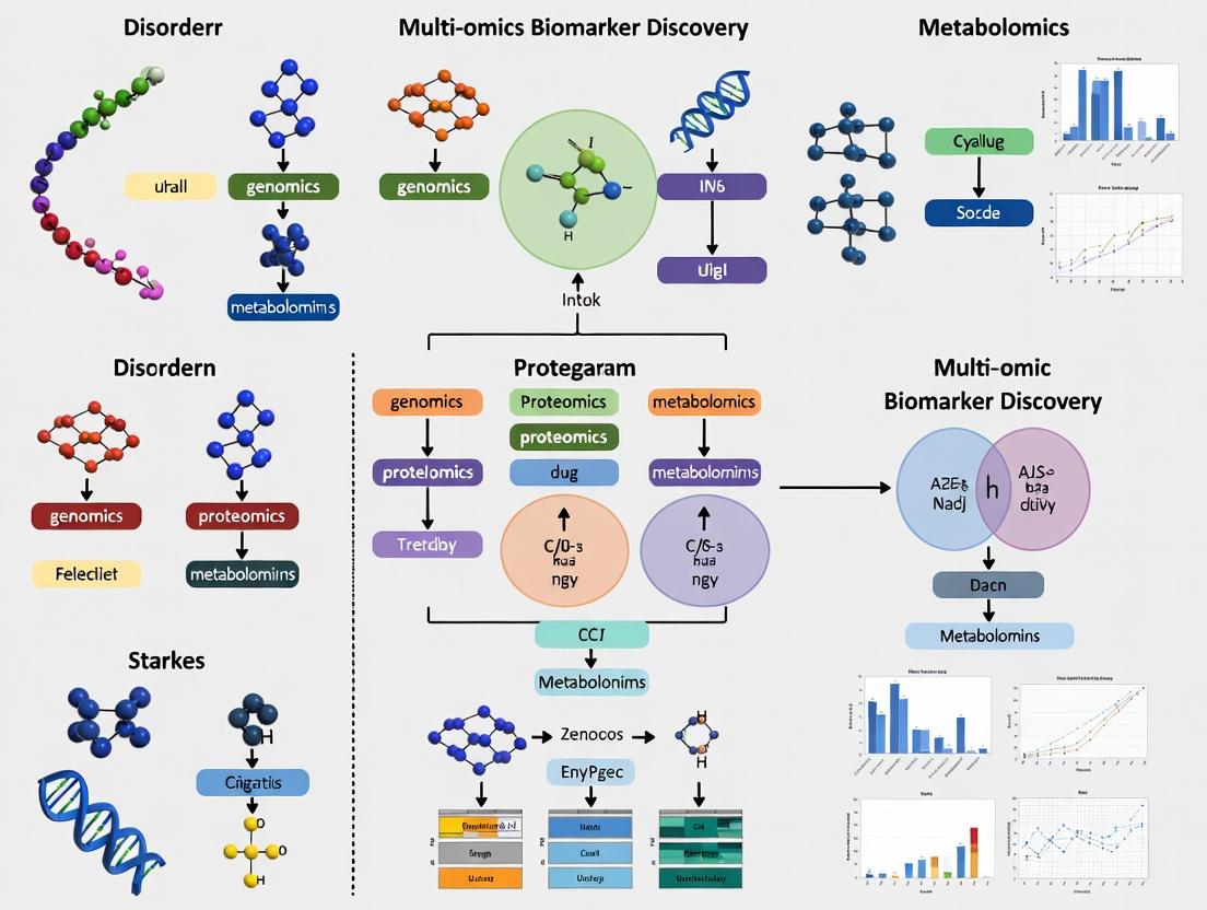
Visualizing Multi-Omics Workflows and Biological Networks
Title: Multi-Omics Biomarker Discovery Workflow
Title: Multi-Omics View of Insulin Signaling
The Scientist's Toolkit: Key Research Reagent Solutions
Table 3: Essential Reagents and Kits for Multi-Omics Metabolic Research
| Item & Example Vendor | Function in Multi-Omics Workflow |
|---|---|
| PAXgene Blood RNA Tubes (Qiagen) | Stabilizes intracellular RNA and gene expression profiles in whole blood at collection, enabling reliable transcriptomics from the same draw used for serum/plasma. |
| SeraPrep II Immunodepletion Columns (Thermo Fisher) | Remove high-abundance proteins (e.g., albumin, IgG) from plasma/serum to deepen coverage of the low-abundance proteome critical for biomarker discovery. |
| TMTpro 16-plex Isobaric Label Reagents (Thermo Fisher) | Enable multiplexed quantitative proteomics of up to 16 samples simultaneously, reducing batch effects and increasing throughput for cohort studies. |
| Biocrates MxP Quant 500 Kit (Biocrates) | A targeted metabolomics & lipidomics kit for absolute quantification of ~630 metabolites from a single sample, providing standardized data for integration. |
| RNeasy Plus Micro Kit (Qiagen) | RNA extraction from low-input or microdissected samples (e.g., LCM-captured tissue), ensuring compatibility with downstream spatial or single-cell transcriptomics. |
| Seahorse XFp FluxPak (Agilent) | Measures real-time cellular metabolic phenotypes (glycolysis, OXPHOS) in live cells, providing functional validation for omics-derived hypotheses. |
| Cell Signaling PathScan Intracellular Signaling Kits (CST) | Multiplex ELISA-based arrays for quantifying phosphorylation states of key signaling nodes (e.g., AKT, mTOR, AMPK), bridging proteomics to functional pathways. |
This whitepaper delineates the core omics layers—genomics, transcriptomics, proteomics, and metabolomics—within the integrative framework of multi-omics biomarker discovery for metabolic disorders. It provides a technical guide to methodologies, data integration, and translational applications, focusing on conditions like type 2 diabetes (T2D), non-alcoholic fatty liver disease (NAFLD), and cardiovascular metabolic syndromes.
Metabolic disorders are characterized by complex, systemic dysregulations that cannot be fully captured by a single analytical lens. A multi-omics approach, integrating vertical data from the genome to the metabolome, is essential for mapping the causal pathways from genetic predisposition to functional phenotypic outcomes. This integrated view accelerates the discovery of diagnostic, prognostic, and theranostic biomarkers, facilitating personalized therapeutic strategies.
Core Omics Layers: Technologies and Applications
Genomics
Objective: To identify heritable genetic variants (SNPs, indels, CNVs) associated with metabolic disease susceptibility and phenotypic variance.
- Key Technologies: Whole-genome sequencing (WGS), whole-exome sequencing (WES), genotyping arrays, epigenomic profiling (bisulfite-seq for DNA methylation).
- Role in Biomarker Discovery: Provides the foundational layer of predisposition. Genome-wide association studies (GWAS) have identified loci linked to insulin resistance, lipid metabolism, and adipogenesis.
Transcriptomics
Objective: To profile the complete set of RNA transcripts (coding and non-coding) to understand gene expression dynamics in metabolic tissues.
- Key Technologies: Bulk and single-cell RNA sequencing (scRNA-seq), spatial transcriptomics, microarrays.
- Role in Biomarker Discovery: Captures active regulatory states. Reveals tissue-specific (liver, adipose, muscle) expression signatures, alternative splicing events, and non-coding RNA networks in response to metabolic stress.
Proteomics
Objective: To identify, quantify, and characterize the full complement of proteins and their post-translational modifications (PTMs).
- Key Technologies: Liquid chromatography-tandem mass spectrometry (LC-MS/MS), affinity-based proteomics (SOMAscan, Olink), phosphoproteomics.
- Role in Biomarker Discovery: Directly assays functional effectors. Detects signaling pathway alterations, secreted biomarkers in biofluids, and drug target engagement.
Metabolomics
Objective: To comprehensively measure small-molecule metabolites (<1500 Da) representing substrates, intermediates, and end-products of metabolic pathways.
- Key Technologies: Mass spectrometry (MS) and nuclear magnetic resonance (NMR) spectroscopy, often coupled with gas or liquid chromatography (GC/LC).
- Role in Biomarker Discovery: Represents the ultimate functional readout of cellular physiology. Identifies dysregulated pathways (e.g., glycolysis, TCA cycle, fatty acid oxidation, bile acid metabolism) and signatures of mitochondrial dysfunction.
Table 1: Exemplary Multi-Omics Biomarker Discoveries in Metabolic Disorders
| Omics Layer | Technology Used | Key Biomarker Candidates | Associated Disorder | Effect Size / Fold-Change | Sample Type | Reference (Year) |
|---|---|---|---|---|---|---|
| Genomics | GWAS Meta-Analysis | GCKR rs1260326 variant | T2D, NAFLD | Odds Ratio: ~1.12 (T2D) | Blood DNA | Vujkovic et al., Nat. Genet. (2020) |
| Transcriptomics | scRNA-seq of Liver | Inflammatory macrophages (TREM2+CD9+) | NASH | 15-20x increase in NASH | Liver biopsy | Xiong et al., Cell Metab. (2019) |
| Proteomics | LC-MS/MS Plasma Profiling | FGF21, ApoE, PIIINP | NAFLD progression | AUC: 0.80-0.90 | Blood Plasma | Mann et al., Nat. Med. (2022) |
| Metabolomics | LC-MS Serum Profiling | Branched-Chain Amino Acids (BCAAs) | Insulin Resistance | 1.5-2.0x increase | Blood Serum | Newgard et al., Cell Metab. (2009) |
| Multi-Omics | Integrative Network | PNPLA3 genotype → lipid species → fibrosis | NAFLD | Combined AUC > 0.92 | Liver Tissue & Plasma | DiStefano et al., Hepatology (2022) |
Table 2: Comparison of Core Omics Methodologies
| Parameter | Genomics | Transcriptomics | Proteomics | Metabolomics |
|---|---|---|---|---|
| Analyte | DNA | RNA | Proteins & Peptides | Metabolites |
| Dynamic Range | Static (except epigenomics) | High (~10⁶) | Very High (>10¹⁰) | High (~10⁶) |
| Primary Technology | NGS | NGS | Mass Spectrometry | MS / NMR |
| Temporal Resolution | Low | Medium-High | Medium | Very High |
| Key Challenge | Functional interpretation | RNA-to-Protein correlation | Depth, PTM coverage | Annotation, ID |
| Sample Prep Time | Days | 1-2 Days | 1-3 Days | Hours-1 Day |
Detailed Experimental Protocols
Protocol: Single-Nuclei RNA Sequencing (snRNA-seq) from Frozen Human Adipose Tissue
Purpose: To profile cell-type-specific transcriptomic alterations in metabolic tissues without requiring fresh dissociation.
- Nuclei Isolation: Cryopreserved adipose tissue (~50-100 mg) is minced in lysis buffer (10 mM Tris-HCl, 10 mM NaCl, 3 mM MgCl2, 0.1% IGEPAL). Homogenize with a Dounce homogenizer (15-20 strokes). Filter through a 40-µm strainer and pellet nuclei at 500g for 5 min at 4°C.
- Fluorescence-Activated Nuclei Sorting (FANS): Resuspend nuclei in PBS with DAPI (1 µg/mL). Sort intact nuclei (DAPI-positive) using a 100-µm nozzle to remove debris and cytoplasmic RNA.
- Library Preparation: Use a commercial snRNA-seq kit (e.g., 10x Genomics Chromium Next GEM). Adjust nuclei concentration to ~1000/µL. Aim for 5,000-10,000 nuclei recovery. Perform GEM generation, RT, cDNA amplification, and library construction per manufacturer's instructions.
- Sequencing & Analysis: Sequence on an Illumina NovaSeq (PE150). Align reads (STARsolo or Cell Ranger). Downstream analysis includes clustering (Seurat), differential expression (MAST), and trajectory inference (Monocle3).
Protocol: Untargeted LC-MS Metabolomics of Human Plasma
Purpose: For global metabolite profiling to identify dysregulated pathways.
- Sample Extraction: Thaw plasma aliquots (50 µL) on ice. Add 200 µL of cold methanol:acetonitrile (1:1, v/v) with internal standards (e.g., isotopically labeled amino acids, fatty acids). Vortex vigorously, incubate at -20°C for 1 hr, centrifuge at 16,000g for 15 min at 4°C.
- Liquid Chromatography: Transfer supernatant for HILIC (polar metabolites) and C18 (lipids) separation on a UPLC system. Use a BEH Amide column (HILIC) and a C18 BEH column, with gradients from 5% to 95% organic phase (acetonitrile/water with 0.1% formic acid or ammonium acetate).
- Mass Spectrometry: Analyze using a high-resolution Q-TOF or Orbitrap mass spectrometer in both positive and negative electrospray ionization (ESI) modes. Data-Dependent Acquisition (DDA) mode: Full MS scan (m/z 50-1200) followed by MS/MS on top ions.
- Data Processing: Convert raw files (mxML/mzXML). Process with XCMS or MS-DIAL for peak picking, alignment, and annotation against public databases (HMDB, METLIN, LipidMaps). Statistical analysis (PCA, PLS-DA) performed in R.
Visualizations
Title: Integrated Multi-Omics Workflow for Biomarker Discovery
Title: Omics-Relevant Insulin Resistance Signaling Pathway
The Scientist's Toolkit: Research Reagent Solutions
Table 3: Essential Reagents and Kits for Multi-Omics Experiments
| Category | Product/Kit Name | Function in Workflow | Key Application |
|---|---|---|---|
| Nucleic Acid Isolation | Qiagen AllPrep DNA/RNA/miRNA | Simultaneous co-isolation of genomic DNA and total RNA from a single sample. | Preserves molecular relationships for genomics/transcriptomics integration. |
| Single-Cell Genomics | 10x Genomics Chromium Next GEM Single Cell 3' Kit | Creates barcoded GEMs for high-throughput 3' transcriptome profiling of thousands of single cells/nuclei. | scRNA-seq of liver, pancreatic islets, or adipose tissue. |
| Proteomics Sample Prep | PreOmics iST-BCT Kit | All-in-one workflow: lysis, reduction, alkylation, digestion in a single cartridge. Ideal for precious clinical samples. | Rapid, reproducible proteomic prep from tissue or cell pellets. |
| Metabolite Extraction | Biocrates AbsoluteIDQ p400 HR Kit | Targeted metabolomics kit for quantitative analysis of ~400 metabolites across multiple pathways. | High-throughput validation of biomarker panels in plasma/serum. |
| Multiplex Immunoassay | Olink Target 96 or 384 Panels | Proximity extension assay (PEA) technology for high-sensitivity, multiplex quantification of proteins in low sample volumes. | Discovery/validation of inflammatory or cardiometabolic plasma protein biomarkers. |
| Data Integration Software | Thermo Fisher Scientific Compound Discoverer / Omics Studio | Unified platform for processing and correlating MS-based proteomics and metabolomics data. | Integrative pathway analysis across omics layers. |
The pathophysiological overlap between Non-Alcoholic Fatty Liver Disease (NAFLD)/Non-Alcoholic Steatohepatitis (NASH), Type 2 Diabetes (T2D), Atherosclerosis, and Cardiometabolic Syndrome represents a paradigm of metabolic interconnectivity. Research within a multi-omics biomarker discovery framework is essential to deconvolute shared molecular pathways, identify predictive and diagnostic signatures, and facilitate the development of targeted, multi-disease therapeutic strategies.
Core Pathogenic Mechanisms and Interconnections
Insulin Resistance as the Central Driver
Chronic caloric excess and adipose tissue dysfunction lead to systemic insulin resistance, disrupting glucose and lipid homeostasis across liver, muscle, and vasculature.
Lipotoxicity and Ectopic Fat Deposition
Excessive free fatty acids (FFAs) spill over into non-adipose tissues, driving steatosis in the liver (NAFLD/NASH), beta-cell dysfunction in the pancreas (T2D), and foam cell formation in arterial walls (Atherosclerosis).
Chronic Low-Grade Inflammation
Activation of innate immune signaling (e.g., NLRP3 inflammasome) and pro-inflammatory cytokine release (TNF-α, IL-1β, IL-6) from adipose tissue and liver creates a systemic inflammatory milieu that exacerbates all target disorders.
Endothelial Dysfunction and Oxidative Stress
Metabolic insults impair nitric oxide bioavailability, increase reactive oxygen species (ROS), and promote a pro-thrombotic, pro-atherogenic vascular phenotype central to cardiometabolic syndrome.
Table 1: Core Biomarker Categories Across Targeted Metabolic Disorders
| Omics Layer | Biomarker Examples | Associated Disorder(s) | Typical Change vs. Healthy | Potential Clinical Utility |
|---|---|---|---|---|
| Genomics | PNPLA3 (rs738409), TM6SF2, GCKR variants | NAFLD/NASH, T2D | SNP presence increases risk | Risk stratification |
| Transcriptomics | SCD1, ChREBP, SREBP-1c (Lipogenesis genes) | NAFLD, T2D | Upregulated | Disease activity |
| Proteomics | FGF21, CK-18 (M30/M65 fragments), Adiponectin | NASH, T2D | FGF21↑, CK-18↑, Adiponectin↓ | Diagnostic (NASH), Prognostic |
| Metabolomics | Branched-Chain Amino Acids (BCAAs), Diacylglycerols (DAGs), Ceramides | T2D, Cardiometabolic Syndrome | Elevated | Predictive of insulin resistance |
| Lipidomics | Specific Phosphatidylcholine (PC) species, Free Cholesterol, Oxidized LDL | Atherosclerosis, NAFLD | PC↓, Free Cholesterol↑, oxLDL↑ | Cardiovascular risk assessment |
| Microbiomics | Firmicutes/Bacteroidetes ratio, Akkermansia muciniphila abundance | All | Ratio ↑, Akkermansia ↓ | Indicator of dysbiosis severity |
Table 2: Key Systemic Quantitative Parameters
| Parameter | NAFLD/NASH | T2D | Atherosclerosis | Cardiometabolic Syndrome |
|---|---|---|---|---|
| HOMA-IR | >2.5 | >2.5 | Often Elevated | Defining Feature (>2.5) |
| HbA1c (%) | May be normal/elevated | ≥6.5 | Correlates with risk | Often 5.7-6.4 (Prediabetes) |
| ALT (U/L) | >30 (M), >19 (F) | May be elevated | Normal | May be elevated |
| HDL-C (mg/dL) | Low | Low | Low | Low (<40 M, <50 F) |
| Triglycerides (mg/dL) | Elevated | Elevated | Elevated | Elevated (≥150) |
| hs-CRP (mg/L) | >2.0 | >2.0 | >2.0 | >2.0 |
| FIB-4 Score | >1.3 (concern) | N/A | N/A | N/A |
Experimental Protocols for Multi-Omics Biomarker Discovery
Integrated Serum Metabolomics and Lipidomics Profiling
Objective: Identify circulating metabolic signatures predictive of NAFLD progression to NASH or T2D onset. Sample Preparation: 100 µL of fasting serum + 400 µL ice-cold methanol:acetonitrile (1:1) containing internal standards. Vortex, centrifuge (14,000g, 15min, 4°C). Dry supernatant under nitrogen. LC-MS/MS Analysis: Re-constitute in 100 µL solvent. Use reversed-phase C18 column. Gradient: Water/ACN with 0.1% formic acid. Full scan (m/z 70-1050) in positive/negative ESI modes. Data Processing: Use XCMS, MS-DIAL for peak alignment, annotation via HMDB/LipidMaps databases. Statistical analysis (PLS-DA, ROC curves) in R.
Hepatic and Adipose Tissue Transcriptomics (RNA-Seq)
Objective: Map dysregulated pathways across tissues in a metabolic syndrome model. Tissue Lysis & RNA Extraction: Homogenize tissue in TRIzol. Chloroform phase separation. RNA precipitation with isopropanol. Wash with 75% ethanol. DNase I treatment. Library Prep & Sequencing: Poly-A selection. Fragment RNA. Synthesize cDNA. Ligate adapters. Amplify (12-15 cycles). Sequence on Illumina NovaSeq (150bp paired-end, 30M reads/sample). Bioinformatics: Align to reference genome (STAR). Quantify gene expression (featureCounts). Differential expression (DESeq2). Pathway enrichment (GSEA, KEGG).
Protocol for Validating Candidate Biomarkers via ELISA/MSD
Objective: Quantify candidate protein biomarkers (e.g., FGF21, CK-18) in a clinical cohort. Multiplex Immunoassay (MSD): Coat MSD plate with capture antibodies overnight. Block with Blocker A. Add serum samples/calibrators (1:2 dilution). Incubate 2h. Add detection antibody with SULFO-TAG. Read on MSD SECTOR Imager. Data Analysis: Generate standard curve (4-parameter logistic fit). Calculate sample concentrations. Correlate with clinical phenotypes.
Signaling Pathway and Workflow Visualizations
Multi-Omics Discovery Workflow
The Scientist's Toolkit: Key Research Reagent Solutions
Table 3: Essential Reagents for Metabolic Disorder Research
| Reagent/Category | Supplier Examples | Primary Function in Research |
|---|---|---|
| Human/Mouse Metabolic Syndrome Array Kits | Meso Scale Discovery (MSD), Luminex | Multiplex quantification of cytokines, adipokines, and metabolic hormones (e.g., leptin, adiponectin, resistin). |
| Phospho-/Total Antibody Panels (AKT, IRS1, AMPK) | Cell Signaling Technology, Abcam | Assess insulin signaling pathway activity in tissue lysates via Western blot or ELISA. |
| Activity Assay Kits (Caspase-1, NLRP3 Inflammasome) | Cayman Chemical, Abcam | Quantify inflammasome activation, a key inflammatory driver in NASH and T2D. |
| Lipid Extraction & Profiling Kits | Avanti Polar Lipids, Cayman Chemical | Standardized extraction and analysis of ceramides, DAGs, and other lipotoxic species. |
| Seahorse XFp/XFe96 Analyzer Reagents | Agilent Technologies | Measure real-time mitochondrial respiration (OCR) and glycolysis (ECAR) in live cells. |
| PNPLA3 Genotyping Assays | Thermo Fisher (TaqMan), IDT | Determine genetic risk variants for NAFLD progression in patient cohorts. |
| Recombinant Proteins (FGF21, GLP-1) | R&D Systems, PeproTech | Use as therapeutic controls or for in vitro mechanistic studies. |
| Stable Isotope-Labeled Metabolites (13C-Glucose, 15N-AA) | Cambridge Isotope Laboratories | Enable flux analysis to track metabolic pathway dynamics in vitro and in vivo. |
| 3D Spheroid/Organoid Culture Kits (Hepatocytes, Adipocytes) | STEMCELL Technologies, Corning | Model human tissue interactions and disease pathology in a more physiologically relevant system. |
| Next-Generation Sequencing Library Prep Kits | Illumina, NEB | Prepare high-quality libraries for transcriptomic, epigenomic, and genomic profiling. |
The research and clinical diagnosis of metabolic disorders, such as type 2 diabetes (T2D), non-alcoholic fatty liver disease (NAFLD), and cardiovascular disease (CVD), have long relied on the identification of single biomarkers. Classic examples include hemoglobin A1c (HbA1c) for glycemic control, LDL-cholesterol for cardiovascular risk, and alanine aminotransferase (ALT) for liver injury. While invaluable, this reductionist approach often fails to capture the complex, multifactorial etiology of these diseases, leading to incomplete risk stratification, heterogeneous treatment responses, and a limited understanding of underlying pathophysiology.
The advent of high-throughput technologies in genomics, transcriptomics, proteomics, and metabolomics (collectively, multi-omics) has catalyzed a paradigm shift from single-molecule biomarkers to network biology. This conceptual framework views disease not as a consequence of a single defective molecule but as a perturbation within a complex, interconnected biological system. This whitepaper provides an in-depth technical guide to this transition, detailing the core principles, methodologies, and applications of network-based biomarker discovery within the specific context of multi-omics research in metabolic disorders.
The Conceptual and Technical Evolution
The Limitations of Single Biomarkers
Single biomarkers are typically identified through univariate statistical analyses correlating the level of a single molecule with a disease state. Key limitations include:
- Low Specificity & Sensitivity: Many single biomarkers are influenced by unrelated physiological or pathological processes.
- Lack of Mechanistic Insight: They indicate association, not causation or pathway involvement.
- Inability to Handle Heterogeneity: They cannot explain the diverse subtypes (endotypes) within a common disease diagnosis (e.g., lean NAFLD vs. obese NAFLD).
The Rise of Network Biology
Network biology integrates multi-omics data to construct models of biological systems as graphs or networks, where nodes represent biomolecules (genes, proteins, metabolites) and edges represent interactions (physical binding, metabolic conversion, co-expression). This systems-level approach allows for:
- Identification of Network Biomarkers: Dysregulated modules or sub-networks that are more robust and disease-specific than individual molecules.
- Discovery of Master Regulators: Key hub nodes whose perturbation disproportionately impacts the entire network.
- Elucidation of Crosstalk: Understanding how pathways across different molecular layers (e.g., genomics and metabolomics) interact to drive disease.
Table 1: Comparison of Single Biomarker vs. Network Biology Paradigms
| Feature | Single Biomarker Paradigm | Network Biology Paradigm |
|---|---|---|
| Analytical Unit | Single molecule (e.g., Glucose) | Interacting modules of molecules |
| Primary Analysis | Univariate statistics | Multivariate, graph theory, machine learning |
| Data Type | Single-omics (e.g., clinical chemistry) | Integrated multi-omics |
| Disease Model | Linear cause-effect | System perturbation |
| Output | Diagnostic/Prognostic value | Mechanistic understanding, stratified subtypes |
| Example in T2D | HbA1c level | Inflammatory-metabolic network signature |
Core Methodologies for Network-Based Discovery
Multi-Omics Data Generation & Preprocessing
High-quality, integrated data is the foundation. Key technologies include:
- Genomics/Epigenomics: Whole-genome sequencing (WGS), methylation arrays (e.g., Illumina EPIC).
- Transcriptomics: RNA-Sequencing (RNA-Seq), single-cell RNA-Seq (scRNA-Seq).
- Proteomics: Mass spectrometry (LC-MS/MS), Olink or SomaScan platforms.
- Metabolomics/Lipidomics: LC-MS or GC-MS platforms.
Experimental Protocol: Plasma Metabolomics for NAFLD Study
- Sample Collection: Collect fasting plasma in EDTA tubes from NAFLD patients and healthy controls. Centrifuge at 2,000g for 10 min at 4°C. Aliquot and store at -80°C.
- Metabolite Extraction: Thaw aliquots on ice. Mix 50 µL plasma with 200 µL ice-cold methanol containing internal standards (e.g., isotopically labeled amino acids, lipids). Vortex vigorously for 30 sec.
- Protein Precipitation: Incubate at -20°C for 1 hour. Centrifuge at 15,000g for 15 min at 4°C.
- LC-MS Analysis: Transfer 150 µL of supernatant to an LC vial. Analyze using a reversed-phase C18 column coupled to a high-resolution tandem mass spectrometer (e.g., Q-Exactive HF). Use both positive and negative electrospray ionization modes.
- Data Processing: Use software (e.g., MS-DIAL, XCMS) for peak picking, alignment, and annotation against public databases (HMDB, METLIN). Normalize data using internal standards and quality control (QC) samples.
Data Integration and Network Construction
This is the critical technical step. Common approaches include:
- Correlation Networks: Constructed based on pairwise correlations (e.g., Spearman, Pearson) between molecular features across samples. A threshold (e.g., |r| > 0.7, p-adjusted < 0.01) defines an edge.
- Knowledge-Based Networks: Utilize prior interaction databases (e.g., STRING for protein-protein interactions, KEGG/Reactome for pathways, Recon3D for metabolism) as a scaffold, onto which omics data (e.g., gene expression) is mapped.
- Bayesian Networks: Probabilistic graphical models that can infer causal directionality from observational data.
- Multi-Layer Networks: Integrate different omics layers into a single network, where edges can exist within and between layers (e.g., a transcription factor node in the proteomic layer connected to its target gene in the transcriptomic layer).
Experimental Protocol: Weighted Gene Co-expression Network Analysis (WGCNA) for Transcriptomic Data
- Input Data: A normalized gene expression matrix (rows=genes, columns=samples).
- Similarity Matrix: Calculate a matrix of pairwise correlations (e.g., Pearson) between all genes across all samples.
- Adjacency Matrix: Transform the similarity matrix into an adjacency matrix using a soft-power threshold (β) to emphasize strong correlations: a_ij = |cor(x_i, x_j)|^β. β is chosen based on scale-free topology criterion.
- Topological Overlap Matrix (TOM): Calculate TOM to measure network interconnectedness, reducing noise from spurious correlations.
- Module Detection: Use hierarchical clustering with dynamic tree cutting to identify modules (clusters) of highly co-expressed genes.
- Module-Trait Association: Correlate the module eigengene (first principal component of the module) with clinical traits (e.g., HOMA-IR, liver fat percentage) to identify disease-relevant modules.
- Downstream Analysis: Perform enrichment analysis on key modules and identify hub genes (genes with high intramodular connectivity).
Network Analysis and Biomarker Identification
Key analytical tasks include:
- Topological Analysis: Identifying hub nodes (high degree/centrality), bottlenecks, and network communities/modules.
- Differential Network Analysis: Comparing network properties (e.g., connectivity, module composition) between disease and control states.
- Master Regulator Inference: Using algorithms like VIPER or MARINA to infer transcription factor/protein activity from network models and expression data.
- Machine Learning Integration: Using network-derived features (e.g., module activity scores) as input for classifiers (e.g., Random Forest, SVM) to build predictive models for disease subtyping or progression.
Application in Metabolic Disorders: A Case Study
Study Goal: To identify a network-based biomarker for stratifying NAFLD patients into progressive vs. non-progressive steatohepatitis (NASH).
Workflow & Results:
- Cohort: 150 biopsy-proven NAFLD patients (75 non-progressive steatosis, 75 progressive NASH with fibrosis).
- Multi-Omics Profiling: Plasma metabolomics/lipidomics and hepatic transcriptomics (RNA-Seq) were performed.
- Integration: WGCNA was applied to the transcriptomic data. A specific "mito-inflammatory" module was strongly associated with fibrosis stage (cor = 0.82, p = 1e-12). This module was enriched for oxidative phosphorylation and TNF-α signaling pathways.
- Multi-Layer Network: The hub genes from this module (e.g., ACSL1, CPT1A) were used as seeds to connect to altered plasma metabolites (e.g., specific long-chain acylcarnitines, bile acids) via a knowledge-based metabolic network (Recon3D).
- Network Biomarker: A small sub-network comprising ACSL1, CPT1A, and three plasma acylcarnitines was extracted. The composite score of this sub-network outperformed ALT alone in predicting progressive NASH (AUC: 0.94 vs. 0.67).
Table 2: Performance of Single vs. Network Biomarkers in NAFLD Progression Prediction
| Biomarker Type | Specific Example | AUC (95% CI) | Sensitivity | Specificity |
|---|---|---|---|---|
| Single Clinical | ALT (>40 U/L) | 0.67 (0.59-0.75) | 65% | 69% |
| Single Omics | Plasma C16:0 Acylcarnitine | 0.78 (0.71-0.85) | 75% | 73% |
| Network/Module | "Mito-inflammatory" Module Eigengene | 0.91 (0.86-0.96) | 88% | 87% |
| Multi-Layer Subnet | ACSL1-CPT1A-Acylcarnitines Score | 0.94 (0.90-0.98) | 92% | 89% |
Diagram 1: Multi-Omics Network Analysis Workflow
Diagram 2: Key Signaling Pathway in Metabolic Inflammation
The Scientist's Toolkit: Key Research Reagent Solutions
Table 3: Essential Reagents & Kits for Multi-Omics Network Studies
| Item Name | Vendor Examples | Function in Research |
|---|---|---|
| Total RNA Isolation Kit | Qiagen RNeasy, Zymo Research | High-yield, pure RNA extraction from tissues/cells for transcriptomics (RNA-Seq). |
| High-Sensitivity Proteomics Kit | Thermo Fisher TMTpro, Bruker timsTOF | Multiplexed protein labeling and preparation for deep-coverage LC-MS/MS proteomics. |
| Metabolite Extraction Solvent | Methanol/ACN/H2O (8:1:1), Biotage | Standardized solvent for reproducible quenching and extraction of polar metabolites. |
| Next-Gen Sequencing Library Prep Kit | Illumina TruSeq, NEB Next Ultra | Prepares RNA/DNA libraries for high-throughput sequencing on platforms like NovaSeq. |
| Pathway & Network Analysis Software | Cytoscape, Gephi, Ingenuity IPA (QIAGEN) | Visualizes and analyzes biological networks, performs enrichment analyses. |
| Single-Cell Dissociation Kit | Miltenyi Biotec, 10x Genomics | Gentle tissue dissociation into viable single-cell suspensions for scRNA-Seq. |
| Multiplex Immunoassay Panels | Olink Target 96, Meso Scale Discovery | Quantifies dozens of proteins simultaneously from low-volume biofluids. |
| Stable Isotope-Labeled Internal Standards | Cambridge Isotopes, Sigma-Aldrich | Enables absolute quantification and quality control in metabolomics/lipidomics. |
The shift from single biomarkers to network biology represents a fundamental evolution in our approach to understanding complex metabolic disorders. By integrating multi-omics data through a network lens, researchers can move beyond mere correlation to uncover causative drivers, define molecularly distinct disease endotypes, and identify robust, system-level biomarkers. This paradigm promises to accelerate the development of personalized diagnostic strategies and targeted therapies. The technical path forward requires continued advancement in bioinformatics tools for data integration, standardization of multi-omics protocols, and validation of network biomarkers in large, longitudinal cohorts. The future of biomarker discovery lies not in finding a single "needle in the haystack," but in comprehensively mapping the entire "haystack" to understand its structure and vulnerabilities.
In the pursuit of multi-omics biomarker discovery for metabolic disorders, public data repositories and consortium resources are indispensable. They provide the large-scale, integrated molecular and phenotypic datasets required to understand the complex interactions between genomics, transcriptomics, proteomics, and metabolomics. This whitepaper provides a technical guide to key resources, their application in metabolic research, and protocols for leveraging them.
The table below summarizes the core characteristics of leading repositories relevant to multi-omics metabolic disorder research.
| Repository/Resource | Primary Data Type(s) | Sample Size (Approx.) | Key Disease Relevance | Data Access Model |
|---|---|---|---|---|
| GTEx (Genotype-Tissue Expression) | Genotype, RNA-Seq (multi-tissue) | 17,000+ samples, 54 tissues | Tissue-specific gene regulation in diabetes, NAFLD | Controlled access (dbGaP) |
| UK Biobank | Genomics, Imaging, Clinical, Biomarkers | 500,000 participants | Type 2 diabetes, CVD, obesity | Application-based access |
| Metabolomics Workbench | Metabolomics (MS, NMR) | 1000+ studies | Metabolic dysregulation, inborn errors | Open / Controlled |
| TOPMed (NHLBI) | Whole Genome Seq, Phenotypes | 180,000+ participants | Cardiometabolic traits | Controlled access (dbGaP) |
| AMP-T2D (Accelerating Medicines Partnership) | Multi-omics (genomic, epigenomic, transcriptomic) | Varied by cohort | Type 2 Diabetes mechanisms | Application-based portal |
| Metabolights | Metabolomics | 1000+ studies | Broad metabolic phenotypes | Open access |
Detailed Methodologies for Key Analysis Workflows
Protocol 1: Integrating GTEx eQTLs with GWAS Loci for Metabolic Traits
Objective: Identify putative causal genes for metabolic disorder GWAS hits using expression Quantitative Trait Loci (eQTLs).
- Data Acquisition: Download latest GTEx v8 summary statistics for eQTLs (all tissues) from the GTEx Portal. Obtain GWAS summary statistics for a trait (e.g., fasting insulin) from public sources like GWAS Catalog or consortia.
- Locus Definition: For each independent GWAS lead SNP (p < 5e-8), define a genomic window (e.g., ±1 Mb).
- Colocalization Analysis: Use statistical tools (e.g.,
colocin R,fastENLOC) to compute posterior probabilities that the GWAS signal and tissue-specific eQTL signal share a single causal variant. Prioritize genes with P(Coloc) > 0.80. - Metabolic Tissue Focus: Filter results for key metabolic tissues: subcutaneous/adipose, liver, pancreas, skeletal muscle.
- Functional Validation Curation: Cross-reference prioritized genes with knockout mouse phenotypes (e.g., IMPC) and in vitro perturbation studies.
Protocol 2: Utilizing UK Biobank for Biomarker Discovery & Validation
Objective: Discover and validate circulating metabolomic biomarkers for incident Type 2 Diabetes (T2D).
- Cohort Definition (in UK Biobank): Using linked health records, define a nested case-control cohort: cases = participants diagnosed with T2D after baseline assessment; controls = matched participants remaining disease-free.
- Data Extraction: Apply for and extract NMR metabolomics data (Nightingale Health panel), baseline clinical chemistry, genetic data, and lifestyle factors.
- Statistical Modelling:
- Perform conditional logistic regression for each log-transformed metabolite, adjusting for age, sex, BMI, and genetic principal components.
- Apply multiple testing correction (FDR < 0.05).
- Conduct Mendelian Randomization (MR) using published genetic instruments for metabolites to assess potential causal relationships with T2D risk.
- Multi-omics Integration: Overlap MR results with GTEx eQTLs in relevant tissues to propose gene-metabolite-disease pathways.
Protocol 3: Targeted Metabolomic Query and Meta-Analysis via Metabolomics Workbench
Objective: Investigate the consistency of a specific metabolite (e.g., 2-hydroxybutyrate) across studies of insulin resistance.
- Advanced Query: On the Metabolomics Workbench, use the "Study Search" with keywords: "insulin resistance", "human plasma/serum", "MS".
- Study Filtering: Select studies with raw data (mzML, mzXML) and sample-level metadata available.
- Data Harmonization: Download peak area or concentration tables. Map metabolite identifiers to a common ontology (e.g., HMDB ID). Apply ComBat or similar batch correction for cross-study analysis.
- Meta-Analysis: For the metabolite of interest, calculate standardized mean difference (SMD) between insulin-resistant vs. control groups across studies using a random-effects model (e.g.,
metaforpackage in R). - Pathway Enrichment: Combine all dysregulated metabolites from the meta-analysis into a pathway over-representation analysis (using MetaboAnalyst) to identify disturbed pathways (e.g., branched-chain amino acid catabolism).
Visualizing the Integrated Multi-Omics Workflow
(Diagram 1: Multi-Omics Integration Workflow for Biomarker Discovery)
The Scientist's Toolkit: Essential Research Reagent Solutions
| Item / Resource | Function in Multi-Omics Metabolic Research | Example Vendor/Platform |
|---|---|---|
| NMR Metabolomics Panels | High-throughput, quantitative profiling of ~250 circulating metabolites for cohort phenotyping. | Nightingale Health, Bruker IVDr |
| LC-MS/MS Assay Kits | Targeted, sensitive quantification of specific metabolite classes (e.g., bile acids, eicosanoids). | Biocrates, Cayman Chemical |
| Proximity Extension Assay (PEA) | High-multiplex protein quantification from minimal sample volume for proteomic integration. | Olink Explore, Somalogic SomaScan |
| scRNA-Seq Kits | Single-cell transcriptomic profiling of pancreatic islets, liver, or adipose tissue. | 10x Genomics Chromium, Parse Biosciences |
| CRISPR Screening Libraries | Functional genomics validation of candidate genes in metabolic cell models. | Dharmacon, Horizon Discovery |
| Stable Isotope Tracers (e.g., 13C-Glucose) | For flux analysis experiments to trace metabolic pathways in vitro or in vivo. | Cambridge Isotope Laboratories |
| Bioinformatics Pipelines (Nextflow/Snakemake) | Reproducible processing of raw multi-omics data (FASTQ, mzML). | nf-core, custom workflows |
| Colocalization & MR Software | Statistical analysis for causal inference from genetic and molecular QTL data. | coloc, TwoSampleMR, MendelianRandomization (R packages) |
From Samples to Signatures: Cutting-Edge Methodologies in Multi-Omics Workflows
Within multi-omics biomarker discovery for metabolic disorders, the integrity of the research thesis is fundamentally determined by upstream study design. Robust cohort selection, precise phenotyping, and strategic multi-layer sampling are critical to generating biologically relevant, statistically powered, and reproducible omics data. This guide details technical considerations for these foundational elements.
Cohort Selection for Metabolic Phenotyping
Cohort selection must balance biological relevance with practical constraints. Key quantitative considerations are summarized below.
Table 1: Quantitative Considerations for Cohort Selection in Metabolic Disorders Research
| Design Parameter | Target Range/Consideration | Rationale |
|---|---|---|
| Sample Size (Discovery) | 500 - 2000 participants | Provides 80-90% power to detect modest effect sizes (e.g., fold change >1.5) in untargeted omics, accounting for multiple testing. |
| Case:Control Ratio | 1:1 to 1:2 | Optimal for statistical power in most analyses. 1:2 can enhance power for rare phenotypes. |
| Age Stratification | Decade-based bins (e.g., 40-49, 50-59) | Controls for age-related metabolic drift (e.g., declining insulin sensitivity). |
| BMI Matching | ± 2.0 kg/m² between groups | Critical to isolate metabolic dysfunction independent of adiposity. |
| Fasting Duration | 10-12 hours minimum | Standardizes metabolomic and lipidomic measurements. |
| Ethnic Heterogeneity | ≥3 distinct populations (if feasible) | Enhances generalizability of discovered biomarkers. |
| Confounder Data Capture | Medication (30+ classes), Diet (FFQ), Activity (IPAQ) | Essential for covariate adjustment in models. |
Protocol 1: Deep Metabolic Phenotyping Protocol
- Pre-visit: Participants maintain usual diet/activity for 3 days, fast 12 hours overnight, abstain from alcohol/strenuous exercise for 24h.
- Day of Visit:
- Baseline Samples: Phlebotomy for plasma, serum, PBMCs. Aliquot and snap-freeze in liquid N₂.
- Oral Glucose Tolerance Test (OGTT): Administer 75g glucose. Collect blood at 0, 30, 60, 90, 120 min for insulin, glucose, metabolomics.
- Anthropometrics: DEXA scan (lean/fat mass), waist/hip circumference, BP.
- Biomarker Panel: Clinical chemistry (HbA1c, lipids, liver enzymes), hs-CRP, adiponectin, leptin.
- Storage: All biospecimens at -80°C within 2 hours of collection.
High-Resolution Phenotyping Strategies
Phenotyping extends beyond clinical diagnostics to capture continuous metabolic gradients.
Table 2: Tiered Phenotyping Approach for Metabolic Syndrome
| Tier | Phenotype Level | Assessment Tools | Omics Integration |
|---|---|---|---|
| Tier 1: Clinical | Diabetes, NAFLD, CVD status | EHR, ICD codes, medication lists | Stratification variable |
| Tier 2: Quantitative | HOMA-IR, Matsuda Index, liver fat % | OGTT, MRI-PDFF, NMR lipidomics | Continuous variable for correlation |
| Tier 3: Dynamic | Metabolic flexibility, β-cell function | Euglycemic-hyperinsulinemic clamp, mixed-meal test | Paired pre-/post-perturbation omics |
| Tier 4: Molecular | Oxidative stress, inflammation | 8-iso-PGF2α, oxLDL, cytokine multiplex assays | Covariates or integration targets |
Multi-Layer Sampling for Multi-Omics
Coordinated sampling across biological layers is non-negotiable for integrated analysis.
Protocol 2: Multi-Layer Biospecimen Collection from a Single Blood Draw
- Blood Collection: Draw into vacutainers: EDTA (plasma), serum separator, PAXgene (RNA), sodium heparin (PBMCs).
- Processing (within 30 min):
- Plasma: Centrifuge 2000g, 10min, 4°C. Aliquot for metabolomics (50µL), proteomics (100µL), biobank.
- Serum: Allow clot, centrifuge 2000g, 10min. Aliquot for clinical chemistry, cytokine panels.
- PBMCs: Isolate via Ficoll density gradient. Aliquot for DNA extraction (genomics), chromatin assays (ATAC-seq), and culture.
- PAXgene RNA: Invert 10x, store at -20°C then -80°C.
- Stool & Urine: Collect concurrent stool (for gut microbiome) and first-morning urine (for metabolomics) using standardized kits.
Integrated Experimental Workflow
Title: Multi-Omics Biomarker Discovery Workflow
Key Signaling Pathways in Metabolic Dysfunction
Title: Insulin & Inflammatory Signaling Cross-Talk
The Scientist's Toolkit: Essential Research Reagent Solutions
Table 3: Key Reagents for Metabolic Multi-Omics Studies
| Item | Function & Application | Key Consideration |
|---|---|---|
| PAXgene Blood RNA Tubes | Stabilizes intracellular RNA at collection for transcriptomics. | Eliminates need for immediate processing; critical for field studies. |
| Stabilized EDTA Plasma Tubes | Contains protease/phosphatase inhibitors for proteomics/phosphoproteomics. | Preserves labile post-translational modifications. |
| Ficoll-Paque PREMIUM | Density gradient medium for high-yield, viable PBMC isolation. | Consistency is vital for downstream cell-based assays (e.g., Seahorse). |
| C18 SPE Plates | Solid-phase extraction for LC-MS metabolomics/lipidomics sample prep. | Removes salts and proteins; enriches non-polar metabolites. |
| Olink Target 96/Explore | Proximity extension assay for high-sensitivity multiplex proteomics (1µL plasma). | Detects low-abundance cytokines/adipokines without immunoaffinity depletion. |
| Macronutrient-Standardized Meal | For dynamic postprandial metabolic challenge tests (e.g., mixed-meal test). | Enables study of metabolic flexibility; must be identical across cohort. |
| D₂O (Deuterium Oxide) | Tracer for in vivo measurement of hepatic de novo lipogenesis (DNL) via NMR/GC-MS. | Safe, non-radioactive method to quantify lipid turnover. |
| Seahorse XFp Flux Pak | Cartridge and media for measuring mitochondrial respiration/glycolysis in PBMCs/ adipocytes. | Functional phenotyping of cellular metabolism. |
In the pursuit of robust multi-omics biomarker discovery for metabolic disorders (e.g., type 2 diabetes, NAFLD, obesity), the integration of Next-Generation Sequencing (NGS), Mass Spectrometry (MS), and High-Throughput Screening (HTS) platforms forms the technological cornerstone. This whitepaper details the core methodologies, protocols, and data integration strategies essential for generating actionable biological insights.
Next-Generation Sequencing (NGS) in Transcriptomics and Epigenomics
NGS enables comprehensive profiling of the genome, transcriptome, and epigenome, crucial for understanding genetic predispositions and regulatory mechanisms in metabolic diseases.
Core Protocols
Protocol 1: Single-Cell RNA Sequencing (scRNA-seq) for Pancreatic Islet or Adipose Tissue Analysis
- Objective: To profile cell-type-specific transcriptional alterations in metabolic tissues.
- Workflow:
- Tissue Dissociation: Fresh tissue is dissociated into a single-cell suspension using collagenase IV/DNase I.
- Viability & Concentration: Assessed via trypan blue and counted.
- Library Preparation: Using 10x Genomics Chromium Controller for droplet-based partitioning and barcoding (v3.1 chemistry).
- Sequencing: On Illumina NovaSeq 6000, targeting 50,000 reads per cell.
- Data Analysis: Cell Ranger pipeline for demultiplexing, alignment (to GRCh38), and UMI counting. Downstream analysis with Seurat for clustering (graph-based), differential expression, and trajectory inference.
Protocol 2: Whole-Genome Bisulfite Sequencing (WGBS) for DNA Methylation Profiling
- Objective: To map genome-wide methylation patterns in patient-derived samples.
- Workflow:
- DNA Extraction & Fragmentation: Sonicate high-quality DNA to 200-300bp.
- Bisulfite Conversion: Use EZ DNA Methylation-Lightning Kit (Zymo Research) for >99% conversion efficiency.
- Library Prep & Amplification: Repair, A-tailing, adaptor ligation, and PCR amplification with methylation-aware polymerase.
- Sequencing: Paired-end 150bp sequencing on Illumina platform.
- Data Analysis: Trim Galore for adapter/bias trimming, Bismark for alignment, and MethylKit for differential methylation region (DMR) calling.
Table 1: Key NGS Performance Metrics for Metabolic Disorder Studies
| Application | Recommended Platform | Typical Read Depth/ Coverage | Key QC Metric | Primary Output |
|---|---|---|---|---|
| scRNA-seq | 10x Genomics + Illumina NovaSeq | 50,000 reads/cell | Median genes/cell > 1,000; Mitochondrial reads < 20% | Digital gene expression matrix |
| WGBS | Illumina NovaSeq 6000 | 30X genome coverage | Bisulfite conversion rate > 99% | Methylation ratio per CpG site |
| Whole Exome Seq | Illumina HiSeq 4000 | 100X mean coverage | >95% target bases covered at 20X | Variant Call Format (VCF) file |
Diagram 1: Generic NGS Data Generation Workflow
Mass Spectrometry (MS) for Proteomics and Metabolomics
MS provides precise quantification of proteins, metabolites, and lipids, offering a direct readout of functional states in metabolic pathways.
Core Protocols
Protocol 3: Data-Independent Acquisition (DIA) Proteomics for Plasma/Serum Profiling
- Objective: To identify and quantify thousands of proteins for biomarker candidacy.
- Workflow:
- Sample Prep: Deplete top 14 high-abundance proteins. Reduce, alkylate, and digest with trypsin (1:50 w/w, 37°C, overnight).
- Chromatography: Online fractionation using a 90-min gradient on a C18 column (Thermo Easy-Spray, 75µm x 25cm).
- MS Acquisition: On a Thermo Exploris 480 with FAIMS Pro interface. DIA method: 40x 4 m/z isolation windows covering 400-1000 m/z, 3 FAIMS CVs (-45V, -60V, -75V).
- Data Analysis: Spectral library generation from gas-phase fractionated runs. DIA data processed with Spectronaut (Biognosys) or DIA-NN.
Protocol 4: Untargeted Lipidomics via LC-MS/MS
- Objective: Global profiling of lipid species from tissue homogenates (e.g., liver, muscle).
- Workflow:
- Lipid Extraction: Methyl-tert-butyl ether (MTBE) method. Homogenize tissue in MeOH/MTBE, vortex, centrifuge, collect organic layer.
- LC-MS: Reverse-phase chromatography (C8 column) coupled to Q-Exactive HF-X in positive/negative switching mode.
- Acquisition: Full MS scan (m/z 200-2000) at 120k resolution, followed by top-10 data-dependent MS/MS at 15k resolution.
- Data Analysis: Lipid identification/alignment with MS-DIAL. Quantification via peak area.
Table 2: Key MS Platform Performance Metrics
| Omics Type | MS Platform | Resolution | Mass Accuracy | Dynamic Range | Identifications per Run |
|---|---|---|---|---|---|
| DIA Proteomics | Thermo Exploris 480 + FAIMS | 120,000 @ m/z 200 | < 3 ppm | > 5 orders | 6,000-8,000 proteins |
| Untargeted Lipidomics | Q-Exactive HF-X | 240,000 @ m/z 200 | < 1 ppm | > 4 orders | 1,500-2,000 lipid species |
| Targeted Metabolomics | SCIEX 6500+ QTRAP | Unit (Q1/Q3) | NA | > 5 orders | 200-300 metabolites |
Diagram 2: MS Profiling in Insulin Signaling Pathway
High-Throughput Screening (HTS) Platforms
HTS enables functional validation of omics-derived targets in cellular models of metabolic dysfunction.
Core Protocol
Protocol 5: High-Content Screening (HCS) for Lipid Droplet Phenotypes
- Objective: To screen siRNA or compound libraries for modulators of hepatic lipid accumulation.
- Workflow:
- Cell Model: Seed HepG2 or primary hepatocytes in 384-well imaging plates.
- Treatment: Transfer siRNA (library) or compounds via acoustic dispensing. Incubate 72h.
- Staining: Fix, permeabilize, stain nuclei (Hoechst), neutral lipids (BODIPY 493/503), and actin (Phalloidin-647).
- Imaging & Analysis: Image on PerkinElmer Operetta CLS. Automated analysis using Harmony software: segment nuclei/cells, quantify lipid droplet count, size, and total intensity per cell.
Table 3: HTS/HCS Platform Specifications
| Parameter | siRNA Screening | Small Molecule Screening | Phenotypic Readout |
|---|---|---|---|
| Plate Format | 384-well | 384 or 1536-well | High-content imaging |
| Library Size | 5,000 genes (kinome) | 100,000 compounds | N/A |
| Replicates | n=3 technical, n=2 biological | n=2 technical | N/A |
| Key QC Metric | Z'-factor > 0.5 | Signal-to-Noise > 10 | CV of controls < 20% |
| Primary Data | Lipid droplet area/cell | Viability % & lipid content | Multiparametric cell data |
The Scientist's Toolkit: Research Reagent Solutions
Table 4: Essential Reagents and Kits for Multi-Omics Experiments
| Item | Vendor (Example) | Function | Key Application |
|---|---|---|---|
| Chromium Next GEM Single Cell 3' Kit v3.1 | 10x Genomics | Partitioning, barcoding, and RT for scRNA-seq | Transcriptomics |
| Nextera DNA Flex Library Prep Kit | Illumina | Fast, integrated library preparation for WGS/WES | Genomics |
| EpiNext High-Sensitivity Bisulfite Kit | Epicentre | Efficient bisulfite conversion for low-input samples | Epigenomics (WGBS) |
| S-Trap Micro Spin Columns | Protifi | Efficient protein digestion and clean-up for proteomics | Bottom-up Proteomics |
| Piero BODIPY 493/503 | Thermo Fisher | Selective neutral lipid staining for fixed cells | Lipid droplet HCS |
| MTBE, LC-MS Grade | Sigma-Aldrich | Organic solvent for comprehensive lipid extraction | Lipidomics |
| Seahorse XFp FluxPak | Agilent | Cartridge and media for real-time metabolic analysis | Cellular Bioenergetics HTS |
| Magnetic Bead-based Depletion Kit (Human 14) | Thermo Fisher | Removal of high-abundance plasma proteins | Plasma Proteomics |
Diagram 3: Multi-Omics Integration for Biomarker Discovery
Data Preprocessing & Normalization Strategies for Each Omics Layer
Thesis Context: This guide details essential preprocessing and normalization methodologies for individual omics layers, framed within a multi-omics integration pipeline for biomarker discovery in metabolic disorders (e.g., Type 2 Diabetes, NAFLD). Consistent data refinement at each layer is critical for robust downstream integration and biological interpretation.
Genomics (DNA-seq, SNP arrays)
Objective: To accurately identify genetic variants (SNPs, indels) and correct for technical artifacts. Key Challenges: Batch effects, GC bias, library size differences.
Experimental Protocol (Typical GATK Best Practices Workflow):
- Raw Data (FASTQ): Adapter trimming using Trimmomatic or Cutadapt.
- Alignment: Map reads to reference genome (e.g., GRCh38) using BWA-MEM or STAR.
- Post-Alignment Processing:
- Duplicate Marking: Identify PCR duplicates using Picard Tools
MarkDuplicates. - Base Quality Score Recalibration (BQSR): Correct systematic errors in base quality scores using GATK
BaseRecalibratorandApplyBQSR. - Variant Calling: Call variants using GATK
HaplotypeCallerfor germline variants.
- Duplicate Marking: Identify PCR duplicates using Picard Tools
- Variant Normalization: Left-align and trim variants using
bcftools normto ensure consistent representation.
Normalization for Downstream Analysis: For SNP array data used in GWAS, common steps include:
- Genotype Quality Control: Filter samples based on call rate (<98%), heterozygosity outliers, and gender mismatch. Filter variants based on call rate (<95%), Hardy-Weinberg equilibrium p-value (e.g., <1e-6), and minor allele frequency (MAF <1% for rare variant studies).
- Batch/Chip Effect Correction: Use methods like Principal Component Analysis (PCA) to identify and adjust for batch-associated population stratification, often implemented in PLINK.
Transcriptomics (RNA-seq, Microarrays)
Objective: To obtain accurate gene expression estimates comparable across samples. Key Challenges: Library size, gene length, compositional bias, batch effects.
Experimental Protocol (RNA-seq Quantification):
- Raw Read Processing: Adapter trimming, quality filtering (FastQC, Trimmomatic).
- Alignment & Quantification:
- Pseudo-alignment: Use
kallistoorSalmonfor fast transcript-level quantification against a reference transcriptome. - Alignment-based: Align reads with HISAT2 or STAR to a reference genome, then count reads per gene using
featureCounts.
- Pseudo-alignment: Use
- Normalization: Choose method based on data characteristics and assumption validity.
Table 1: Common RNA-Seq Normalization Methods
| Method | Formula/Principle | Use Case | Key Consideration for Metabolic Disorders |
|---|---|---|---|
| Counts Per Million (CPM) | Count_gene / Total_Counts * 1e6 |
Within-sample comparison. Not for between-sample. | Simple but fails to correct for library composition. |
| Transcripts Per Million (TPM) | (Reads_gene / Gene_length_kb) / (Σ(Reads_gene / Gene_length_kb)) * 1e6 |
Within-sample, comparable across samples. | Accounts for gene length; preferred for expression level comparison. |
| DESeq2's Median of Ratios | Counts scaled by sample-specific size factors (median ratio of counts to geometric mean per gene). | Between-sample comparison for differential expression. | Robust to composition bias; assumes few genes are differentially expressed. |
| EdgeR's TMM | Trimmed Mean of M-values. Scales libraries based on a subset of stable genes. | Between-sample comparison for differential expression. | Similar assumptions to DESeq2; performs well in most cases. |
| Upper Quartile (UQ) | Count_gene / 75th_percentile_count * 1e6 |
Alternative when many genes are zero or lowly expressed. | Less sensitive to highly expressed genes, but may be unstable. |
Pathway Analysis Workflow: Differential expression results are typically fed into tools like GSEA or Ingenuity Pathway Analysis to identify perturbed pathways in metabolic tissues.
Figure 1: Core RNA-Seq Preprocessing & Analysis Pipeline
Proteomics (LC-MS/MS)
Objective: To transform raw spectral data into quantitative protein abundances. Key Challenges: Missing values, dynamic range, sample loading, batch effects.
Experimental Protocol (Label-Free Quantification - LFQ):
- Raw File Conversion: Convert .raw files to open formats (e.g., .mzML) using MSConvert.
- Feature Detection & Quantification: Use software like MaxQuant or DIA-NN.
- MaxQuant Workflow: Perform peptide identification via Andromeda search against a protein database, match-between-runs (MBR) to transfer IDs, and compute intensity-based absolute quantification (iBAQ) or LFQ intensities.
- Post-Processing: Filter for contaminants, reverse hits, and proteins only identified by site.
Normalization: Applied to the peptide or protein intensity matrix.
- Median/MAD Normalization: Center log2 intensities by subtracting the median and scaling by the median absolute deviation (MAD).
- Quantile Normalization: Forces intensity distributions across samples to be identical. Can be too aggressive if global changes are expected.
- VSN (Variance Stabilizing Normalization): Models and removes the mean-variance dependence in MS data. Often implemented in
limma. - ComBat: Removes known batch effects using an empirical Bayes framework.
Metabolomics (LC/GC-MS, NMR)
Objective: To correct for systematic variation in metabolite peak intensities or areas. Key Challenges: Peak misalignment, instrumental drift, batch effects, high missingness.
Experimental Protocol (Untargeted LC-MS):
- Raw Data Processing: Use XCMS, MS-DIAL, or MZmine for:
- Peak Picking: Identify chromatographic peaks.
- Peak Alignment: Align peaks across samples based on m/z and retention time.
- Peak Grouping & Gap Filling: Group features and fill in missing peaks.
- Feature Table Generation: Output is a matrix of samples (rows) × metabolite features (columns) with intensity values.
Normalization & Correction Strategy:
- Internal Standard (IS) Normalization: Divide intensity of each feature by the intensity of a spiked-in IS (e.g., stable isotope-labeled compound) to correct for sample preparation variation.
- Probabilistic Quotient Normalization (PQN): Assumes most metabolite concentrations are constant. Normalizes based on the median fold change of all metabolites. Excellent for urine metabolomics.
- Batch Effect Correction: Use Quality Control-based Robust LOESS Signal Correction (QCRLSC) or batch-specific IS.
- Drift Correction: Use QC samples injected at regular intervals to model and correct for temporal intensity drift.
Table 2: Key Normalization Methods Across Omics Layers
| Omics Layer | Primary Normalization Goal | Common Methods | Tool/Software Examples |
|---|---|---|---|
| Genomics (SNP) | Remove population stratification & batch effects. | PCA-based correction, Genomic Control. | PLINK, GCTA, SAIGE |
| Transcriptomics | Make expression counts comparable across samples. | TMM, Median of Ratios, TPM, Upper Quartile. | edgeR, DESeq2, kallisto |
| Proteomics | Correct systematic bias in protein intensities. | Median Normalization, VSN, LFQ, ComBat. | MaxQuant, limma, Perseus |
| Metabolomics | Correct for dilution, drift, & preparation variation. | PQN, Internal Std. Normalization, QCRLSC. | XCMS, MetaboAnalyst, in-house R scripts |
The Scientist's Toolkit: Research Reagent Solutions
Table 3: Essential Materials for Multi-Omics Preprocessing
| Item | Function in Preprocessing Context | Example Product/Brand |
|---|---|---|
| SPRIselect Beads | Size-selective magnetic bead-based cleanup for NGS libraries (cDNA, amplicons). Adjustable bead-to-sample ratio for size selection. | Beckman Coulter SPRIselect |
| KAPA HyperPrep Kit | Library preparation for RNA/DNA sequencing. Provides reagents for end-repair, A-tailing, adapter ligation, and PCR amplification. | Roche KAPA HyperPrep |
| Pierce Quantitative Colorimetric Peptide Assay | Accurately measure peptide concentration before MS analysis to enable equal sample loading, a critical pre-normalization step. | Thermo Fisher Scientific 23275 |
| Stable Isotope-Labeled Internal Standards (SIL IS) | Spiked into metabolomics/proteomics samples pre-extraction to correct for losses during preparation and ionization variability in MS. | Cambridge Isotope Laboratories (CIL), Sigma-Aldrich MSK-AAPE-1 |
| Pooled Quality Control (QC) Sample | Aliquoted from a pool of all study samples. Run repeatedly throughout the MS batch to monitor stability, correct drift, and filter unreliable features. | N/A (Study-specific) |
| Universal Human Reference RNA (UHRR) | Control for transcriptomics platform performance and batch alignment in microarrays or RNA-seq. | Agilent Technologies 740000 |
| NAD/NADH & NADP/NADPH Assay Kits | Critical for validating metabolic pathway perturbations suggested by omics data in metabolic disorder research (e.g., redox state). | Abcam ab65313, Colorimetric assays |
| BCA Protein Assay Kit | Standard method for determining total protein concentration for sample loading normalization in proteomics (e.g., before western blot or MS). | Thermo Fisher Scientific 23225 |
Conclusion: Effective preprocessing and layer-specific normalization are non-negotiable first steps in multi-omics biomarker discovery for metabolic disorders. The choice of method must be guided by the technology's inherent biases, the study design, and the biological question. Consistent application of these protocols ensures data quality, enabling meaningful integration across omics layers to uncover robust, systems-level insights into disease mechanisms.
In the pursuit of robust biomarkers for metabolic disorders such as type 2 diabetes, NAFLD, and cardiovascular disease, multi-omics integration has become indispensable. This whitepaper examines three principal computational paradigms for integrating genomics, transcriptomics, proteomics, and metabolomics data within a multi-omics biomarker discovery pipeline. The selection of an integration approach directly impacts the biological interpretability, statistical power, and translational potential of discovered biomarkers.
Core Integration Paradigms
Concatenation-Based (Early) Integration
This method merges multiple omics datasets into a single, high-dimensional matrix prior to analysis.
- Principle: All datasets (e.g., SNP arrays, RNA-seq counts, LC-MS peaks) are combined horizontally (sample-wise) into a unified feature space.
- Typical Use: Input for machine learning models (e.g., PLS-DA, Random Forest, Deep Neural Networks) predicting disease state or clinical outcomes.
- Key Challenge: The "curse of dimensionality" (p >> n) and significant technical noise variation between platforms.
Table 1: Quantitative Comparison of Core Integration Approaches
| Aspect | Concatenation-Based | Multi-Stage | Model-Based |
|---|---|---|---|
| Data Structure | Single combined matrix (n x ∑pᵢ) | Multiple matrices analyzed sequentially | Joint model on multiple matrices |
| Dimensionality | Very High (∑pᵢ features) | Moderate (analyzed per dataset) | Controlled via latent variables |
| Handles Noise | Poor (requires extensive pre-processing) | Good (per-dataset normalization) | Very Good (explicit noise models) |
| Interpretability | Low (black-box models) | High (clear per-omics contributions) | Moderate (via factor loadings) |
| Example Algorithms | SVM, Random Forest, MLP | MOFA, iCluster, Pattern Discovery | JIVE, SMIDA, BNMM, mixOmics |
| Typical Runtime | Fast to Moderate | Moderate | Slow (MCMC, iterative) |
| Suitability for Biomarkers | Predictive Classifiers | Mechanistic & Candidate Discovery | Holistic Pathway & Subtype Discovery |
Multi-Stage (Intermediate) Integration
Analyses are performed on each omics dataset separately, with results (statistics, selected features) integrated in a subsequent stage.
- Principle: Independent analyses (e.g., differential expression) generate p-values or effect sizes for each omics layer. These results are combined statistically or via priority rules.
- Typical Use: Identification of consensus molecular signatures (e.g., genes with both SNP association and differential expression) or pathway enrichment across omics layers.
Model-Based (Late) Integration
Joint statistical models are constructed to infer latent structures that explain covariation across all omics datasets simultaneously.
- Principle: Models like Factor Analysis, Bayesian Networks, or Multivariate Regression decompose the data into shared and dataset-specific components.
- Typical Use: Discovery of latent patient subtypes driven by multi-omics patterns and identification of core regulatory networks in metabolic dysfunction.
Experimental Protocols for Benchmarking
Protocol 1: Benchmarking Pipeline for Metabolic Disorder Data
- Data Acquisition: Obtain matched multi-omics datasets (e.g., from a cohort like Human Liver Atlas or in-house NAFLD study). Minimum: Whole blood or tissue-derived DNA (Genotyping Array), RNA (RNA-seq), Serum (LC-MS Metabolomics).
- Pre-processing & Normalization:
- Genomics: Quality control, imputation, normalization for population stratification.
- Transcriptomics: Alignment (STAR), quantification (featureCounts), TPM normalization, batch correction (ComBat).
- Metabolomics: Peak alignment, missing value imputation (k-NN), probabilistic quotient normalization, log-transformation.
- Concatenation Workflow: Scale each dataset (mean=0, variance=1). Horizontally merge by sample ID. Apply dimensionality reduction (PCA or UMAP). Train a Random Forest/ElasticNet classifier on 70% training set to predict disease vs. control. Validate on 30% hold-out set. Report AUC, precision, recall.
- Multi-Stage Workflow: Perform per-dataset differential analysis (limma for RNA, linear model for metabolites). Select top features (FDR < 0.05). Intersect enriched pathways (KEGG, Reactome) from separate analyses using Fisher's combined probability test. Generate a ranked biomarker list.
- Model-Based Workflow: Apply Joint & Individual Variation Explained (JIVE) using the
r.jivepackage (R). Determine rank of shared/individual structures via permutation. Interpret shared loadings to identify multi-omics driver features for patient stratification. - Evaluation: Compare approaches by: (a) Predictive performance (AUC), (b) Biological coherence of biomarkers in known metabolic pathways (e.g., insulin signaling, fatty acid oxidation), (c) Stability on bootstrap resampling.
Visualizing Workflows and Relationships
Title: Three Multi-Omics Integration Workflow Paths
Title: Multi-Omics Data Flow in Metabolic Dysregulation
The Scientist's Toolkit: Research Reagent Solutions
Table 2: Essential Reagents & Tools for Multi-Omics Integration Experiments
| Item | Function in Workflow | Example Product/Platform |
|---|---|---|
| High-Throughput DNA/RNA Extraction Kit | Simultaneous, high-purity nucleic acid isolation from precious biospecimens (e.g., liver biopsy). | Qiagen AllPrep, MagMAX mirVana |
| Multiplex Immunoassay Panel | Quantify dozens of protein biomarkers (cytokines, adipokines, hormones) from low-volume serum. | Luminex xMAP, Olink Target 96 |
| LC-MS/MS Metabolomics Kit | Standardized extraction and analysis of polar/non-polar metabolites for cohort profiling. | Biocrates MxP Quant 500, Cayman Metabolon |
| UMI-based RNA-seq Library Prep | Reduces technical noise in transcriptomics data, crucial for concatenation methods. | Illumina Stranded Total RNA with UMIs |
| Bioinformatics Pipeline Suites | Containerized, reproducible workflows for each omics data type normalization. | nf-core/rnaseq, nf-core/sarek, MS-DIAL |
| Multi-Omics Integration Software | Key platforms implementing the three core approaches. | mixOmics (R), MOFA2 (Python/R), OmicsPLS (R) |
| Pathway & Network Analysis DB | Databases for biological interpretation of integrated biomarker lists. | KEGG, Reactome, STRING, WikiPathways |
Network Analysis and Pathway Enrichment to Derive Biological Insight
In the realm of metabolic disorders research—such as obesity, type 2 diabetes, and non-alcoholic fatty liver disease (NAFLD)—multi-omics integration (genomics, transcriptomics, proteomics, metabolomics) generates vast, high-dimensional datasets. The core challenge is transforming these data into actionable biological insight and candidate biomarkers. Network Analysis and Pathway Enrichment are pivotal computational techniques that address this challenge. They move beyond single-gene or single-metabolite lists to interpret data in the context of interconnected biological systems. This guide details the technical application of these methods to derive mechanistic understanding and prioritize biomarkers within a multi-omics biomarker discovery pipeline.
Foundational Concepts and Workflow
Network Analysis models biological entities (e.g., genes, proteins) as nodes and their interactions (e.g., physical binding, co-expression, metabolic conversion) as edges. This reveals modules, hubs, and interaction patterns.
Pathway Enrichment Analysis statistically evaluates whether a set of differentially expressed molecules is over-represented in known biological pathways, providing functional context.
The integrated workflow is as follows:
Diagram Title: Core Workflow for Network & Pathway Analysis
Detailed Experimental Protocols
Protocol 3.1: Weighted Gene Co-Expression Network Analysis (WGCNA) for Transcriptomic Data
Objective: Identify modules of highly correlated genes from RNA-seq data and associate them with clinical traits of metabolic disorders.
Materials & Methods:
- Input Data: Normalized gene expression matrix (e.g., FPKM, TPM) from RNA-seq of patient (e.g., NAFLD vs. control) samples.
- Network Construction: Calculate pairwise correlations between all genes. Raise the correlation matrix to a soft-thresholding power (β) to achieve scale-free topology (R² > 0.8). The resulting adjacency matrix defines connection strength.
- Module Detection: Convert adjacency to a Topological Overlap Matrix (TOM). Use hierarchical clustering with dynamic tree cutting to identify gene modules, each assigned a color label (e.g., "turquoise module").
- Trait Association: Correlate module eigengenes (first principal component of a module) with clinical traits (e.g., HOMA-IR, liver fat percentage). Identify modules highly associated with the disease phenotype.
- Downstream Analysis: Export genes from significant modules for pathway enrichment and hub gene identification (intramodular connectivity > 0.8).
Protocol 3.2: Over-Representation Analysis (ORA) for Pathway Enrichment
Objective: Determine if a list of differentially expressed genes (DEGs) is statistically enriched for genes involved in specific metabolic pathways.
Materials & Methods:
- Input Data: A query list of significant DEGs (e.g., adjusted p-value < 0.05, |log2FC| > 1). A background list (typically all genes detected in the experiment).
- Database Selection: Use curated pathway databases (KEGG, Reactome) specific to Homo sapiens.
- Statistical Test: Perform a hypergeometric test or Fisher's exact test. The 2x2 contingency table is:
- a = Genes in query list AND in pathway
- b = Genes in query list NOT in pathway
- c = Genes in background (not in query) AND in pathway
- d = Genes in background (not in query) NOT in pathway
- Multiple Testing Correction: Apply Benjamini-Hochberg procedure to control False Discovery Rate (FDR). Pathways with FDR < 0.05 are considered significantly enriched.
- Visualization: Generate dot plots or bar charts of -log10(FDR) and enrichment ratio.
Key Research Reagent Solutions
| Item | Function in Analysis |
|---|---|
| R/Bioconductor Packages (WGCNA, limma, clusterProfiler) | Core open-source software for performing statistical analysis, network construction, and enrichment in a reproducible environment. |
| Cytoscape with StringApp, cytoHubba | Visualization platform for biological networks. Enables custom layout, integration with PPI databases, and identification of hub nodes. |
| KEGG & Reactome Pathway Databases | Curated repositories of manually drawn pathway maps and molecular interaction networks used as reference for enrichment testing. |
| STRING Database | Resource of known and predicted Protein-Protein Interactions (PPIs), essential for constructing prior-knowledge interaction networks. |
| MetaboAnalyst 5.0 | Web-based platform for comprehensive metabolomic data analysis, including pathway analysis (via MSEA) for metabolite sets. |
Data Presentation: Example Results from a Simulated NAFLD Study
Table 1: Top WGCNA Modules Associated with Liver Fat Percentage
| Module Color | # Genes | Module-Trait Correlation (r) | p-value | Key Enriched Pathways (FDR<0.05) |
|---|---|---|---|---|
| Turquoise | 1,245 | 0.87 | 3.2e-08 | Oxidative Phosphorylation, TCA Cycle, Fatty Acid Degradation |
| Blue | 892 | 0.72 | 5.1e-05 | Inflammatory Response, TNF-α Signaling, Complement Cascade |
| Brown | 543 | -0.68 | 2.3e-04 | Insulin Signaling Pathway, Adipocytokine Signaling |
Table 2: Pathway Enrichment Results for the 'Turquoise' Module (ORA)
| Pathway ID (KEGG) | Pathway Name | Enrichment Ratio | p-value | FDR (q-value) | Leading Edge Genes |
|---|---|---|---|---|---|
| hsa00190 | Oxidative Phosphorylation | 8.2 | 1.5e-12 | 4.2e-10 | ATP5F1B, COX5A, NDUFV2, SDHB |
| hsa00020 | Citrate Cycle (TCA) | 6.5 | 7.8e-07 | 1.1e-04 | IDH3A, SDHA, SUCLG2, MDH2 |
| hsa00071 | Fatty Acid Degradation | 5.1 | 2.4e-05 | 2.2e-03 | ACADM, HADHA, EHHADH, CPT1A |
Visualization of Integrated Insights
The integrated network-pathway view reveals the interplay between mitochondrial dysfunction and inflammation in metabolic disease.
Diagram Title: Network-Pathway Crosstalk in Metabolic Disorder
Network and pathway enrichment analysis are non-negotiable components of the modern multi-omics toolkit. By applying the protocols outlined—from WGCNA to ORA—researchers can systematically move from lists of molecules to a coherent, testable systems-level narrative. In metabolic disorders, this consistently reveals the central axis of mitochondrial bioenergetics failure intertwined with chronic inflammation, as visualized. This integrated insight directly informs the prioritization of hub genes like CPT1A as high-confidence biomarker candidates or therapeutic targets for further validation in preclinical and clinical studies.
Machine Learning for Feature Selection and Predictive Biomarker Panel Identification
The identification of robust, predictive biomarker panels from high-dimensional multi-omics data is a central challenge in modern metabolic disorders research (e.g., Type 2 Diabetes, NAFLD, metabolic syndrome). Machine learning (ML) provides a critical toolbox for navigating this complexity, moving beyond single-molecule biomarkers to multivariate panels that capture systemic pathophysiological states. This technical guide details contemporary ML methodologies for feature selection and panel identification, framed within the integrative analysis of genomics, transcriptomics, proteomics, and metabolomics data to elucidate actionable insights for diagnosis, prognosis, and therapeutic targeting.
Core Machine Learning Paradigms for Feature Selection
Feature selection methods reduce dimensionality, mitigate overfitting, and enhance model interpretability. They are categorized as filter, wrapper, and embedded methods.
Table 1: Quantitative Comparison of Feature Selection Methods
| Method Category | Example Algorithms | Avg. % Features Retained (Typical Range) | Computational Cost | Interpretability | Model-Specific? |
|---|---|---|---|---|---|
| Filter | ANOVA F-test, Mutual Information, mRMR | 10-20% | Low | High | No |
| Wrapper | Recursive Feature Elimination (RFE), Boruta | 5-15% | Very High | Medium | Yes |
| Embedded | LASSO, Elastic Net, Random Forest Importance | 2-10% | Medium | Medium-High | Yes |
| Advanced ML | Autoencoder-based, Stability Selection | 1-5% | High | Low-Medium | Varies |
Experimental Protocol for Stability Selection with Randomized LASSO:
- Input: Normalized multi-omics matrix
X(samples x features), clinical outcome vectory. - Subsampling: Perform
B=1000random subsamples of the data (e.g., 80% of samples). - Randomized LASSO: On each subsample, apply LASSO regression with a randomly per-iteration penalty parameter
λdrawn from a uniform distribution[λ_min, λ_max]. - Selection Probability: Calculate the probability of each feature being selected across all
Bruns. - Thresholding: Retain features with a selection probability above a predefined threshold (e.g., 0.8). This set forms a stable biomarker candidate panel.
Workflow for Predictive Biomarker Panel Identification
Diagram 1: Predictive Biomarker Panel Identification Workflow.
Signaling Pathway Analysis for Candidate Validation
Validating biomarker panels involves mapping selected features to dysregulated pathways in metabolic disorders, such as insulin signaling and inflammation.
Diagram 2: Insulin Signaling Pathway with Inflammatory Crosstalk.
The Scientist's Toolkit: Key Research Reagent Solutions
Table 2: Essential Materials for Multi-Omics Biomarker Discovery Experiments
| Item / Reagent Solution | Function in Biomarker Discovery | Example Vendor/Product |
|---|---|---|
| High-Throughput LC-MS/MS Kit | Untargeted and targeted metabolomics/proteomics profiling for biomarker candidate discovery. | Thermo Fisher Orbitrap Exploris, Agilent 6495 LC/TQ |
| Multi-Omic Data Integration Software | Statistical and ML-driven integration of disparate omics data layers. | MOFA2 (R/Python), OmicsNet, Symphony |
| NGS Library Prep Kit (scRNA-seq) | Single-cell transcriptomic profiling of metabolic tissues (liver, adipose). | 10x Genomics Chromium, Parse Biosciences |
| Proximity Extension Assay (PEA) | High-sensitivity, high-specificity multiplex protein quantification from low-volume serum. | Olink Target 96/384 Panels |
| Stable Isotope Tracer (e.g., 13C-Glucose) | Flux analysis to measure dynamic metabolic pathway activity in vivo or in models. | Cambridge Isotope Laboratories |
| Automated Feature Selection Pipeline | Integrated code environment for reproducible filter-wrapper-embedded selection. | Scikit-learn, MLJAR, AutoGluon |
Experimental Protocol for a Multi-Omics Validation Study
Protocol: Cross-Platform Validation of a Serum Metabolite-Protein Panel for NAFLD Progression
Cohort & Sample Prep:
- Discovery Cohort:
N=300(Healthy, Steatosis, NASH). Collect fasting serum. - Aliquoting: Split each sample for parallel metabolomics (Oligo) and proteomics (PEA) analysis.
- Blinding: Randomize sample run order within and across platforms.
- Discovery Cohort:
Multi-Omics Data Acquisition:
- Metabolomics: Using LC-QTOF-MS. Normalize to internal standards and QC pool. Perform peak picking, alignment, and compound identification via reference libraries.
- Proteomics: Using Olink Inflammation Panel (92 proteins). Data delivered as Normalized Protein eXpression (NPX) values.
Integrated Analysis & ML Pipeline:
- Preprocessing: Log-transform, center, and scale each omics dataset. Handle missing values via KNN imputation.
- Concatenation: Merge metabolomics and proteomics data matrices by sample ID.
- Feature Selection: Apply a two-stage approach:
- Stage 1 (Filter): Retain features with ANOVA p<0.01 across disease states.
- Stage 2 (Embedded): Apply L1-regularized logistic regression (LASSO) to the filtered set. Use 10-fold cross-validation on the discovery cohort to determine the optimal penalty (λ).
- Model Training: Train a support vector machine (SVM) with a radial basis function kernel using only the LASSO-selected features.
Validation & Reporting:
- Independent Cohort: Test the locked model and fixed feature panel on an independent validation cohort (
N=150). - Performance Metrics: Report AUC-ROC, sensitivity, specificity, and positive/negative predictive values.
- Pathway Enrichment: Input final panel features into KEGG or Reactome for over-representation analysis.
- Independent Cohort: Test the locked model and fixed feature panel on an independent validation cohort (
Navigating the Challenges: Troubleshooting and Optimizing Multi-Omics Biomarker Pipelines
Managing Batch Effects, Technical Noise, and Platform-Specific Variability
In the pursuit of robust, translatable biomarkers for complex metabolic disorders—such as type 2 diabetes, NAFLD/NASH, and metabolic syndrome—integrated multi-omics approaches (genomics, transcriptomics, proteomics, metabolomics) have become indispensable. However, the analytical power of these high-dimensional datasets is critically undermined by non-biological variation introduced at every stage of the workflow. Batch effects (systematic biases between experimental runs), technical noise (stochastic measurement error), and platform-specific variability (differences between instruments, reagents, or protocols) can obfuscate true biological signals, leading to irreproducible findings and failed validation. This technical guide provides a comprehensive framework for identifying, diagnosing, and mitigating these confounders within the specific context of metabolic biomarker discovery.
Non-biological variation manifests differently across omics layers. The table below categorizes primary sources and their typical impact.
Table 1: Sources of Technical Variation in Multi-Omics for Metabolic Research
| Omics Layer | Primary Sources of Batch Effects | Primary Sources of Technical Noise | Key Platform-Specific Variables |
|---|---|---|---|
| Transcriptomics | RNA extraction kit lot, sequencing lane, library prep date, technician. | Library amplification bias, stochastic sampling in low-count genes. | Sequencing platform (Illumina vs. MGI), read length, flow cell chemistry. |
| Proteomics | LC column age, mass spectrometer calibration, digestion efficiency, sample prep day. | Ion counting stochasticity, dynamic range limitations, peptide ionization efficiency. | MS instrument (Orbitrap vs. Q-TOF), acquisition mode (DDA vs. DIA), labeling method (TMT vs. label-free). |
| Metabolomics | Extraction solvent batch, derivatization time/temp, LC-MS column conditioning. | Ion suppression/enhancement in ESI, detector drift, metabolite instability. | Platform (GC-MS vs. LC-MS vs. NMR), column chemistry, ionization source (ESI vs. APCI). |
Diagnostic and Exploratory Data Analysis
Prior to any formal analysis, identifying technical confounders is essential.
Experimental Design
- Randomization: Distribute biological groups of interest (e.g., healthy vs. NASH) evenly across processing batches and instrument runs.
- Balancing: Ensure age, sex, and BMI are balanced across batches for case-control studies.
- Replication: Include technical replicates (aliquots of the same sample) and biological replicates. Incorporate pooled quality control (QC) samples—a mixture of aliquots from all study samples—analyzed repeatedly throughout the batch sequence.
Visualization Techniques
- Principal Component Analysis (PCA): Color samples by batch, processing date, or platform. Strong clustering by these technical factors, rather than biological phenotype, indicates a dominant batch effect.
- Hierarchical Clustering & Heatmaps: Reveal sample similarities driven by technical parameters.
- Relative Log Expression (RLE) Plots: For transcriptomics/proteomics, assess deviation from median profile; increased spread indicates batch-driven dispersion.
Title: Diagnostic Workflow for Detecting Technical Variation
Statistical Testing for Batch Effects
Protocol: PERMANOVA Test for Batch Influence
- Input: Normalized, but not batch-corrected, data matrix (e.g., gene counts, metabolite intensities).
- Distance Matrix: Calculate a distance matrix (e.g., Euclidean, Bray-Curtis) between all samples.
- Model: Run PERMANOVA (adonis function in R/vegan) using the formula
distance_matrix ~ Batch + Phenotype. - Interpretation: A significant p-value for the
Batchterm (p < 0.05) indicates a statistically significant batch effect on the overall data structure. Report the R² to estimate effect size.
Mitigation Strategies: From Wet Lab to Dry Lab
Pre-Analytical & Wet-Lab Protocols
Detailed Protocol: Sample Processing for Multi-Omic Metabolic Studies Objective: Minimize pre-analytical variation in plasma/serum and liver tissue for metabolomics and proteomics.
- Standardized Collection: For blood, use consistent anticoagulant (e.g., EDTA), draw time (fasting), and processing delay (<30 mins). Centrifuge at 2000xg, 4°C for 10 mins. Aliquot plasma immediately.
- Snap-Freezing: For liver biopsies, rinse in saline, blot dry, snap-freeze in liquid N₂ within <5 mins of excision. Store at -80°C.
- Randomized Batch Processing: Thaw samples in a randomized order on ice. For extraction:
- Metabolomics: Use a single lot of methanol:acetonitrile:water (e.g., 40:40:20) for protein precipitation. Include a pooled QC in every extraction batch.
- Proteomics: Perform single-pot, solid-phase-enhanced sample preparation (SP3) or FASP digestion using the same enzyme lot. Use isobaric labeling (e.g., TMT 16-plex) to combine samples from multiple batches into a single MS run, thereby confounding batch with the plex.
- Instrument QC: Run system suitability tests (e.g., standard metabolite mix, HeLa protein digest) at the start of each sequence. For LC-MS, monitor retention time stability, peak width, and intensity of QC standards.
Computational Correction Methods
Table 2: Comparison of Computational Batch Correction Algorithms
| Method | Principle | Best For | Key Considerations for Metabolic Data |
|---|---|---|---|
| ComBat | Empirical Bayes adjustment of mean and variance per feature per batch. | Medium-to-large batch sizes, when batch is known. | Can over-correct if biological signal correlates with batch. Use prior.plots=TRUE to check. |
| Remove Unwanted Variation (RUV) | Uses control genes/features (e.g., housekeepers or ERCC spikes) or replicates to estimate factors. | Datasets with known negative controls or replicates. | Critical to choose appropriate negative controls; challenging in metabolomics. |
| Surrogate Variable Analysis (SVA) | Identifies latent factors (surrogate variables) capturing unmodeled variation, including batch. | Complex designs where batch is unknown or confounded. | May capture biological variation; must interpret SVs cautiously. |
| ANOVA-Based Correction | Simple linear model subtracting batch means per feature. | Simple, known batch effects in balanced designs. | Assumes additive effect; can be too aggressive. |
| Quality Control-Based Robust Spline Correction (QC-RSC) | Uses repeated measures of pooled QC samples to model and correct temporal drift. | LC-MS metabolomics/proteomics data with intensive QC sampling. | Gold standard for untargeted omics. Relies on high-quality, representative QCs. |
Protocol: QC-RSC Correction for LC-MS Metabolomics Data
- Data Preparation: Aggregate raw peak areas/height for all samples and QC injections.
- LOESS Fitting: For each metabolite, fit a LOESS regression model of intensity vs. injection order using only the pooled QC samples.
- Drift Modeling: The fitted LOESS curve models the systematic instrumental drift over time.
- Sample Correction: For each biological sample, correct the metabolite intensity by applying the inverse of the drift predicted by the QC-based model at that injection order.
- Validation: Post-correction, QC samples should cluster tightly in PCA, demonstrating removal of temporal noise.
Title: Computational Batch Correction & Validation Pipeline
The Scientist's Toolkit: Research Reagent Solutions
Table 3: Essential Reagents and Materials for Technical Variation Control
| Item | Function in Managing Variability | Example Product/Category |
|---|---|---|
| Pooled Reference Materials | Serves as a continuous, identical QC sample across batches/runs to monitor and correct for technical drift. | NIST SRM 1950 (Metabolites in Plasma), BioreclamationIVT pooled human plasma/liver homogenate. |
| Stable Isotope-Labeled Internal Standards | Corrects for ion suppression/enhancement and recovery variability in targeted metabolomics/proteomics. | CIL (Cambridge Isotope Labs) kits for bile acids, SCFAs, amino acids. Heavy peptide standards for PRM assays. |
| ERCC RNA Spike-In Mix | Exogenous RNA controls at known concentrations to diagnose and normalize for technical noise in RNA-seq. | Thermo Fisher Scientific ERCC RNA Spike-In Mix. |
| Single-Lot Enzyme/Kit Consumables | Using a single manufacturing lot for all samples minimizes reagent-driven batch effects (e.g., digestion efficiency). | Trypsin/Lys-C protease (single lot), Qiagen RNeasy kit (single lot), Waters Ostro plate (single lot). |
| Isobaric Labeling Reagents | Allows multiplexing of samples from different batches into a single MS run, physically confounding batch with the plex. | TMTpro 16-plex (Thermo), DiLeu (commercial variants). |
| Retention Time Index Standards | Mixture of compounds spanning the chromatographic window to align LC-MS features across runs and correct RT shifts. | Waters ESI Positive/Negative Ion Calibration Solution, Fiehn RI standards mix. |
Validation and Reporting Standards
After correction, rigorous validation is mandatory.
- Visual Inspection: PCA must show tight clustering of QC samples and improved separation by biological phenotype.
- Statistical Confirmation: PERMANOVA should no longer show a significant batch effect. The variance explained (R²) by batch should be minimized.
- Signal Preservation: Known biological truths (e.g., difference between extreme phenotypes in a validation set) should be retained or enhanced.
- Reporting: Fully document all steps: preprocessing, normalization, correction algorithm with parameters, and software versions (e.g.,
sva_3.46.0,ComBatwithmod = model.matrix(~phenotype)). This enables reproducibility.
In multi-omics biomarker discovery for metabolic disorders, technical variation is not an artifact to be ignored but a systematic error to be experimentally designed against, rigorously diagnosed, and meticulously corrected. A layered strategy combining robust wet-lab protocols, strategic use of QC materials, and informed application of computational tools is essential to distill the true biological signal of metabolic dysregulation from the noise of measurement. Only through such disciplined practice can we generate biomarker candidates with the robustness required for clinical translation.
Addressing Missing Data and Heterogeneous Data Scales Across Omics Layers
Within multi-omics biomarker discovery for metabolic disorders (e.g., type 2 diabetes, NAFLD), integrating genomic, transcriptomic, proteomic, and metabolomic data presents a formidable computational challenge. The raw data are characterized by high dimensionality, abundant missing values (Missing Not At Random, MNAR, and Missing Completely At Random, MCAR), and severe heterogeneity in measurement scales and variances. Failure to address these issues systematically leads to biased integration, spurious correlations, and non-reproducible biomarkers. This guide details contemporary, robust methodologies for preprocessing and harmonizing multi-omics data to enable downstream integrative analysis.
Quantitative Landscape of Missing Data in Omics
Table 1: Prevalence and Nature of Missing Data Across Omics Layers
| Omics Layer | Typical Assay | Approx. Missing Rate | Primary Mechanisms | Example in Metabolic Research |
|---|---|---|---|---|
| Genomics | Whole-Genome Sequencing | <1% | Low coverage regions, alignment issues. | Rare variant calling in PPARG gene. |
| Transcriptomics | RNA-Seq, Microarrays | 5-15% | Low-expression genes, dropout events (scRNA-seq). | Undetected low-abundance inflammatory cytokines in adipose tissue. |
| Proteomics | Mass Spectrometry (LC-MS/MS) | 15-30% (DDA), <5% (DIA) | Stochastic ion selection, low-abundance proteins, limit of detection. | Missing data for key regulatory phospho-proteins in insulin signaling. |
| Metabolomics | LC-MS, GC-MS | 10-25% | Concentrations below detection limit, ion suppression, sample instability. | Missing low-concentration lipid species or bile acids in serum. |
Methodologies for Handling Missing Data
Diagnosis and Imputation Strategies
- Diagnosis: Visualize missing patterns using heatmaps (e.g.,
ggplot2::geom_raster(),seaborn.heatmap). Perform statistical tests (Little's MCAR test) to identify the missingness mechanism. Imputation Protocols:
A. k-Nearest Neighbors (kNN) Imputation
- Protocol: For a sample with a missing value in feature j, find the k most similar samples (Euclidean distance computed using non-missing features). Impute using the weighted average of feature j from these neighbors.
- Application: Suitable for transcriptomics and metabolomics data where biological replicates exist.
- Tools:
impute::impute.knnin R,sklearn.impute.KNNImputerin Python.
B. MissForest (Random Forest-based Imputation)
- Protocol: An iterative imputation method. Initializes missing values with mean/mode. Then, in each iteration, a random forest is trained for each feature with missing values, using all other features as predictors, to predict and update the missing values. Iterates until convergence or a set number of rounds.
- Application: Powerful for mixed data types (continuous, categorical) and non-linear relationships common in multi-omics.
- Tools:
missForestpackage in R.
C. Bayesian Principal Component Analysis (BPCA)
- Protocol: Models the data matrix using a probabilistic PCA framework. The posterior distribution of the model parameters is estimated using variational Bayes, and missing values are inferred conditional on the observed data and the posterior.
- Application: Effective for proteomics data with complex covariance structures.
- Tools:
pcaMethods::bpcain R.
D. Quantification-based (MNAR-specific)
- Protocol: For left-censored MNAR data (e.g., metabolomics). Impute using a minimum value (e.g., 1/2 of minimum detected value), or model-based methods like
quantile regression imputation of left-censored data (QRILC)orimpute.MinDetinPerseus/NPARC. - Application: Essential for LC-MS metabolomics data where missing = below detection limit.
Imputation Performance Evaluation Workflow
Diagram Title: Workflow for Imputation Algorithm Evaluation
Harmonizing Heterogeneous Data Scales and Distributions
Table 2: Normalization and Scaling Techniques for Multi-Omics Integration
| Technique | Mathematical Formulation | Primary Use Case | Caveats for Metabolic Data |
|---|---|---|---|
| Z-score Scaling | ( X_{\text{scaled}} = \frac{X - \mu}{\sigma} ) | Within-omics normalization for methods assuming equal variance (PCA). | Sensitive to outliers (common in metabolomics). Distorts original data structure. |
| Quantile Normalization | Forces all sample distributions to be identical. | Microarray transcriptomics, large batch corrections. | Assumes most features are non-differential; can remove true biological signal. |
| ComBat (Batch Correction) | Empirical Bayes framework to adjust for batch. | Removing technical batch effects across sequencing runs or MS batches. | Requires known batch variable. Can over-correct if batches confound with biology (e.g., case/control split by batch). |
| Variance Stabilizing Normalization (VSN) | ( f(X) = \text{arsinh}(a + bX) ) | Proteomics and metabolomics count-like data where variance depends on mean. | Assumes a specific mean-variance relationship. |
| Probabilistic Quotient Normalization (PQN) | Normalizes by most probable dilution factor based on reference (e.g., median sample). | NMR/LC-MS metabolomics to correct for urinary or serum concentration dilution. | Requires a sensible reference spectrum. May not suit tissues. |
| Log/Power Transformation | (\log(X+1)), (X^{1/2}) | Reducing right-skewness in count data (RNA-seq, spectral counts). | Choice of pseudocount or power is arbitrary and influences downstream results. |
Multi-Omics Integration Workflow
Diagram Title: Data Harmonization Pipeline for Multi-Omics Integration
The Scientist's Toolkit: Research Reagent Solutions
Table 3: Essential Tools & Reagents for Multi-Omics Data Processing
| Item/Reagent | Function & Application | Example Product/Software |
|---|---|---|
| Reference Metabolite Standards | For retention time alignment and peak identification in LC-MS metabolomics; crucial for cross-study integration. | IROA Mass Spectrometry Metabolite Library (IROA Technologies), Mass Spectrometry Metabolite Library (Sigma-Aldrich). |
| Batch-Specific Internal Standards | Corrects for technical variation and signal drift within and across MS runs, aiding in normalization. | Stable Isotope-Labeled Internal Standards for proteomics (SILAC peptides) and metabolomics (e.g., C13-labeled amino acids). |
| Universal Human Reference (UHR) Samples | Serves as a technical control across omics platforms (RNA-seq, MS) to monitor batch effects and enable cross-platform calibration. | Universal Human Reference RNA (Agilent), Standard Reference Material (SRM) 1950 (NIST). |
| Multi-Omics Data Integration Software | Implements advanced algorithms for joint analysis of processed, harmonized data. | MOFA+ (R/Python), mixOmics (R), DIABLO (R), Omics Notebook (Python). |
| Containerization & Workflow Tools | Ensures computational reproducibility of preprocessing pipelines across research groups. | Docker/Singularity containers, Nextflow/Snakemake workflows with versioned software environments. |
Application in Metabolic Disorder Biomarker Discovery: A Protocol
Protocol: Integrated Analysis of Adipose Tissue in Insulin Resistance
- Data Acquisition: Obtain paired RNA-seq (poly-A selected), LC-MS/MS proteomics (TMT-labeled), and LC-MS metabolomics (lipidomics) data from human subcutaneous adipose tissue biopsies (n=100, case/control).
- Missing Data Handling:
- RNA-seq: Filter genes with >70% zeros. Use
kNNimputation onlog2(CPM+1)values. - Proteomics: Filter proteins quantified in <50% of samples. Impute remaining MNAR values using
QRILC(quantile regression). - Metabolomics: Impute MNAR values with
MinDet(minimum value detection algorithm).
- RNA-seq: Filter genes with >70% zeros. Use
- Normalization & Scaling:
- RNA-seq: TMM normalization (
edgeR) followed byvoomtransformation. - Proteomics: Median-centering within TMT batches, then
vsnnormalization. - Metabolomics: PQN normalization using the median sample as reference, followed by
log2transformation. - Cross-omics scaling: Apply
pareto scaling(mean-centered, divided by sqrt(sd)) to each omics block prior to integration to give lower abundance features higher weight.
- RNA-seq: TMM normalization (
- Integration & Biomarker Identification: Use
DIABLO(Data Integration Analysis for Biomarker discovery using Latent cOmponents) from themixOmicspackage to identify a multi-omics biomarker signature discriminating insulin resistant from sensitive individuals, with tuning parameters selected via repeated cross-validation.
Robust preprocessing is the non-negotiable foundation of successful multi-omics integration for metabolic biomarker discovery. A principled, stepwise approach—combining MNAR-aware imputation, distribution-aware normalization, and systematic batch correction—transforms raw, disparate data layers into a coherent dataset. This enables advanced integrative models to uncover biologically interpretable, systems-level biomarkers and therapeutic targets for complex disorders like type 2 diabetes and NAFLD with greater fidelity and translational potential.
Optimizing Statistical Power and Mitigating False Discovery in High-Dimensional Searches
In multi-omics biomarker discovery for metabolic disorders (e.g., Type 2 Diabetes, NAFLD), researchers face the significant challenge of extracting meaningful biological signals from high-dimensional datasets. These datasets, integrating genomics, transcriptomics, proteomics, and metabolomics, offer unprecedented resolution but introduce severe statistical complexities. The core dilemma is the trade-off between statistical power (the probability of detecting true associations) and the false discovery rate (FDR; the proportion of false positives among declared discoveries). This guide details the methodological framework essential for navigating this landscape, ensuring robust and replicable findings.
Core Statistical Challenges in High-Dimensional Multi-Omics
The simultaneous testing of thousands to millions of molecular features drastically increases the likelihood of false positives. Traditional p-value thresholds (e.g., p < 0.05) become wholly inadequate. Key challenges include:
- Multiple Testing Burden: Testing m hypotheses at a significance level α yields an expected m × α false positives.
- Correlation Structure: Omics features are not independent (e.g., co-expressed genes, metabolites in a pathway), violating assumptions of many correction methods and complicating error rate control.
- Heterogeneous Data Types: Integrating continuous (metabolite levels), discrete (SNP counts), and count (RNA-seq) data requires specialized models.
- Low Signal-to-Noise Ratio: True effect sizes for complex metabolic disease biomarkers are often small.
Quantitative Framework for Error Control
The following table summarizes the primary error rates and corresponding control methods used in high-dimensional searches.
Table 1: Error Rates and Control Methods in Multiple Hypothesis Testing
| Error Rate | Definition | Control Method | Typical Threshold | Use Case Context |
|---|---|---|---|---|
| Family-Wise Error Rate (FWER) | Probability of ≥1 false discovery. | Bonferroni, Holm | α = 0.05 | Very stringent validation; limited feature sets. |
| False Discovery Rate (FDR) | Expected proportion of false discoveries among all rejections. | Benjamini-Hochberg (BH), Benjamini-Yekutieli | q = 0.05, 0.10 | Standard for exploratory high-dimensional screening. |
| Local False Discovery Rate (lfdr) | Posterior probability that a specific null hypothesis is true, given its test statistic. | Empirical Bayes, Mixture Models | lfdr < 0.20 | Ranking & prioritizing individual features; incorporates effect sizes. |
| Per Family Error Rate (PFER) | Expected number of false discoveries. | Westfall-Young Permutation | Varies | Power-focused discovery when some false positives are tolerable. |
Methodologies for Optimizing Power and Controlling FDR
Pre-Processing and Dimensionality Reduction
Aim: Reduce the multiplicity burden a priori without discarding signal.
- Variance-Based Filtering: Remove features with near-zero variance across samples.
- Correlation-Based Clustering: Group highly correlated features (e.g., from the same metabolic pathway) and select a representative.
- Biological Knowledge Integration: Restrict analyses to genes/proteins/metabolites in disease-relevant pathways (e.g., insulin signaling, fatty acid oxidation) using curated databases (KEGG, Reactome).
Two-Stage and Adaptive Designs
- Discovery-Validation Split: Reserve a significant portion (e.g., 30-50%) of samples for an independent validation cohort where no further multiple testing correction is needed.
- Adaptive FDR Procedures (STAR, ADF): Use data-dependent weighting or feature pre-selection from an independent assay to increase power for promising hypotheses.
Regularized Regression and Machine Learning
These methods perform variable selection and model fitting simultaneously, inherently controlling for overfitting.
- LASSO (L1 Regularization): Shrinks coefficients of non-informative features to zero. Stability Selection with LASSO provides FDR control.
- Elastic Net: Combines L1 and L2 regularization, effective for correlated omics features.
- Random Forest: Provides feature importance scores. The Boruta algorithm uses a permutation-based shadow feature approach for all-relevant feature selection with statistical control.
Empirical Bayesian and Hierarchical Modeling
Leverage information sharing across all tested features to stabilize variance estimates, enhancing power for weak signals.
- limma-voom (Transcriptomics): Uses an empirical Bayes method to shrink feature-specific variances towards a pooled estimate.
- MAPE (Multi-omics): Models omics layers within a hierarchical framework, borrowing strength across data types.
Experimental Protocols for Multi-Omics Biomarker Discovery in Metabolic Disorders
Protocol 1: Integrated Multi-Omics Discovery Workflow
- Cohort Design: Recruit well-phenotyped cohorts (Case/Control or longitudinal). Sample Size Calculation must account for high-dimensional testing (see Table 2).
- Sample Collection & Multi-Omics Profiling:
- Genomics: GWAS or WES from blood-derived DNA.
- Transcriptomics: RNA-seq from relevant tissue (e.g., liver biopsy, adipose) or peripheral blood mononuclear cells (PBMCs).
- Metabolomics/Proteomics: LC-MS/MS on plasma/serum and tissue lysates.
- Pre-processing & Normalization: Apply platform-specific normalization (e.g., RLE for RNA-seq, Probabilistic Quotient Normalization for metabolomics).
- Feature-Level Integration & Analysis: Perform association testing (linear/logistic regression adjusted for covariates) for each omics layer separately.
- Multiple Testing Correction: Apply the Benjamini-Hochberg procedure within each layer to control the FDR at q < 0.10.
- Cross-Omics Validation: Require significant features from one layer (e.g., genetic variant) to have supporting evidence in another (e.g., cis-eQTL/gene expression correlation).
- Pathway & Network Analysis: Use tools like MultiOmics Factor Analysis (MOFA) or Integrative NMF (iNMF) to identify latent factors driving variation across all omics datasets.
Protocol 2: Independent Validation Using Orthogonal Assays
- Candidate Selection: Prioritize top integrated biomarker candidates from the discovery workflow.
- Targeted Assay Development: Develop and validate highly specific, quantitative assays (e.g., MRM-MS for proteins/peptides, TQ-MS for metabolites).
- Blinded Analysis: Measure candidate biomarkers in the held-out validation cohort under blinded conditions.
- Statistical Validation: Test association using simple statistics (t-test, AUC). Significance is declared at p < 0.05 without further multiple testing correction, as the hypothesis was pre-specified.
Table 2: Example Sample Size and Power Considerations for Multi-Omics Studies
| Omics Layer | Typical Features Tested | Recommended FDR Threshold (q) | Effect Size (Cohen's f²) | Minimum Sample Size (Power=0.8) |
|---|---|---|---|---|
| GWAS | 1M - 10M SNPs | 5 × 10⁻⁸ (FWER) | Very Small (0.005) | 10,000+ |
| Transcriptomics (Bulk) | 20,000 Genes | 0.05 - 0.10 | Small (0.02) | 50-100 per group |
| Metabolomics (Untargeted) | 1,000 - 10,000 Features | 0.05 - 0.10 | Moderate (0.15) | 30-50 per group |
| Proteomics (DIA) | 5,000 - 10,000 Proteins | 0.05 - 0.10 | Moderate (0.10) | 40-70 per group |
Visualizations
Diagram 1: Multi-omics biomarker discovery workflow.
Diagram 2: FDR control vs statistical power strategies.
The Scientist's Toolkit: Research Reagent Solutions
Table 3: Essential Reagents & Kits for Multi-Omics Biomarker Discovery
| Item / Kit Name | Vendor Examples | Function in Workflow |
|---|---|---|
| PAXgene Blood RNA Tubes | Qiagen, BD | Stabilizes intracellular RNA in whole blood for transcriptomic studies from blood, critical for longitudinal human studies. |
| Streck Cell-Free DNA BCT Tubes | Streck | Preserves blood samples for cell-free DNA/RNA analysis, enabling liquid biopsy approaches in metabolic disease. |
| RNeasy Lipid Tissue Mini Kit | Qiagen | Efficient RNA isolation from fatty tissues (liver, adipose), a key step for tissue-specific transcriptomics in metabolic disorders. |
| High-Select Top14 Abundant Protein Depletion Spin Columns | Thermo Fisher | Depletes high-abundance plasma proteins (e.g., albumin) to deepen proteome coverage in plasma/serum proteomics. |
| Biocrates MxP Quant 500 Kit | Biocrates | A targeted metabolomics kit for absolute quantification of ~630 metabolites across key pathways, ideal for validation. |
| Seahorse XFp Cell Mito Stress Test Kit | Agilent | Measures live-cell mitochondrial function (OCR, ECAR), a functional validation assay for biomarkers linked to metabolic flux. |
| TruSeq Stranded Total RNA Library Prep Kit | Illumina | Prepares RNA-seq libraries, including ribosomal RNA depletion, for comprehensive transcriptional profiling. |
| Olink Target 96 or Explore Panels | Olink | High-specificity, multiplex immunoassays for protein biomarker validation using Proximity Extension Assay (PEA) technology. |
| Cell Signaling Technology (CST) Antibody Panels | CST | Validated antibodies for phosphorylated and total proteins for Western blot validation of signaling pathway hits (e.g., insulin signaling). |
Computational Resource Management and Pipeline Reproducibility (FAIR Principles)
Within multi-omics biomarker discovery for metabolic disorders (e.g., NAFLD, Type 2 Diabetes), the volume and complexity of data from genomics, transcriptomics, proteomics, and metabolomics necessitate robust computational strategies. This guide details the implementation of FAIR (Findable, Accessible, Interoperable, Reusable) principles to manage computational resources and ensure pipeline reproducibility, a cornerstone for generating translatable, clinically relevant findings.
The FAIR Principles in Multi-Omics Computation
Effective data stewardship is critical for cross-study validation and biomarker robustness.
Table 1: FAIR Principles Applied to Computational Resource Management
| FAIR Principle | Computational & Pipeline Implementation in Multi-Omics |
|---|---|
| Findable | Persistent identifiers (DOIs) for pipelines, versioned code repositories (Git), rich metadata in standardized formats (CWL, Nextflow). |
| Accessible | Pipeline code in public repositories (GitHub, GitLab), containerized environments (Docker, Singularity) for access protocols. |
| Interoperable | Use of community standards (MIAME, MIAPE, ISA-Tab), common data models (OMOP), and workflow languages (WDL, Snakemake). |
| Reusable | Comprehensive documentation (README, CITATION.cff), licensed code, and detailed provenance tracking of all analysis steps. |
Efficient management of hardware and software resources prevents bottlenecks in large-scale multi-omics analyses.
Table 2: Quantitative Resource Benchmarks for Typical Multi-Omics Pipelines
| Analysis Stage | Typical Data Volume (per sample) | Recommended Compute | Estimated Runtime* | Primary Memory Demand |
|---|---|---|---|---|
| WGS Alignment & Variant Calling | ~90 GB FASTQ | 16-32 CPU cores | 18-24 hours | High (32-64 GB) |
| RNA-Seq Quantification | ~5 GB FASTQ | 8-16 CPU cores | 2-4 hours | Medium (16-32 GB) |
| LC-MS Proteomics (DIA) | ~2 GB .raw | 12-24 CPU cores | 3-6 hours | High (32+ GB) |
| NMR Metabolomics | ~50 MB | 4-8 CPU cores | <1 hour | Low (8 GB) |
| Multi-Omics Integration | Varies | 16+ CPU cores, GPU optional | 1-8 hours | High (64+ GB) |
*Runtime varies based on infrastructure and pipeline optimization.
Reproducible Pipeline Architecture: A Detailed Protocol
The following methodology ensures a fully reproducible analysis pipeline for metabolic disorder biomarker discovery.
Experimental Protocol: Implementing a FAIR-Compliant Multi-Omics Pipeline
Objective: To create a reusable, containerized pipeline for the integrative analysis of transcriptomic and metabolomic data from liver tissue of NAFLD patients.
Materials: High-performance computing (HPC) cluster or cloud instance (AWS, GCP), Git, Docker/Singularity, Nextflow, and relevant public datasets (e.g., from GEO or Metabolomics Workbench).
Procedure:
- Version Control & Repository Setup:
- Initialize a Git repository with a structured layout:
workflow/(main Nextflow script),modules/(individual process definitions),conf/(configuration profiles for local/cluster/cloud),docs/, andtest/. - Commit all changes with descriptive messages. Host the repository on a public platform like GitHub.
- Initialize a Git repository with a structured layout:
Containerization of Analysis Environments:
- Create separate Dockerfiles for each distinct software environment (e.g., one for R-based statistical analysis, another for Python-based machine learning).
- Build images and push to a public registry (Docker Hub, Quay.io) or use Singularity images for HPC.
- Example Dockerfile for an R environment:
Workflow Definition with Nextflow:
- Write the main pipeline (
main.nf) using Nextflow DSL2. Define each analytical step (quality control, alignment, quantification, normalization, integration) as a separate process. - Processes pull from the defined container images, ensuring environmental consistency.
- Use the
publishDirdirective to organize outputs systematically.
- Write the main pipeline (
Configuration for Portability:
- Create configuration files (
nextflow.config,conf/cloud.config) specifying compute parameters (cpus, memory), container paths, and executor (slurm, awsbatch, local).
- Create configuration files (
Provenance and Metadata Capture:
- Enable Nextflow's built-in reporting features (
-with-report,-with-trace,-with-timeline) to generate execution logs. - Use the
-with-dagoption to render the workflow graph (see Diagram 1). - Embed sample metadata (ISA-Tab format) within the repository.
- Enable Nextflow's built-in reporting features (
Execution and Validation:
- Run the pipeline on a test dataset:
nextflow run main.nf -profile test,docker. - Use MD5 checksums or
nf-core/testpipelines to verify output consistency across runs on different systems.
- Run the pipeline on a test dataset:
Diagram 1: FAIR Multi-Omics Pipeline DAG
The Scientist's Toolkit: Research Reagent Solutions
Table 3: Essential Computational "Reagents" for Reproducible Multi-Omics
| Item/Category | Specific Example(s) | Function in Pipeline |
|---|---|---|
| Workflow Manager | Nextflow, Snakemake, WDL/Cromwell | Defines, orchestrates, and executes complex, multi-step computational pipelines with built-in parallelism and provenance tracking. |
| Containerization | Docker, Singularity (Apptainer) | Packages software, libraries, and dependencies into isolated, portable units, guaranteeing identical execution environments. |
| Version Control | Git (GitHub, GitLab) | Tracks all changes to code and documentation, enabling collaboration, rollback, and attribution. |
| Data Standards | ISA-Tab, MIAME, MXDF | Structured metadata frameworks that make multi-omics datasets Findable and Interoperable. |
| Omics Analysis Suites | nf-core pipelines, QIIME 2, MaxQuant, OpenMS | Community-vetted, versioned pipelines providing gold-standard analysis for specific omics modalities. |
| Integration Libraries | mixOmics (R), MOFA (Python/R), Galaxy-P | Specialized statistical and machine learning toolkits for joint analysis of multiple omics data layers. |
| Provenance Tools | YesWorkflow, RO-Crate, Nextflow Reports | Captures and visualizes the data lineage, parameters, and environment of an analysis run. |
Key Signaling Pathways & Computational Workflow
Diagram 2: Multi-Omics Integration in Metabolic Dysfunction
Adherence to FAIR principles through meticulous computational resource management and reproducible pipeline design is non-negotiable for biomarker discovery in metabolic disorders. It transforms isolated analyses into a cumulative, collaborative, and clinically verifiable scientific endeavor. By implementing containerized workflows, comprehensive provenance tracking, and standardized data practices, research teams can accelerate the translation of multi-omics insights into actionable diagnostic and therapeutic strategies.
In multi-omics biomarker discovery for metabolic disorders (e.g., NAFLD, Type 2 Diabetes), a primary bottleneck is moving from high-throughput correlative associations to actionable causal understanding. Observed alterations in the transcriptome, proteome, metabolome, and microbiome are often intertwined, making it difficult to discern drivers from passengers in disease pathogenesis. This guide outlines a structured, experimental framework to transition from correlation to causation.
Foundational Principles: Establishing Causal Evidence
Causality requires evidence beyond statistical association. Key criteria include:
- Temporality: The cause must precede the effect.
- Strength & Consistency: The association should be robust and replicable.
- Biological Gradient: A dose-response relationship.
- Experimental Evidence: Manipulation of the putative cause alters the effect.
- Plausibility & Coherence: Consistency with known biological mechanisms.
Integrative Analytical & Experimental Framework
A multi-stage pipeline is required to nominate and validate causal candidates from multi-omics data.
Diagram: Multi-Omics Causal Inference Pipeline
Key Experimental Protocols for Causal Validation
Mendelian Randomization (MR) for Human Genetic Evidence
Protocol: Two-Sample MR using GWAS and Phenome-Wide Data
- Instrument Selection: Identify strong (p < 5e-8), independent genetic variants (SNPs) associated with the exposure (e.g., plasma metabolite level) from a Genome-Wide Association Study (GWAS).
- Outcome Data: Obtain SNP-outcome associations (e.g., with NAFLD risk) from a non-overlapping GWAS consortium.
- Harmonization: Align effect alleles for exposure and outcome datasets.
- Statistical Analysis: Perform inverse-variance weighted (IVW) regression as primary analysis. Use MR-Egger, weighted median, and MR-PRESSO methods to assess pleiotropy and robustness.
- Sensitivity Testing: Calculate Cochran's Q statistic for heterogeneity. Perform leave-one-out analysis.
Data Table: Example MR Results for a Putative Causal Metabolite in T2D
| Exposure (Metabolite) | Method | Beta (Causal Estimate) | 95% CI | P-value | Pleiotropy P-value (MR-Egger) |
|---|---|---|---|---|---|
| Plasma Glutamate | IVW | 0.32 | [0.21, 0.43] | 2.1e-08 | 0.15 |
| Plasma Glutamate | MR-Egger | 0.29 | [0.10, 0.48] | 3.0e-03 | - |
| Plasma Glutamate | Weighted Median | 0.30 | [0.16, 0.44] | 4.5e-05 | - |
Functional Validation Using CRISPR/Cas9 in Cell Models
Protocol: Gene Knockout in HepG2 Cells to Test Causal Role
- Guide RNA Design: Design 2-3 sgRNAs targeting the gene of interest (e.g., PEMT) using established algorithms (e.g., CRISPick). Include a non-targeting control sgRNA.
- Lentiviral Transduction: Clone sgRNAs into a lentiCRISPRv2 vector. Package lentivirus in HEK293T cells.
- Cell Line Generation: Transduce HepG2 cells with virus and select with puromycin (2 µg/mL) for 72 hours.
- Validation of Knockout: Assess editing efficiency via T7E1 assay or NGS. Confirm loss of protein via Western Blot.
- Phenotypic Assay: Measure downstream metabolites (via LC-MS) and lipid accumulation (via Oil Red O staining and quantification).
- Rescue Experiment: Re-express a codon-optimized, sgRNA-resistant cDNA of PEMT in knockout cells to confirm phenotype specificity.
Diagram: CRISPR-Cas9 Functional Validation Workflow
The Scientist's Toolkit: Research Reagent Solutions
| Item / Reagent | Function / Application in Causal Validation |
|---|---|
| LentiCRISPRv2 Vector | All-in-one plasmid for expression of Cas9, sgRNA, and puromycin resistance. |
| Validated sgRNA Libraries | Pre-designed, high-efficiency sgRNA sets for gene knockout or activation (e.g., Brunello library). |
| Recombinant Human Proteins | For rescue experiments or exogenous treatment to mimic elevated biomarker levels. |
| Stable Isotope Tracers (e.g., 13C-Glucose) | To trace metabolic flux and establish precursor-product relationships in perturbed systems. |
| Magnetic Bead-based Metabolite Kits | For standardized, high-recovery extraction of metabolites from serum or cell lysates for LC-MS. |
| Phenotype-Specific Assay Kits | Quantitative kits for lipid accumulation (Oil Red O), β-oxidation, insulin signaling (p-AKT ELISA). |
| Human Primary Hepatocytes | More physiologically relevant cell model for metabolic studies compared to immortalized lines. |
| Organ-on-a-Chip (Liver-chip) | Microphysiological system for testing causality in a tissue-context with flow and multiple cell types. |
Elucidating Mechanism: From Causal Node to Pathway
Once causality is established, detailed mechanistic pathways must be mapped.
Diagram: Example Causal Pathway in NAFLD Progression
Proposed causal mechanism linking a genetic/metabolomic finding (PEMT/PC) to NAFLD pathogenesis.
Overcoming the correlation-causation hurdle in multi-omics research demands a sequential, hypothesis-driven integration of computational causal inference and direct experimental perturbation. By systematically applying frameworks like Mendelian Randomization and functional genomics, researchers can identify and validate drivers of metabolic disorders, transforming biomarker lists into targets for therapeutic intervention.
Establishing Credibility: Validation Frameworks and Comparative Analysis of Multi-Omics Biomarkers
Within multi-omics biomarker discovery for metabolic disorders (e.g., NAFLD, type 2 diabetes), analytical validation is the critical bridge from exploratory research to clinical utility. It establishes that a measurement procedure is reliable for its intended purpose. This guide details the core pillars of validation—assay development, sensitivity, specificity, and reproducibility—in the context of complex multi-omic workflows (genomics, transcriptomics, proteomics, metabolomics).
Foundational Concepts and Definitions
Analytical Sensitivity (Limit of Detection, LoD): The lowest concentration of an analyte that can be reliably distinguished from zero. Analytical Specificity/Selectivity: The ability to measure the target analyte accurately in the presence of potential interferents (e.g., isobaric metabolites, homologous proteins). Reproducibility: The precision of an assay under varied conditions (inter-day, inter-operator, inter-laboratory).
Assay Development Workflow for Multi-Omic Biomarkers
A structured development phase precedes formal validation.
Title: Multi-omics Assay Development Workflow
Core Validation Parameters: Methodologies & Protocols
Sensitivity (LoD & LoQ) Determination
Protocol: Serial dilution of a purified, matrix-matched analyte standard.
- Prepare a calibrator sample with known high concentration of target analyte in relevant biological matrix (e.g., human plasma for metabolomics).
- Perform serial dilution (e.g., 1:2) in analyte-free matrix to create samples spanning expected low range.
- Analyze each dilution with ≥5 replicates across 3 separate runs.
- LoD Calculation: Typically, mean signal of blank + 3*(standard deviation of blank). Alternatively, from dilution series where signal-to-noise (S/N) ≥ 3.
- LoQ Calculation: Lowest concentration where precision (CV) ≤ 20% and accuracy (80-120%). S/N ≥ 10 is often used.
Specificity/Selectivity Assessment
Protocol: Interference and spike-recovery testing.
- Interference: Analyze samples containing potential endogenous interferents (e.g., structurally similar metabolites, lipids, hemoglobin in hemolyzed samples). Compare measured target analyte concentration to control.
- Cross-reactivity: For multiplex immunoassays or targeted panels, test each analyte against high concentrations of all other analytes in the panel.
- Spike-Recovery in Diverse Matrices: Spike a known amount of pure analyte into different donor samples (n≥10) with varying pathologies. Calculate % Recovery = (Measured Concentration – Endogenous Concentration) / Spiked Concentration * 100.
Reproducibility (Precision) Evaluation
Protocol: CLSI EP15-A3 guideline-based experiment.
- Prepare three pools of QC samples (Low, Mid, High concentration) in the relevant biological matrix.
- Over 5 days, with 2 operators and 2 instrument calibrations, analyze each QC sample in duplicate.
- Use nested ANOVA to partition total variance into components: between-run, between-day, between-operator.
- Calculate total CV as √(CVrun² + CVday² + CV_operator²).
Table 1: Representative Validation Parameters for Multi-Omic Assays in Metabolic Research
| Validation Parameter | Metabolomics (LC-MS) | Proteomics (Immunoassay) | Transcriptomics (qPCR) | Acceptance Criteria |
|---|---|---|---|---|
| LoD (Typical Range) | 0.1-10 nM | 1-100 pg/mL | 10-100 copies/µL | S/N ≥ 3, CV < 25% |
| LoQ (Typical Range) | 1-50 nM | 10-500 pg/mL | 100-1000 copies/µL | CV ≤ 20%, Recovery 80-120% |
| Within-Run Precision (CV%) | < 10% | < 8% | < 5% | CV ≤ 15% |
| Total Precision (CV%) | < 15% | < 12% | < 10% | CV ≤ 20% |
| Specificity/Recovery | 85-115% | 90-110% | 90-110% | Mean Recovery 85-115% |
| Linear Dynamic Range | 3-4 orders | 2-3 orders | 6-8 orders | R² > 0.99 |
Table 2: Sources of Variability in Multi-Omic Reproducibility Studies
| Source of Variability | Impact on Metabolomics | Impact on Proteomics | Mitigation Strategy |
|---|---|---|---|
| Pre-analytical | Sample collection tube, hemolysis, freeze-thaw cycles | Protease activity, exosome lysis | Standardized SOPs, protease inhibitors, PAXgene tubes |
| Analytical | Chromatographic drift, ion suppression | Lot-to-lot antibody variation, plate washing | Internal standards (SIL), randomized sample order, QC samples |
| Post-analytical | Peak integration algorithm, database matching | Normalization method, imputation of missing data | Consistent software/parameters, manual review, MIAME/MIAPE compliance |
The Scientist's Toolkit: Research Reagent Solutions
Table 3: Essential Reagents & Materials for Multi-Omic Assay Validation
| Item | Function & Importance in Validation |
|---|---|
| Stable Isotope-Labeled (SIL) Internal Standards | Corrects for matrix effects and extraction losses in MS-based assays; critical for accurate quantification. |
| Certified Reference Materials (CRMs) | Provides a metrological traceable value for analyte concentration; used to establish accuracy and calibrate assays. |
| Biologically Relevant QC Pools | Pooled patient samples used to monitor long-term assay performance and reproducibility across runs. |
| Artificial Matrices (e.g., Dialyzed Serum) | Used in preparation of calibration standards to minimize background interference from endogenous analytes. |
| Multiplex Bead Kits (e.g., Luminex) | Enable validation of multi-analyte protein panels efficiently, assessing cross-reactivity within the panel. |
| Synthetic DNA/RNA Spike-ins (e.g., ERCC for RNA-Seq) | External controls for NGS and qPCR to assess sensitivity, dynamic range, and technical variability. |
| Processed Sample Banks | Aliquots of extracted DNA, RNA, or protein from well-characterized samples for longitudinal reproducibility testing. |
Integrated Validation in a Multi-Omic Workflow
Validation of a single assay is insufficient for integrated multi-omics. A systems-level approach is required.
Title: Integrated Multi-Omics Analytical Validation Loop
Rigorous analytical validation is non-negotiable for translating multi-omics discoveries in metabolic disorders into reliable tools for patient stratification, drug target engagement assessment, or companion diagnostics. A methodical, parameter-driven approach encompassing sensitivity, specificity, and reproducibility builds the foundation for subsequent clinical validation and, ultimately, trust in the data driving precision medicine.
This technical guide examines the critical role of biological validation within a multi-omics biomarker discovery pipeline for metabolic disorders. It details the in vitro and in vivo models and functional assays required to transition from high-confidence omics-derived candidates to mechanistically understood biomarkers with therapeutic relevance.
The integration of genomics, transcriptomics, proteomics, and metabolomics generates high-dimensional data, pinpointing numerous candidate biomarkers for conditions like NAFLD, type 2 diabetes, and metabolic syndrome. Biological validation is the essential step that tests the causal or consequential role of these candidates in disease pathophysiology, moving beyond correlation.
In Vitro Model Systems
In vitro models provide a controlled environment for initial functional characterization and mechanistic dissection.
Primary Cell Cultures
- Primary Human Hepatocytes: Gold standard for studying hepatic glucose/lipid metabolism, insulin signaling, and toxicity.
- Primary Adipocytes (from stromal vascular fraction): Key for investigating adipokine secretion, lipolysis, and insulin sensitivity.
- Primary Myotubes: Differentiated from human skeletal muscle satellite cells for studies on glucose uptake and mitochondrial function.
Immortalized Cell Lines
Widely used for high-throughput screening and genetic manipulation.
| Cell Line | Origin | Key Metabolic Applications |
|---|---|---|
| HepG2 | Human hepatocellular carcinoma | Lipid accumulation, gluconeogenesis, lipoprotein secretion. |
| C2C12 | Mouse myoblast | Differentiation into myotubes for insulin-stimulated glucose uptake assays. |
| 3T3-L1 | Mouse embryonic fibroblast | Differentiation into adipocytes for studies on adipogenesis and lipid storage. |
| INS-1/832-13 | Rat insulinoma | Glucose-stimulated insulin secretion (GSIS) beta-cell function. |
Advanced Co-culture and 3D Models
These systems better mimic tissue-tissue crosstalk (e.g., liver-pancreas axis) and microenvironment.
- Spheroids & Organoids: Patient-derived hepatic or pancreatic organoids model complex tissue architecture and disease phenotypes.
- Microfluidic "Organs-on-Chips": Enable mechanistic study of inter-organ communication (e.g., gut-liver axis) under dynamic flow.
Key Functional Assays In Vitro
- Glucose Uptake Assay: Using fluorescent 2-NBDG or radioactive tracers in muscle/adipocyte cells.
- Lipid Accumulation Analysis: Oil Red O staining and quantification in hepatocytes or adipocytes.
- Mitochondrial Respiration: Real-time assessment via Seahorse Extracellular Flux Analyzer.
- Glucose-Stimulated Insulin Secretion (GSIS): In beta-cell lines using ELISA/RIA.
- Gene Perturbation: CRISPR-Cas9 knockout or siRNA knockdown of candidate biomarker genes to observe phenotypic consequences.
Detailed Protocol: Glucose Uptake Assay in Differentiated C2C12 Myotubes
- Differentiation: Culture C2C12 myoblasts to confluence in growth medium (DMEM + 10% FBS). Switch to differentiation medium (DMEM + 2% horse serum) for 5-7 days to form myotubes.
- Serum Starvation: Incubate cells in low-glucose, serum-free DMEM for 3-5 hours.
- Insulin Stimulation: Treat cells with 100 nM insulin (or vehicle) in Krebs-Ringer-Phosphate-HEPES (KRPH) buffer for 20 min.
- Uptake Phase: Add 100 µM 2-NBDG (a fluorescent glucose analog) to each well for 20 min at 37°C.
- Wash & Lysis: Wash cells 3x with ice-cold PBS. Lyse cells in RIPA buffer.
- Measurement: Quantify fluorescence in lysates (Ex/Em ~465/540 nm) using a plate reader. Normalize to total protein content (BCA assay).
In Vivo Model Systems
In vivo models are indispensable for validating biomarker function within integrated physiology.
Diet-Induced Models
Most clinically relevant for common metabolic disorders.
| Model | Induction Method | Phenotype | Relevance to Human Disease |
|---|---|---|---|
| High-Fat Diet (HFD) Mouse | 45-60% kcal from fat for 12+ weeks | Obesity, insulin resistance, hepatic steatosis. | Common metabolic syndrome. |
| High-Fat High-Sucrose (HFHS) / Western Diet Mouse | High fat + fructose/sucrose | Accelerated steatosis, progression to NASH with inflammation. | NAFLD/NASH progression. |
| Diet-Induced Obese (DIO) Rat | Long-term high-fat feeding | Robust obesity, hyperglycemia, hypertension. | Metabolic syndrome with comorbidities. |
Genetic & Chemically-Induced Models
Used to study specific pathways or accelerate disease.
| Model | Genetic/Chemical Basis | Key Metabolic Features |
|---|---|---|
| ob/ob Mouse | Leptin gene mutation | Severe obesity, hyperphagia, insulin resistance, fatty liver. |
| db/db Mouse | Leptin receptor mutation | Obesity, severe diabetes, steatosis. |
| KK-Ay Mouse | Ectopic agouti expression | Moderate obesity, insulin resistance, hyperinsulinemia. |
| STZ-induced NASH Mouse | Low-dose streptozotocin + HFD | Beta-cell dysfunction combined with HFD induces rapid NASH fibrosis. |
Key Functional Readouts In Vivo
- Metabolic Phenotyping: Indirect calorimetry (energy expenditure, RER), locomotor activity, food/water intake.
- Glucose Homeostasis: Intraperitoneal/ oral glucose tolerance test (IPGTT/OGTT), insulin tolerance test (ITT).
- Insulin Signaling Assessment: Tissue collection post-insulin injection for p-AKT/AKT immunoblotting.
- Hyperinsulinemic-Euglycemic Clamp: Gold standard for measuring whole-body insulin sensitivity (requires surgical catheterization).
- In vivo Biomarker Modulation: Using AAVs or antisense oligonucleotides (ASOs) to overexpress or knockdown candidate genes in specific tissues.
Detailed Protocol: Intraperitoneal Glucose Tolerance Test (IPGTT) in Mice
- Preparation: Fast mice for 6 hours (typically 8 AM - 2 PM) with free access to water.
- Baseline Blood Glucose: Measure blood glucose from tail nick using a glucometer (T=0).
- Glucose Injection: Administer a sterile D-glucose solution (2 g/kg body weight, 10-20% w/v in PBS) intraperitoneally.
- Time-Point Measurements: Measure blood glucose at T=15, 30, 60, 90, and 120 minutes post-injection.
- Analysis: Plot glucose vs. time. Calculate area under the curve (AUC).
Integration with Multi-Omics Discovery
The validation loop informs and refines the discovery process.
- Candidate Selection: Prioritize targets from integrative omics networks (e.g., proteins/metabolites mapping to dysregulated pathways in NAFLD).
- Perturbation: Modulate target in relevant model (e.g., knockdown in primary hepatocyte).
- Phenotypic Readout: Assess functional outcome (e.g., lipid accumulation, glucose output).
- Omics Re-profiling: Post-perturbation, perform targeted transcriptomics/proteomics to map mechanistic pathways.
- Biomarker Correlation: Measure candidate levels in model biofluids (plasma, urine) and correlate with phenotypic severity.
The Scientist's Toolkit: Research Reagent Solutions
| Item | Function & Application in Metabolic Research |
|---|---|
| 2-NBDG (2-(N-(7-Nitrobenz-2-oxa-1,3-diazol-4-yl)Amino)-2-Deoxyglucose) | Fluorescent glucose analog for real-time, non-radioactive measurement of cellular glucose uptake. |
| Oil Red O Stain | Lipophilic dye used to stain and quantify neutral lipid droplets in fixed cells (hepatocytes, adipocytes). |
| Seahorse XF Analyzer Kits (e.g., Mito Stress Test, Glycolysis Stress Test) | Pre-optimized reagent kits for live-cell analysis of mitochondrial respiration and glycolytic function. |
| Dextrose (D-Glucose), Sterile Solution | For in vivo tolerance tests (GTT, ITT) and in vitro high-glucose challenge experiments. |
| Human/Mouse Insulin | Key reagent for stimulating insulin signaling pathways in both cell-based assays and in vivo studies. |
| CRISPR-Cas9 Systems (e.g., lentiviral sgRNA, RNP complexes) | For stable or transient gene knockout in cell lines to validate biomarker function. |
| AAV Vectors (serotype 8 for liver) | For tissue-specific overexpression or knockdown of candidate genes in rodent models. |
| ELISA/RIA Kits for Metabolic Hormones (Insulin, Glucagon, Leptin, Adiponectin, FGF21) | Quantitative measurement of key metabolic biomarkers in cell supernatants, serum, or plasma. |
| High-Fat Diet Rodent Pellets (e.g., D12492, 60% kcal fat) | Standardized diet for inducing obesity and insulin resistance in mice and rats. |
Visualizations
Biological Validation Workflow in Multi-Omics Research
Insulin Signaling & Inflammatory Inhibition
Within the framework of multi-omics biomarker discovery for metabolic disorders, achieving robust clinical validation is the pivotal step that translates putative biomarkers into clinically actionable tools. This whitepaper details the technical and methodological principles for independent cohort testing and establishing association with hard clinical endpoints, a non-negotiable requirement for regulatory acceptance and clinical implementation in conditions such as non-alcoholic steatohepatitis (NASH), type 2 diabetes (T2D), and cardiovascular disease (CVD).
The Imperative for Independent Validation in Multi-omics Research
Multi-omics integration (genomics, transcriptomics, proteomics, metabolomics) generates high-dimensional candidate biomarkers. The "training-testing-validation" paradigm mandates that models developed in a discovery cohort must be locked and then tested in a fully independent cohort with no sample overlap. This prevents over-optimism and assesses generalizability across different populations, instrumentation, and clinical sites.
Defining "Hard Endpoints" for Metabolic Disorders
Hard endpoints are clinically meaningful, patient-centric outcomes less susceptible to measurement bias than surrogate markers.
Table 1: Hard Endpoints in Metabolic Disorder Trials
| Metabolic Disorder | Hard Endpoints (Primary) | Hard Endpoints (Secondary/Composite) |
|---|---|---|
| NASH / MASLD | Liver-related mortality, Liver transplantation, Cirrhosis progression (histological) | CV events, All-cause mortality, Decompensation events (ascites, variceal bleed) |
| Type 2 Diabetes | CV mortality, Major Adverse CV Events (MACE: MI, stroke, CV death), End-stage renal disease | Heart failure hospitalization, Amputation, Severe retinopathy leading to vision loss |
| Atherosclerotic CVD | CV mortality, Non-fatal MI, Non-fatal stroke | Coronary revascularization, Hospitalization for unstable angina |
| Obesity | All-cause mortality, CV mortality, Incidence of T2D or CVD |
Core Methodological Framework: From Discovery to Validation
Cohort Design and Sample Sizing
Independent validation cohorts must be prospectively designed or utilize well-characterized, archival biobanks. Key considerations:
- Population Relevance: Matched to the intended-use population (ethnicity, disease stage, comorbidities).
- Statistical Power: Sample size calculated based on the expected effect size, endpoint incidence rate, and desired precision (confidence intervals) for the association metric (e.g., hazard ratio).
- Blinding: Testing must be performed blinded to clinical endpoint data.
Protocol: Sample Size Estimation for Cox Proportional Hazards Model
- Objective: Determine the number of subjects required in the validation cohort to detect a significant association between a biomarker and a time-to-event endpoint.
- Input Parameters:
- Hazard Ratio (HR): The expected effect size (e.g., HR=2.0 for high vs. low biomarker).
- Proportion in "High" group: Based on expected biomarker distribution.
- Significance level (α): Typically 0.05.
- Power (1-β): Typically 80% or 90%.
- Accrual time & Follow-up time: For estimating event rates.
- Expected event rate: In the control/reference group.
- Tool: Use validated software (e.g.,
powerSurvEpiin R, PASS, or Schoenfeld formula). - Output: Minimum total number of subjects and/or number of events required.
Analytical Validation Prerequisites
Prior to clinical validation, the assay must be analytically validated per guidelines (e.g., CLSI, FDA). Table 2: Minimum Analytical Performance Requirements
| Parameter | Target Performance | Example Method for Mass Spectrometry Assay |
|---|---|---|
| Precision (CV%) | Intra-run <15%, Inter-run <20% | Repeated analysis of QC samples (low, mid, high) |
| Accuracy (%) | ±15% of true value | Spike-and-recovery, comparison to reference method |
| Linearity | R² > 0.98 across dynamic range | Serial dilution of analyte in matrix |
| Lower Limit of Quantification (LLOQ) | Sufficient for biological range | Signal-to-noise >10, precision & accuracy <20% |
| Stability | Documented under storage/handling conditions | Bench-top, freeze-thaw, long-term storage studies |
Statistical Analysis for Association with Hard Endpoints
Primary Analysis: Time-to-event analysis using Cox proportional hazards regression.
- Model:
λ(t|X) = λ₀(t) * exp(β₁*Biomarker + β₂*Age + β₃*Sex + ...) - Key Output: Hazard Ratio (HR) per unit increase (or per SD) of the biomarker, with 95% Confidence Interval (CI) and p-value.
- Assumption Checking: Proportional hazards assumption tested using Schoenfeld residuals.
Secondary Analyses:
- Discrimination: Ability to separate those with and without the event. Measured by Time-dependent AUC or C-index.
- Reclassification: Improvement in risk stratification using metrics like Net Reclassification Improvement (NRI) and Integrated Discrimination Improvement (IDI) when adding the biomarker to a baseline clinical model (e.g., adjusted for age, sex, BMI, diabetes status).
- Calibration: Agreement between predicted and observed event rates (Hosmer-Lemeshow test, calibration plots).
Table 3: Example Validation Results for a Hypothetical NASH Biomarker Panel
| Analysis | Baseline Model (Clinical Factors) | Baseline Model + Biomarker Panel | Statistical Test | P-value |
|---|---|---|---|---|
| C-index (95% CI) | 0.72 (0.65-0.79) | 0.81 (0.75-0.87) | DeLong's test | 0.008 |
| Hazard Ratio per SD | N/A | 1.92 (1.45-2.54) | Cox Regression | <0.001 |
| NRI (Event) | Reference | +0.25 | Bootstrap CI | 0.03 |
| NRI (Non-event) | Reference | +0.15 | Bootstrap CI | 0.04 |
| IDI | Reference | 0.08 (0.03-0.14) | Bootstrap CI | 0.002 |
The Scientist's Toolkit: Research Reagent Solutions
Table 4: Essential Reagents for Multi-omics Validation Studies
| Reagent / Material | Function / Purpose | Key Considerations |
|---|---|---|
| Stable Isotope-Labeled Internal Standards (SIL IS) | Absolute quantification in mass spectrometry; corrects for matrix effects & ion suppression. | Use ( ^{13}C ), ( ^{15}N )-labeled analogs of target analytes for identical chromatographic behavior. |
| Multiplex Immunoassay Panels (e.g., Olink, SomaScan) | High-throughput, simultaneous quantification of hundreds of proteins from minimal sample volume. | Validate against orthogonal methods (e.g., ELISA, MS) for key targets; assess dynamic range. |
| Next-Generation Sequencing (NGS) Kits (RNA/DNA) | For validating transcriptomic signatures or genetic variants from discovery. | Select kits with high reproducibility and low input requirements for archived samples. |
| Biobanked Human Serum/Plasma (Characterized) | Independent validation cohort samples with linked clinical endpoint data. | Ensure consistent collection protocols (tube type, time-to-process, freeze-thaw history). |
| Quality Control (QC) Pools | Monitor assay precision and stability across all validation batch runs. | Create large-volume pools from study matrix; aliquot and freeze for long-term use. |
| Automated Nucleic Acid/Protein Extractors | Ensure reproducible, high-throughput sample preparation for omics assays. | Reduces manual variability, crucial for large validation studies (n > 500). |
Critical Pathways in Metabolic Dysfunction and Endpoint Association
Validated biomarkers often reside in key pathological pathways linking metabolic dysfunction to hard endpoints.
Pathways Linking Metabolism to Hard Endpoints
Integrated Validation Workflow
A robust validation study follows a strict, pre-specified sequence.
Biomarker Clinical Validation Workflow
Clinical validation through independent cohort testing and demonstration of association with hard endpoints is the definitive proof of utility for a multi-omics-derived biomarker in metabolic disorders. It requires meticulous planning, rigorous analytical science, and appropriate statistical evaluation of clinical outcomes. Success at this stage bridges the gap between research discovery and applications in patient stratification, therapeutic monitoring, and accelerated drug development.
Within metabolic disorders research, biomarker discovery is pivotal for early diagnosis, patient stratification, and monitoring therapeutic response. This whitepaper, framed within a broader thesis on multi-omics integration, provides a technical comparison of three paradigms: traditional clinical biomarkers, single-omics approaches, and multi-omics strategies. We evaluate their performance in terms of diagnostic accuracy, prognostic value, mechanistic insight, and translational potential.
Quantitative Performance Comparison
The following tables synthesize key performance metrics from recent studies in metabolic disorders (e.g., NAFLD/NASH, Type 2 Diabetes, Atherosclerosis).
Table 1: Diagnostic & Prognostic Performance Metrics
| Biomarker Class | AUC Range (Diagnosis) | Hazard Ratio (Prognosis) | Typical Sample Type | Time-to-Result |
|---|---|---|---|---|
| Traditional Clinical (e.g., ALT, LDL-C) | 0.60 - 0.75 | 1.2 - 2.5 | Blood Serum/Plasma | Minutes-Hours |
| Single-Omics (e.g., Transcriptomics) | 0.70 - 0.85 | 2.0 - 4.0 | Tissue, Blood (cfRNA) | Days |
| Single-Omics (e.g., Metabolomics) | 0.75 - 0.90 | 2.5 - 5.0 | Plasma, Urine | Hours-Days |
| Integrated Multi-Omics | 0.85 - 0.95+ | 3.0 - 8.0+ | Multi-tissue/Blood | Days-Weeks |
Table 2: Capabilities and Limitations
| Aspect | Traditional Clinical | Single-Omics | Multi-Omics |
|---|---|---|---|
| Mechanistic Insight | Low | Medium-High | Very High |
| Throughput | High | Medium | Low-Medium |
| Cost per Sample | Low | High | Very High |
| Data Complexity | Low | High | Very High |
| Identifies Novel Pathways | No | Yes | Yes, with interconnectivity |
| Clinical Adoption | Widespread | Emerging | Pre-clinical/Research |
Detailed Methodological Protocols
Protocol 1: Multi-Omics Integration for NAFLD Progression
Objective: To identify a composite biomarker panel predictive of fibrosis progression in Non-Alcoholic Fatty Liver Disease (NAFLD).
Workflow:
- Cohort: 150 patients stratified by NAFLD Activity Score (NAS) and fibrosis stage (F0-F4).
- Sample Collection: Paired liver biopsy (snap-frozen), fasting plasma, serum.
- Multi-Omics Data Generation:
- Transcriptomics: RNA-Seq on liver tissue. Poly-A selection, Illumina NovaSeq, 50M paired-end reads.
- Proteomics: LC-MS/MS on plasma using TMTpro 16-plex isobaric labeling.
- Metabolomics: Targeted LC-MS for lipids and bile acids in serum.
- Data Processing:
- Transcriptomics: STAR alignment, DESeq2 for differential expression.
- Proteomics: MaxQuant search, Limma for differential abundance.
- Metabolomics: Peak integration, normalization to internal standards.
- Integration & Modeling: Use multi-block sPLS-DA (MixOmics R package) to identify correlated features across omics layers. Train a Random Forest classifier using integrated features for fibrosis prediction.
Protocol 2: Single-Omics (Metabolomics) Discovery for Diabetic Kidney Disease
Objective: To discover plasma metabolites associated with rapid eGFR decline.
Workflow:
- Cohort: 300 T2D patients with longitudinal eGFR measurements over 5 years.
- Sample: Baseline fasting EDTA plasma.
- Metabolomic Profiling: Untargeted metabolomics via HILIC/Q-TOF-MS (positive/negative ionization).
- Data Analysis: Progenesis QI for peak picking/alignment. Univariate (linear mixed models) and multivariate (OPLS-DA) analyses to select metabolites associated with eGFR slope.
- Validation: Top hits validated via targeted LC-MS/MS in an independent cohort.
Visualizations
Title: Multi-Omics Discovery Workflow
Title: Multi-Omics Reveals Causal Pathway
The Scientist's Toolkit: Key Research Reagent Solutions
Table 3: Essential Reagents & Kits for Multi-Omics Biomarker Discovery
| Reagent/Kits | Provider Examples | Primary Function in Workflow |
|---|---|---|
| PAXgene Blood RNA Tubes | Qiagen, BD | Stabilizes intracellular RNA in whole blood for transcriptomic analysis. |
| TMTpro 16-plex Isobaric Labels | Thermo Fisher | Multiplexed quantification of up to 16 proteomic samples in a single LC-MS run. |
| MagMAX mirVana Total RNA Isolation Kit | Thermo Fisher | Simultaneous purification of total RNA (including small RNAs) and proteins from a single sample. |
| Biocrates MxP Quant 500 Kit | Biocrates | Absolute quantification of ~630 metabolites from multiple pathways via targeted LC-MS/MS. |
| TruSeq Stranded Total RNA Library Prep Kit | Illumina | Prepares RNA-Seq libraries from total RNA, preserving strand information. |
| Seahorse XFp FluxPak | Agilent | Measures real-time cellular metabolic fluxes (glycolysis, OXPHOS) in live cells. |
| Olink Target 96 or Explore 1536 | Olink | High-specificity, multiplex immunoassays for proteomics in minute sample volumes. |
| Cytiva AKTA pure system & Columns | Cytiva | For high-performance protein purification prior to structural or functional proteomics. |
Regulatory and Translational Considerations for Diagnostic and Theranostic Applications
The integration of multi-omics data—encompassing genomics, transcriptomics, proteomics, and metabolomics—is revolutionizing the discovery of biomarkers for metabolic disorders such as type 2 diabetes, NAFLD, and obesity. This paradigm generates vast candidate biomarker panels with potential for diagnostic, prognostic, and theranostic (therapy-guiding) applications. However, translating these discoveries from the research bench to clinically approved assays involves navigating a complex landscape of regulatory science, validation rigor, and strategic development. This guide details the critical pathway for transforming multi-omics discoveries into regulated diagnostic and theranostic tools.
Regulatory Frameworks: FDA and EU IVDR
The regulatory pathway is dictated by the intended use, risk classification, and geographic market. Key frameworks are compared below.
Table 1: Comparison of Key Regulatory Pathways for Diagnostic and Theranostic Devices
| Aspect | U.S. Food and Drug Administration (FDA) | EU In Vitro Diagnostic Regulation (IVDR) |
|---|---|---|
| Governing Regulation | Food, Drug, and Cosmetic Act; CLIA | Regulation (EU) 2017/746 (IVDR) |
| Risk-Based Classification | Class I, II, III (increasing risk) | Class A, B, C, D (increasing risk) |
| Premarket Pathway | 510(k) (substantial equivalence), De Novo (novel low/moderate risk), PMA (high risk) | Conformity Assessment involving a Notified Body for Class B-D |
| Theranostic Companion Dx | Typically approved under PMA, linked to a specific therapeutic product | Class C (highest risk for IVDs), requiring notified body review |
| Key Evidence | Analytical Validation; Clinical Validation; CLIA compliance for LDTs | Performance Evaluation (Analytical & Clinical); Post-Market Surveillance |
| Turnaround Time (Approx.) | 510(k): 90-150 days; De Novo: ~1 year; PMA: 1-3 years | Varies; Notified Body review can take 12+ months |
Analytical and Clinical Validation for Multi-Omics Biomarkers
Validation is a multi-stage process essential for regulatory submission and clinical trust.
Analytical Validation
This confirms the assay reliably measures the analyte.
Table 2: Core Analytical Performance Parameters & Target Criteria
| Parameter | Description | Example Target for a Quantitative LC-MS/MS Metabolite Assay |
|---|---|---|
| Precision | Repeatability (within-run) and Reproducibility (between-run, day, operator). | CV < 15% (20% at LLOQ) |
| Accuracy | Closeness to true value, assessed via spike/recovery or reference materials. | Mean recovery 85-115% |
| Sensitivity | Limit of Detection (LOD) and Lower Limit of Quantification (LLOQ). | LLOQ with CV <20% and accuracy 80-120% |
| Specificity | Ability to measure analyte without interference from matrix or similar molecules. | No significant interference (<20% bias) from listed compounds |
| Linearity/Range | Range over which results are directly proportional to analyte concentration. | R² > 0.99 across clinical range |
| Robustness | Resilience to deliberate, small variations in method conditions. | Method meets all criteria with intentional variations |
Clinical Validation
This establishes the clinical significance of the biomarker-claim relationship.
Experimental Protocol: Case-Control Study for a Diagnostic Biomarker Panel
- Objective: Validate a 5-metabolite panel for distinguishing NAFLD from NASH.
- Cohort Design: Retrospective or prospective collection of serum samples from three well-phenotyped cohorts: Healthy controls (n=150), NAFLD (n=150), NASH (n=150). Biopsy confirmation for disease groups is gold standard.
- Sample Analysis: Run all samples in duplicate using the analytically validated LC-MS/MS platform in randomized order, blinded to clinical diagnosis.
- Statistical Analysis:
- Perform logistic regression to build a classifier model (Metabolite Score).
- Assess diagnostic performance using ROC analysis, reporting AUC, sensitivity, specificity, PPV, and NPV at an optimized cut-off.
- Perform internal validation via bootstrapping (e.g., 1000 iterations) to correct for over-optimism.
Clinical Validation Workflow for a Diagnostic Biomarker Panel
The Scientist's Toolkit: Key Research Reagent Solutions
Table 3: Essential Materials for Multi-Omics Biomarker Translation
| Item | Function in Development/Validation |
|---|---|
| Certified Reference Materials (CRMs) | Provides a traceable standard for assay calibration and accuracy assessment, crucial for regulatory submissions. |
| Stable Isotope-Labeled Internal Standards | Enables precise quantification in mass spectrometry by correcting for matrix effects and instrument variability. |
| Multiplex Immunoassay Panels | Validated panels (e.g., cytokine, adipokine) allow high-throughput verification of proteomic discoveries across large cohorts. |
| Biobanked Human Specimens | Well-annotated, ethically sourced samples from relevant metabolic disorder cohorts are critical for clinical validation studies. |
| Next-Generation Sequencing Kits | For genomic/transcriptomic biomarker validation (e.g., for polygenic risk scores or miRNA signatures). |
| CLIA-Validated Assay Services | Contract research organizations offering validated testing to generate clinical-grade data under quality frameworks. |
Pathways to Theranostic Application
Theranostics require especially robust evidence of a predictive relationship between the biomarker and therapeutic response.
Development Pathway for a Predictive Theranostic
Experimental Protocol: Retrospective Analysis from a Therapeutic RCT for a Predictive Biomarker
- Objective: Determine if baseline level of biomarker "M" predicts glycemic response to drug "Z".
- Materials: Archived baseline serum samples from all subjects in the Phase III trial of drug Z vs. placebo.
- Method:
- Measure biomarker M levels in all baseline samples using the analytically validated assay.
- Divide the active treatment arm into "M-High" and "M-Low" groups based on a pre-specified or median cut-off.
- Compare the primary clinical endpoint (e.g., change in HbA1c) between "M-High" and "M-Low" groups within the treatment arm using ANCOVA, adjusting for key covariates.
- Test for a significant treatment-by-biomarker interaction in the full trial population (drug Z vs. placebo, across M levels).
- Outcome: A significant interaction and superior response in the "M-High" group supports the predictive value of M for therapy Z.
Commercialization and Reimbursement Strategy
Translational success requires planning for market access. Early engagement with health technology assessment (HTA) bodies (e.g., CMS, NICE) is critical. Evidence must demonstrate clinical utility—that using the test improves patient outcomes or decision-making compared to standard care—not just clinical validity. Economic analyses (cost-effectiveness, budget impact models) are often required for positive reimbursement decisions.
Within the broader thesis on multi-omics biomarker discovery for metabolic disorders, recent technological convergence has enabled unprecedented systems biology insights. This whitepaper presents technical case studies highlighting successful integrations of genomics, transcriptomics, proteomics, and metabolomics, providing a roadmap for researchers and drug development professionals.
Case Study 1: Elucidating Hepatic Steatosis Pathways
Discovery Context
A 2023 study Cell Metabolism sought to identify early predictive biomarkers for non-alcoholic fatty liver disease (NAFLD) progression by integrating multi-omics data from a longitudinal human cohort.
Experimental Protocol
- Cohort: 150 patients with baseline NAFLD, followed over 36 months.
- Sample Collection: Plasma, peripheral blood mononuclear cells (PBMCs), and liver biopsy tissue (subset) at 0, 12, 24, and 36 months.
- Multi-Omics Profiling:
- Genomics: Whole-genome sequencing (Illumina NovaSeq) for germline variant calling.
- Transcriptomics: RNA-seq (Illumina) on PBMCs and liver tissue (poly-A selected, 100bp paired-end).
- Proteomics: High-resolution mass spectrometry (Thermo Fisher Orbitrap Eclipse) on plasma using TMTpro 16-plex labeling.
- Metabolomics: Untargeted LC-MS (Agilent 6546 Q-TOF) and targeted bile acid panel (Sciex QTRAP 6500+) on plasma.
- Data Integration: Canonical correlation analysis (CCA) and Multivariate Kernel Methods were used to fuse datasets and identify cross-omic modules associated with fibrosis stage progression.
Key Quantitative Findings
Table 1: Key Integrated Biomarkers Associated with Fibrosis Progression (F0 to F2+)
| Omics Layer | Biomarker Identifier | Fold-Change (Progression vs Stable) | p-value | Adjusted p-value (FDR) |
|---|---|---|---|---|
| Transcriptomic (Liver) | PNPLA3 (Isoform 2) | +4.2 | 3.2e-8 | 5.1e-6 |
| Proteomic (Plasma) | FGF21 | +8.7 | 1.1e-10 | 4.3e-9 |
| Proteomic (Plasma) | LEAP2 | -3.5 | 6.5e-7 | 8.9e-6 |
| Metabolomic (Plasma) | Glycodeoxycholate sulfate | +12.1 | 2.4e-9 | 1.2e-7 |
| Metabolomic (Plasma) | Diacylglycerol (36:2) | +5.6 | 7.8e-6 | 1.5e-4 |
Integrated Pathway Diagram
The Scientist's Toolkit: Key Research Reagents & Materials
Table 2: Essential Reagents for Multi-Omics NAFLD Studies
| Item | Function | Example Product/Catalog |
|---|---|---|
| TMTpro 16-plex | Isobaric labeling for multiplexed, quantitative proteomics | Thermo Fisher, A44520 |
| Phospholipid Removal Plate | Clean-up for plasma metabolomics; reduces ion suppression | Waters, 186008640 |
| RiboZero Gold Kit | rRNA depletion for liver transcriptomics of FFPE/low-quality RNA | Illumina, 20040526 |
| Single-Cell Multiome ATAC + Gene Exp. | Assay for paired chromatin accessibility and transcriptome in liver nuclei | 10x Genomics, 1000285 |
| Bile Acid Stable Isotope Standards | Internal standards for absolute quantification of bile acids | Cambridge Isotopes, MSK-BA1-1 |
Case Study 2: Subtype Discovery in Type 2 Diabetes
Discovery Context
A 2024 Nature study applied unsupervised clustering to deeply phenotyped, multi-omics data to redefine subtypes of Type 2 Diabetes (T2D), moving beyond glucose-centric definitions.
Experimental Protocol
- Cohort: ~1000 individuals with T2D (All New Diabetics In Uppsala cohort).
- Deep Phenotyping: OGTT, hyperinsulinemic-euglycemic clamps, DEXA scans, MRI for liver fat.
- Omics Profiling from Baseline Serum:
- Proteomics: Olink Explore 3072 panel (proximity extension assay).
- Metabolomics: Nightingale NMR platform (250+ lipids, lipoproteins, glycolysis metabolites).
- Glycomics: LC-MS based serum N-Glycan profiling.
- Integration & Clustering: Similarity Network Fusion (SNF) combined omics matrices. Cluster-Of-Clusters Analysis (COCA) identified stable endotypes.
Key Quantitative Findings
Table 3: Characteristics of Novel T2D Endotypes
| Endotype | Prevalence | Key Omics Features | Clinical Correlation |
|---|---|---|---|
| Severe Insulin Resistance (SIR) | 18% | ↓ Adiponectin, ↑ ApoB, ↑ BCAA, ↑ Large VLDL | High liver fat, highest CVD risk |
| Insulin Deficient (ID) | 24% | ↓ Proinsulin, ↑ Inflammatory Glycans (e.g., α2,6 sialylation) | Low HOMA2-B, higher retinopathy risk |
| Mild Obesity-Related (MOR) | 39% | ↑ Leptin, ↑ GlycA, ↑ Small HDL | High BMI, low fitness, moderate risk |
| Mild Age-Related (MAR) | 19% | ↑ GDF15, ↓ Branched N-Glycans | Older age, relatively benign profile |
Experimental Workflow Diagram
Lessons and Best Practices
- Cohort Depth Over Breadth: Deeply phenotyped, longitudinal cohorts outperformed larger, shallow biobanks for mechanistic discovery.
- Platform Choice: Proximity extension assays (Olink) provided robust, multiplexed protein quantification in large cohorts where conventional MS was impractical.
- Data Fusion: SNF effectively handled the high-dimensional, heterogeneous data structures from different omics platforms.
Case Study 3: Drug Mechanism of Action in NASH
Discovery Context
A multi-omics approach was used to deconstruct the in vivo mechanism of action for a novel ACC1/2 inhibitor (NDI-010976) in a NASH clinical trial, revealing both therapeutic and unexpected adverse effect pathways.
Experimental Protocol
- Trial Design: Phase 2 randomized, placebo-controlled trial in biopsy-confirmed NASH patients.
- Sampling: Plasma and PBMCs at baseline, week 12, and week 26.
- Multi-Omics Analysis:
- Pharmacoproteomics: SOMAscan 7000-plex array on plasma.
- Metabolomics/Lipidomics: Comprehensive targeted LC-MS panels (Biocrates, Avanti).
- Single-Cell RNA-seq: PBMCs from responders vs. non-responders (10x Genomics).
- Integration: Longitudinal differential analysis, followed by weighted gene co-expression network analysis (WGCNA) on proteomic and metabolomic data, linked to scRNA-seq clusters.
Key Quantitative Findings
Table 4: Multi-Omics Changes with ACC Inhibition (Week 26)
| Omics Layer | Key Decrease | Key Increase | Interpretation |
|---|---|---|---|
| Metabolomics | Malonyl-CoA (-92%), Palmitate (-70%) | Serum Triglycerides (+450%), C18:0 Ceramide (+220%) | Inhibited de novo lipogenesis; compensatory dietary lipid absorption & ceramide synthesis |
| Pharmacoproteomics | FASN (-65%), ACLY (-58%) | FGF21 (+8-fold), ANGPTL8 (+5-fold) | Downstream target engagement; hormone signaling feedback |
| Lipidomics | Hepatic DAGs (-40%) | Plasma VLDL-TG (+480%) | Reduced hepatic lipid storage but increased lipid export/steatosis |
| scRNA-seq (PBMC) | N/A | ↑ Pro-inflammatory Trem2+ macrophages | Systemic immune response to elevated lipids |
Signaling Pathway Diagram
These case studies affirm that integrative multi-omics is indispensable for translating molecular measurements into actionable biological insight for metabolic disorders. Success hinges on hypothesis-driven design, appropriate platform selection, and advanced data fusion methods. The future lies in longitudinal sampling, single-cell multi-omics, and digital twin modeling to predict disease trajectories and personalize therapeutic intervention, ultimately validating robust biomarkers for clinical deployment.
Conclusion
Multi-omics biomarker discovery represents a paradigm shift in understanding and intervening in metabolic disorders. This guide has synthesized the journey from foundational concepts through methodological execution, troubleshooting, and rigorous validation. The key takeaway is that integrated multi-omics approaches, despite their complexity, offer unparalleled power to capture the systemic dysfunction underlying metabolic diseases, moving beyond correlation to reveal mechanistic drivers. Future directions must focus on developing more accessible and standardized computational tools, fostering larger, deeply phenotyped cohorts, and establishing clear regulatory pathways for these complex signatures. The ultimate goal is to translate these sophisticated molecular maps into clinically deployable tools for early detection, patient stratification, and the development of targeted therapies, ushering in a new era of precision metabolic medicine. Success hinges on continued collaboration across biology, bioinformatics, and clinical science.