Decoding Tumor Metabolism: A Comprehensive Guide to 13C Metabolic Flux Analysis in the Tumor Microenvironment
This comprehensive guide details the application of 13C Metabolic Flux Analysis (13C MFA) for investigating the complex metabolic reprogramming within the tumor microenvironment (TME).
Decoding Tumor Metabolism: A Comprehensive Guide to 13C Metabolic Flux Analysis in the Tumor Microenvironment
Abstract
This comprehensive guide details the application of 13C Metabolic Flux Analysis (13C MFA) for investigating the complex metabolic reprogramming within the tumor microenvironment (TME). Aimed at researchers, scientists, and drug development professionals, we cover foundational concepts of fluxomics in cancer, including the Warburg effect, nutrient competition, and metabolic symbiosis. We delve into advanced methodological workflows—from tracer selection and experimental design to data acquisition via LC-MS/GC-MS and computational flux estimation. The article provides actionable troubleshooting strategies for common experimental and analytical pitfalls. Furthermore, it validates 13C MFA by comparing it with complementary 'omics' technologies and discusses its crucial role in identifying metabolic vulnerabilities for novel therapeutic strategies, offering a complete roadmap from theory to application in oncology research.
Understanding the Metabolic Battlefield: Core Principles of Tumor Microenvironment Fluxomics
Cancer metabolism represents a cornerstone of oncological research, with the Warburg effect—the propensity of cancer cells to ferment glucose to lactate even in the presence of oxygen—serving as its most iconic hallmark. However, contemporary research has moved beyond aerobic glycolysis to encompass a complex network of nutrient acquisition, utilization, and signaling that fuels tumorigenesis and progression. This metabolic reprogramming is not merely a passive consequence of oncogenic signaling but is a primary driver of malignancy, conferring advantages in biomass production, redox homeostasis, and survival within the dynamic tumor microenvironment (TME). Framed within the broader thesis of applying 13C Metabolic Flux Analysis (13C MFA) fluxomics to deconvolute the TME, this whitepaper elucidates why targeting cancer metabolism remains a paramount research endeavor for therapeutic intervention.
The Metabolic Landscape of Cancer
Cancer cells rewire central carbon metabolism to meet the dual demands of rapid proliferation and environmental adaptation. Key pathways include:
- Glycolysis & The Warburg Effect: Despite its inefficiency in ATP yield per glucose, glycolysis provides rapid ATP generation and critical biosynthetic precursors (e.g., glucose-6-phosphate, 3-phosphoglycerate) for nucleotides, amino acids, and lipids.
- Glutaminolysis: Glutamine serves as a major nitrogen and carbon donor, replenishing the TCA cycle (anaplerosis) and supporting nucleotide and hexosamine biosynthesis.
- Pentose Phosphate Pathway (PPP): Diverting glycolytic flux through the oxidative PPP generates NADPH for redox defense and ribose-5-phosphate for nucleotide synthesis.
- Mitochondrial Metabolism: Contrary to early assumptions, mitochondria remain functional and often essential, engaging in truncated TCA cycles for lipid synthesis and providing metabolites for epigenetic regulation.
The quantitative interplay of these pathways is precisely measurable through 13C MFA, a systems biology approach that uses stable isotope tracers (e.g., [U-13C]glucose, [5-13C]glutamine) to quantify intracellular metabolic reaction rates (fluxes).
Table 1: Key Metabolic Flux Alterations in Cancer Cells vs. Normal Cells
| Metabolic Pathway | Normal Cell Flux (Relative) | Cancer Cell Flux (Relative) | Primary Tumorigenic Function |
|---|---|---|---|
| Glycolysis to Lactate | Low (Oxidative) | High | Rapid ATP, Lactate secretion, Precursor generation |
| Oxidative PPP | Basal | Elevated | NADPH production, Ribose synthesis |
| Glutaminolysis | Anapleurotic | Highly Elevated | TCA cycle replenishment, Biomass synthesis |
| De Novo Lipogenesis | Regulated | Hyperactive | Membrane biosynthesis, Signaling lipids |
| Mitochondrial Pyruvate Oxidation | High | Reduced/Repurposed | Shunting carbon to biosynthesis |
13C MFA Fluxomics as a Decoding Tool for the TME
The true power of 13C MFA lies in its application to the heterogeneous and compartmentalized TME. It allows researchers to dissect metabolic crosstalk between cancer-associated fibroblasts (CAFs), immune cells, endothelial cells, and tumor cells themselves.
Core 13C MFA Experimental Protocol for TME Investigation:
- Tracer Selection & Design: Choose tracers based on the metabolic question. [1,2-13C]glucose traces glycolysis and PPP flux. [U-13C]glutamine is ideal for analyzing TCA cycle and reductive carboxylation.
- Experimental System Incubation: Cells (co-cultures, 3D spheroids, or ex vivo tumor slices) are incubated in media containing the chosen 13C-labeled tracer for a defined period (hours to days) to reach isotopic steady state or dynamic labeling.
- Metabolite Extraction & Quenching: Rapid quenching of metabolism (e.g., cold methanol) followed by extraction of intracellular metabolites.
- Mass Spectrometry (MS) Analysis: Liquid Chromatography-MS (LC-MS) or Gas Chromatography-MS (GC-MS) is used to detect and quantify the mass isotopomer distribution (MID) of metabolites (e.g., lactate, citrate, malate, ribose-5P).
- Computational Flux Modeling: The MID data is integrated into a genome-scale metabolic network model. Using constraint-based modeling software (e.g., INCA, Escher-FBA), an iterative fitting algorithm calculates the set of metabolic fluxes that best reproduce the experimental labeling patterns.
- Statistical Validation: Confidence intervals for each estimated flux are computed to assess the precision and reliability of the model predictions.
Signaling Drivers and Metabolic Targets
Oncogenic pathways (PI3K/Akt/mTOR, HIF-1α, Myc) directly orchestrate metabolic rewiring. These pathways regulate the expression and activity of metabolic enzymes and transporters (e.g., GLUT1, HK2, PKM2, LDHA).
Table 2: Selected Metabolic Targets in Drug Development
| Target Enzyme/Pathway | Mechanism in Cancer | Drug Examples (Phase) | Challenge |
|---|---|---|---|
| LDHA | Final step in aerobic glycolysis; promotes lactate efflux and TME acidification. | GSK2837808A (Preclinical) | Redundancy with LDHB; systemic toxicity. |
| Glutaminase (GLS1) | First step in glutaminolysis; essential for nitrogen metabolism. | CB-839 (Telaglenastat) (Phase II) | Metabolic plasticity & compensation. |
| IDH1/2 Mutant | Neomorphic enzyme produces oncometabolite 2-HG, driving epigenomic dysregulation. | Ivosidenib (AG-120) (Approved) | Specific to mutant-bearing tumors. |
| MCT4 (SLC16A3) | Lactate/H+ exporter; critical for pH regulation and metabolic symbiosis. | AZD3965 (Phase I) | Cardiac toxicity due to MCT1 inhibition. |
| PI3K/Akt/mTOR | Master regulator upstream of metabolic reprogramming. | Numerous inhibitors (Approved/Clinical) | Pathway feedback reactivation, toxicity. |
The Scientist's Toolkit: Key Research Reagent Solutions
Table 3: Essential Reagents for 13C MFA and Cancer Metabolism Studies
| Item | Function in Research | Example/Supplier Note |
|---|---|---|
| 13C-Labeled Substrates | Serve as metabolic tracers to map pathway utilization and flux. | [U-13C]Glucose, [5-13C]Glutamine (Cambridge Isotope Labs, Sigma-Aldrich). |
| Mass Spectrometry Kits | Enable precise quantification of metabolites and their isotopologues. | Central Carbon Metabolism Kit (Agilent), LC-MS grade solvents. |
| Metabolic Inhibitors/Agonists | Pharmacologically modulate specific pathways to test metabolic dependencies. | 2-DG (glycolysis), BPTES (GLS1), UK-5099 (MPC). |
| Seahorse XF Analyzer Consumables | Real-time measurement of glycolytic and mitochondrial respiration rates (ECAR/OCR). | XF Cell Culture Microplates, XF Assay Media (Agilent). |
| Genetically Encoded Biosensors | Live-cell imaging of metabolite levels (e.g., NADH, ATP, lactate). | SoNar (NADH/NAD+), Laconic (lactate). |
| Flux Analysis Software | Platform for computational modeling of metabolic networks from 13C data. | INCA (Venturelli Lab), IsoCor2, OpenFLUX. |
| Conditioned Media Assays | Analyze metabolic crosstalk by profiling secretions/consumptions in co-culture. | BioProfile Analyzers (Nova Biomedical), YSI Biochemistry Analyzer. |
1. Introduction
The Tumor Microenvironment (TME) is a complex and dynamic ecosystem that is critical to cancer progression, therapeutic resistance, and immune evasion. It comprises a diverse array of cellular and acellular components that interact through intricate signaling networks. A central thesis in modern oncology is that the metabolic demands of both malignant and stromal cells create a competitive and often symbiotic metabolic landscape. This guide frames this understanding within the context of 13C Metabolic Flux Analysis (13C MFA) fluxomics, a powerful technique for quantifying intracellular metabolic reaction rates in vivo and in vitro. 13C MFA provides the quantitative rigor needed to move beyond descriptive "metabolomic" snapshots and understand the functional metabolic dependencies and exchanges that define the TME.
2. Key Cellular Players and Their Metabolic Phenotypes
The TME is orchestrated by several key resident and infiltrating cell types, each with distinct metabolic programs that influence tumor biology.
- Cancer Cells: Often exhibit the Warburg effect (aerobic glycolysis), consuming large amounts of glucose and secreting lactate, even in the presence of oxygen. They also upregulate glutaminolysis and fatty acid synthesis to support rapid proliferation.
- Cancer-Associated Fibroblasts (CAFs): Activated CAFs frequently undergo "aerobic glycolysis," metabolizing glucose to lactate and pyruvate, which are then secreted as fuel for cancer cells (the "reverse Warburg effect"). They also engage in collagen and extracellular matrix (ECM) production, requiring high rates of glycine and proline metabolism.
- Tumor-Associated Macrophages (TAMs): Typically, M2-like TAMs are pro-tumorigenic and rely on oxidative phosphorylation (OXPHOS) and fatty acid oxidation. They can also consume arginine via arginase-1, depleting it from the microenvironment and suppressing T-cell function.
- Tumor-Infiltrating Lymphocytes (TILs): Effective cytotoxic T cells require a shift to aerobic glycolysis and increased glutamine metabolism upon activation. In the TME, they often face metabolic suppression due to glucose deprivation, lactate accumulation, and amino acid scarcity.
- Endothelial Cells: Tumor vasculature is often abnormal. Endothelial cells in angiogenic sprouts rely heavily on glycolysis for ATP production, with fatty acid oxidation being important for cellular homeostasis.
Table 1: Core Metabolic Demands of Key TME Cellular Players
| Cell Type | Primary Energy Source | Key Anabolic Pathways | Major Secretory Product(s) | Impact on TME Metabolite Pool |
|---|---|---|---|---|
| Cancer Cell | Glucose, Glutamine | Glycolysis, PPP, Glutaminolysis, FAS | Lactate, CO₂, Kynurenine* | Depletes glucose & glutamine; acidifies via lactate. |
| CAF | Glucose, Glutamine | Glycolysis, Collagen synthesis | Lactate, Pyruvate, CO₂, ECM proteins | Supplies lactate to cancer cells; remodels ECM. |
| M2-like TAM | Fatty Acids, Glucose | OXPHOS, FAO, Arginine metabolism | Ornithine, Polyamines, TGF-β | Depletes arginine; secretes immunosuppressive cytokines. |
| Cytotoxic T Cell | Glucose, Glutamine | Aerobic Glycolysis, Serine metabolism | IFN-γ, Perforin, Granzymes | Function inhibited by low glucose/high lactate. |
| Endothelial Cell | Glucose (Glycolysis) | FAO, Nucleotide synthesis | Nitric Oxide, Angiogenic factors | Maintains vascular niche; sensitive to metabolic stress. |
Abbreviations: PPP, Pentose Phosphate Pathway; FAS, Fatty Acid Synthesis; FAO, Fatty Acid Oxidation; ECM, Extracellular Matrix.
3. Methodological Focus: 13C MFA for Deconvoluting TME Metabolism
3.1 Core Experimental Protocol for In Vitro 13C MFA Co-culture Studies
- System Design: Establish a physiologically relevant co-culture model (e.g., cancer cells + CAFs) in a bioreactor or transwell system.
- 13C Tracer Infusion: Replace standard culture media with media containing a uniformly labeled 13C tracer (e.g., [U-13C]glucose or [U-13C]glutamine). Ensure rapid, complete media exchange.
- Quenching and Extraction: At designated time points (e.g., 24, 48, 72h), rapidly quench metabolism using cold methanol. Extract intracellular metabolites.
- Mass Spectrometry (MS) Analysis: Analyze extracts via Gas Chromatography or Liquid Chromatography coupled to MS (GC-MS/LC-MS) to determine the mass isotopomer distribution (MID) of key metabolites (e.g., lactate, citrate, amino acids).
- Computational Flux Estimation: Use a genome-scale metabolic model (e.g., Recon3D) constrained by the measured MIDs, uptake/secretion rates, and biomass composition. Employ software (e.g., INCA, CellNetAnalyzer) to perform least-squares regression and compute the most probable metabolic flux map.
- Statistical Analysis & Validation: Use goodness-of-fit metrics and perform sensitivity analysis. Validate key predictions via genetic or pharmacological perturbation of identified pathways.
3.2 Protocol for Ex Vivo 13C MFA of Tumor Fragments
- Tissue Acquisition: Obtain fresh tumor samples (e.g., from PDX models or patient biopsies) in cold preservation media.
- Tissue Processing: Slice tissue into precise, thin fragments (200-300 µm) using a vibratome to maintain viability and architecture.
- 13C Tracer Incubation: Incubate fragments in oxygenated media containing 13C tracers under controlled conditions (37°C, 5% CO2) for several hours.
- Metabolite Harvesting: Separate tissue from media. Rapidly freeze tissue in liquid N2 for intracellular metabolite extraction. Retain media for analysis of secreted metabolites.
- Spatial Resolution (Optional): Use laser-capture microdissection or imaging mass spectrometry (MALDI or DESI) to analyze metabolite labeling in specific histological regions.
- Flux Analysis: Apply similar computational modeling as in Step 5 of Section 3.1, but account for potential multiple cell types as separate compartments within the model.
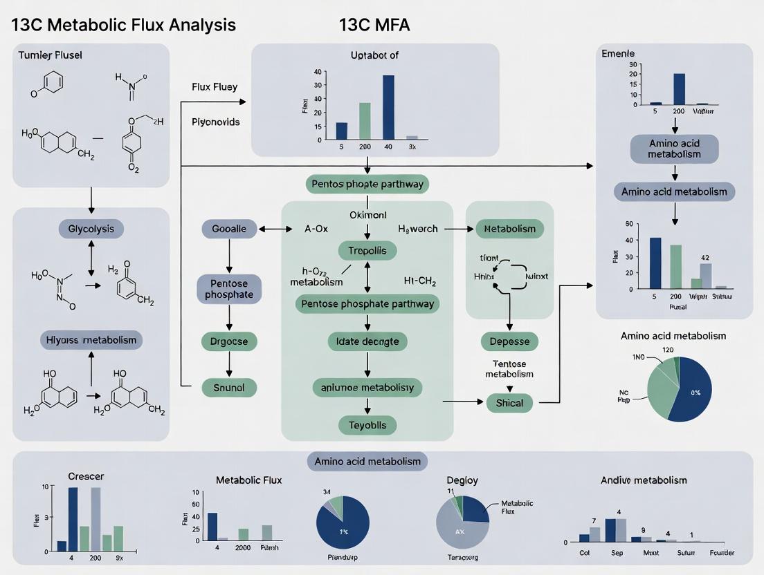
4. Visualizing TME Metabolic Interactions and 13C MFA Workflow
Diagram 1: TME metabolic crosstalk and 13C MFA workflow.
Diagram 2: HIF-1α signaling drives TME metabolic reprogramming.
5. The Scientist's Toolkit: Key Research Reagent Solutions
Table 2: Essential Reagents & Tools for TME 13C Fluxomics
| Item | Function/Application | Example/Note |
|---|---|---|
| 13C-Labeled Tracers | Enable tracking of metabolic fate. Core input for MFA. | [U-13C]Glucose, [U-13C]Glutamine, [1,2-13C]Glucose for pathway resolution. |
| Stable Isotope-Enhanced Media | Chemically defined media for precise 13C tracer studies. | DMEM or RPMI formulations with all glucose/glutamine replaced by 13C versions. |
| Mass Spectrometry Systems | Detect and quantify mass isotopomer distributions (MIDs). | GC-MS for central carbon metabolites; LC-MS for polar/non-volatile compounds. |
| Metabolic Flux Analysis Software | Perform computational flux estimation from MS data. | INCA (Isotopomer Network Compartmental Analysis), CellNetAnalyzer, Escher-FBA. |
| Genome-Scale Metabolic Models | Provide the biochemical reaction network for flux constraints. | Human1, Recon3D. Must be contextualized (e.g., to specific cell types). |
| Ex Vivo Culture Systems | Maintain TME architecture for tracer studies. | Tissue slice culture in air-liquid interface bioreactors. |
| Cell-Type Specific Markers | Isolate or analyze specific TME populations post-experiment. | Antibodies for FACS (CD45, CD3, CD11b, FAP, CD31) or for laser-capture microdissection. |
| Metabolic Inhibitors/Agonists | Validate flux predictions and probe dependencies. | UK5099 (mitochondrial pyruvate carrier inhibitor), BPTES (glutaminase inhibitor), Etomoxir (CPT1 inhibitor). |
6. Conclusion
Defining the TME through the lens of cellular metabolism requires moving beyond cataloging constituents to quantifying the dynamic flow of nutrients. 13C MFA fluxomics is the pivotal methodology for achieving this, offering a systems-level, quantitative map of metabolic interactions between cancer cells, CAFs, immune cells, and endothelial cells. Integrating these flux maps with genomic and proteomic data provides an unparalleled opportunity to identify novel, context-dependent metabolic vulnerabilities for targeted therapeutic intervention. The future of TME research lies in spatially resolved and dynamic 13C MFA, further refining our understanding of this complex metabolic battlefield.
What is Metabolic Flux? From Static Metabolomics to Dynamic Fluxomics
Metabolic flux is the rate of flow of metabolites through a biochemical pathway, quantitatively describing the activity of the cellular metabolic network. Unlike static metabolomics, which provides a snapshot of metabolite concentrations, fluxomics reveals the dynamic in vivo reaction rates, offering a functional readout of cellular physiology. This is critical in tumor microenvironment (TME) research, where metabolic crosstalk and adaptations drive progression and therapy resistance. 13C Metabolic Flux Analysis (13C MFA) is the gold-standard technique for quantifying these fluxes, enabling the mapping of carbon atom fate through central carbon metabolism in cancers and stromal cells.
Core Principles and Quantitative Frameworks
Flux quantification relies on mass balance, isotopomer modeling, and computational simulation. The core equation is the stoichiometric mass balance for each metabolite i in a network of m metabolites and n reactions: S ⋅ v = 0 where S is the m × n stoichiometric matrix and v is the flux vector. In 13C MFA, this is constrained by measurements of isotopic label incorporation from a tracer (e.g., [1,2-13C]glucose) into intracellular metabolites, typically via Mass Spectrometry (MS) or Nuclear Magnetic Resonance (NMR).
Key Quantitative Insights from TME Fluxomics
Table 1: Representative Metabolic Flux Ranges in Cancer Cells vs. Stromal Cells from 13C MFA Studies
| Metabolic Flux (nmol/μg protein/h) | Cancer Cell Range | Cancer-Associated Fibroblast (CAF) Range | Key Implication in TME |
|---|---|---|---|
| Glycolysis | 200 - 800 | 50 - 200 | Warburg effect in cancer; Corrupts milieu |
| Pentose Phosphate Pathway (Oxidative) | 20 - 100 | 5 - 20 | Supports nucleotide synthesis & redox balance |
| TCA Cycle Flux | 50 - 200 | 100 - 300 | CAFs often exhibit more oxidative metabolism |
| Glutamine Anaplerosis | 40 - 180 | 10 - 50 | Fuels cancer cell TCA cycle & biomass |
| Lactate Efflux | 300 - 1000 | 50 - 150 | Cancer cell lactate can fuel CAF metabolism |
| Serine-Glycine-One Carbon | 15 - 60 | 5 - 20 | Critical for methylation & proliferation |
Table 2: Common Tracers and Their Primary Applications in TME Fluxomics
| 13C Tracer | Labeling Pattern | Primary Pathways Probed | Common Application in TME |
|---|---|---|---|
| [U-13C]Glucose | Uniformly labeled | Glycolysis, PPP, TCA, anaplerosis | Mapping overall network activity |
| [1,2-13C]Glucose | 1 & 2 positions labeled | Pentose phosphate pathway (PPP) vs. glycolysis | Quantifying oxidative PPP flux & redox metabolism |
| [5-13C]Glutamine | 5th carbon labeled | Glutaminolysis, reductive TCA metabolism | Tracing glutamine fate in hypoxia |
| [U-13C]Lactate | Uniformly labeled | Cori cycle, lactate utilization | Studying metabolic symbiosis (e.g., lactate shuttle) |
| 13C-Glucose + 13C-Glutamine | Dual tracer | Compartmentalized pathways, crosstalk | Deciphering complex substrate partitioning |
Detailed Experimental Protocol for 13C MFA in TME Models
Aim: To quantify intracellular metabolic fluxes in a 3D co-culture model of cancer cells and fibroblasts.
1. Experimental Design & Tracer Feeding:
- Culture Model: Establish a 3D spheroid co-culture (e.g., ovarian cancer cells + CAFs) in a bioreactor or ultra-low attachment plates.
- Tracer Pulse: Replace media with identically formulated media containing the chosen 13C tracer (e.g., 10 mM [U-13C]glucose). Ensure isotopic steady-state is reached (typically 24-48 hrs for mammalian cells).
- Quenching & Extraction: Rapidly quench metabolism by washing spheroids with cold 0.9% NaCl. Extract metabolites using a cold mixture of 40:40:20 methanol:acetonitrile:water with 0.1% formic acid. Perform repeated freeze-thaw cycles. Centrifuge and collect supernatant.
2. Mass Spectrometry Analysis:
- Platform: LC-MS/MS (e.g., Q-Exactive Orbitrap) coupled to hydrophilic interaction liquid chromatography (HILIC).
- Ionization: Electrospray Ionization (ESI), negative mode for most central carbon metabolites.
- Method: Acquire data in full-scan mode (high resolution >70,000) for mass isotopomer distribution (MID) analysis. Parallel Reaction Monitoring (PRM) for low-abundance metabolites.
- Data Output: Correct raw MIDs for natural isotope abundance using software like IsoCorrection. The final data is the fractional enrichment of each mass isotopologue (M+0, M+1, M+2,...) for key metabolites (e.g., glucose-6-P, lactate, citrate, malate, serine).
3. Computational Flux Estimation:
- Network Construction: Define a stoichiometric model (e.g., core metabolism: glycolysis, PPP, TCA, anaplerosis).
- Simulation & Fitting: Use dedicated software (e.g., INCA, 13CFLUX2, Metran) to simulate MID patterns based on a proposed flux map (vector v). An optimization algorithm minimizes the difference between simulated and measured MIDs.
- Statistical Validation: Perform goodness-of-fit analysis (χ²-test) and Monte Carlo simulations to estimate confidence intervals for each calculated flux.
Title: 13C Metabolic Flux Analysis Core Workflow
Title: Key Metabolic Exchange Fluxes in the Tumor Microenvironment
The Scientist's Toolkit: Essential Research Reagents & Materials
Table 3: Key Reagent Solutions for 13C Fluxomics in TME Research
| Item | Function & Specification | Example Vendor/Cat. No. (Illustrative) |
|---|---|---|
| 13C-Labeled Tracers | Define carbon atom fate through pathways. Purity >99% atom 13C. | Cambridge Isotope Labs (CLM-1396: [U-13C]Glucose) |
| Stable Isotope Media | Chemically defined, tracer-formulated media for controlled feeding. | Gibco DMEM, no glucose, no glutamine (A1443001) |
| Metabolite Extraction Solvent | Rapidly quench enzymes & extract polar metabolites. Cold MeOH:ACN:H2O. | LC-MS grade methanol, acetonitrile (e.g., Sigma 34885, 34851) |
| HILIC LC Columns | Separate polar metabolites for MS analysis. | Waters XBridge BEH Amide Column (186004868) |
| Internal Standards (13C/15N) | Correct for MS instrument variability during extraction. | Isotec/Sigma (e.g., 13C5-15N-Valine for normalization) |
| Flux Estimation Software | Perform isotopomer modeling, simulation, and statistical fitting. | INCA (mfa.vueinnovations.com) / 13CFLUX2 (13cflux.net) |
| LC-MS System | High-resolution mass spectrometer coupled to liquid chromatography. | Thermo Q-Exactive Orbitrap, Agilent 6495 QQQ, Sciex 5500+ |
| 3D Culture Matrix | Model the TME physiologically. | Corning Matrigel (356231), Ultra-Low Attachment Plates (CLS3474) |
Within the tumor microenvironment (TME), cancer cells rewire their metabolic networks to support rapid proliferation, survival, and metastasis. 13C Metabolic Flux Analysis (13C MFA) has emerged as the definitive technique for quantifying the in vivo activity of these pathways. This whitepaper provides an in-depth technical guide on employing 13C tracers to map atomic fate through central carbon metabolism, specifically within the context of tumor metabolism and stromal interactions. We detail current protocols, data analysis frameworks, and essential tools for researchers aiming to uncover novel therapeutic vulnerabilities.
The heterogeneous and often nutrient-poor TME forces dynamic metabolic adaptations. While genomics and proteomics identify parts lists, and metabolomics provides snapshots of pool sizes, only fluxomics—specifically 13C MFA—reveals the functional rates of metabolic reactions. By tracing stable, non-radioactive 13C atoms from a labeled substrate (e.g., [U-13C]glucose) into downstream metabolites, researchers can infer intracellular flux distributions. This is critical for distinguishing between oncogenic driver fluxes (e.g., aerobic glycolysis, glutaminolysis) and compensatory pathways in cancer-associated fibroblasts or immune cells.
Core Principles: From Tracer to Flux Map
The power of 13C MFA lies in measuring isotopic labeling patterns (isotopomer distributions) of metabolic intermediates. Mass spectrometry (GC-MS or LC-MS) detects the mass isotopomer abundances (M+0, M+1, M+2,... M+n). Computational models then simulate these patterns for a given metabolic network and candidate flux set, iteratively optimizing to fit the experimental data.
Key Equation for Flux Estimation:
The system is solved by minimizing the variance-weighted difference between measured (m) and simulated (s) isotopomer data.
Minimize: Φ(v) = Σ [ (m_i - s_i(v)) / σ_i ]²
where v is the flux vector and σ_i is the measurement standard deviation.
Table 1: Common 13C Tracers and Their Application in TME Research
| Tracer | Primary Metabolic Pathways Illuminated | Key Insight in Cancer Metabolism |
|---|---|---|
| [1,2-13C]Glucose | Glycolysis, Pentose Phosphate Pathway (PPP) | Distinguishes oxidative vs. non-oxidative PPP flux; anabolic NADPH production. |
| [U-13C]Glucose | Glycolysis, TCA Cycle, Anaplerosis | Reveals glutamine's anaplerotic contribution and pyruvate carboxylase activity. |
| [U-13C]Glutamine | Glutaminolysis, TCA Cycle, Reductive carboxylation | Quantifies reductive TCA flux (IDH1 reversal) in hypoxia or pseudohypoxia. |
| [3-13C]Lactate | Cori cycle, Gluconeogenesis, TCA cycle | Tracks lactate uptake and utilization by oxidative tumor cells or stromal cells. |
| 13C5-Glutamine | Glutamine metabolism, Nucleotide synthesis | Traces nitrogen and carbon fate into purines/pyrimidines. |
Table 2: Example Flux Results from Murine Tumor Studies (Normalized to Glucose Uptake = 100)
| Metabolic Flux | Aggressive Carcinoma | Slow-Growing Tumor | Cancer-Associated Fibroblasts (CAFs) |
|---|---|---|---|
| Glycolytic Flux (to Lactate) | 85 ± 12 | 45 ± 8 | 95 ± 15 |
| Oxidative PPP Flux | 8 ± 2 | 3 ± 1 | 2 ± 1 |
| TCA Cycle Flux (Vcyc) | 25 ± 5 | 65 ± 10 | 15 ± 4 |
| Glutaminolysis Flux | 18 ± 4 | 5 ± 2 | 1 ± 0.5 |
| Pyruvate Carboxylase Flux | < 2 | 10 ± 3 | 30 ± 7 |
Experimental Protocols
Protocol 4.1:In Vitro13C Tracer Experiment for Adherent Cancer Cells
Objective: To determine central carbon metabolism fluxes in a 2D cancer cell line model.
Materials: See "The Scientist's Toolkit" below.
Procedure:
- Cell Seeding & Quiescence: Seed cells in 6-well plates. Grow to 70-80% confluence in standard medium.
- Tracer Incubation:
- Aspirate medium and wash twice with warm, label-free, serum-free medium.
- Add pre-warmed tracer medium (e.g., DMEM with 10 mM [U-13C]glucose and 2 mM unlabeled glutamine, or vice-versa). Incubate for a time series (e.g., 0, 15, 30, 60, 120 min) to capture isotopic steady-state. For long-term labeling (24h), include dialyzed serum.
- Metabolite Extraction:
- At each time point, quickly aspirate medium and quench metabolism with 1 mL -20°C 80% Methanol.
- Add 400 µL ice-cold H2O, then 400 µL -20°C Chloroform. Vortex vigorously.
- Centrifuge at 13,000g, 15 min, 4°C. Collect the aqueous (top) layer.
- Dry samples in a vacuum concentrator.
- Derivatization & MS Analysis:
- Derivatize with 15 µL Methoxyamine (15 mg/mL in Pyridine, 90 min, RT) followed by 15 µL MSTFA (90 min, RT).
- Analyze by GC-MS (electron impact ionization). Use a standard non-polar column (e.g., Rxi-5Sil MS).
- Data Processing: Correct for natural isotope abundance using software (e.g., IsoCor) and export mass isotopomer distributions (MIDs) for MFA.
Protocol 4.2:Ex Vivo13C Tracing in Tumor Slices
Objective: To preserve the native TME architecture for flux analysis.
Procedure:
- Slice Preparation: Use a vibratome to generate 300-500 µm thick slices from fresh tumor biopsies in ice-cold, oxygenated assay buffer.
- Tracer Incubation: Transfer slices to cell culture inserts in plates with tracer medium. Maintain at 37°C with 95% O2 / 5% CO2 to ensure oxygenation.
- Extraction: After incubation (1-4h), quickly blot slices, snap-freeze in liquid N2, and homogenize in 80% methanol. Proceed with extraction as in 4.1.
Visualizing Metabolic Pathways and Workflows
Diagram 1: 13C MFA Workflow in TME Research
Title: 13C MFA Experimental and Computational Pipeline
Diagram 2: Key Tumor Metabolic Pathways Probed by 13C Tracers
Title: Core Metabolic Network and 13C Tracer Entry Points
The Scientist's Toolkit: Research Reagent Solutions
Table 3: Essential Materials for 13C Tracer Experiments
| Item | Function & Specification | Example Vendor/Cat. No. (Illustrative) |
|---|---|---|
| 13C-Labeled Substrates | High chemical purity (>99%) and isotopic enrichment (≥99% 13C). The core tracer. | Cambridge Isotope Labs (e.g., CLM-1396 for [U-13C]Glucose) |
| Dialyzed Fetal Bovine Serum (dFBS) | Removes small molecules (e.g., glucose, glutamine) to prevent tracer dilution in long-term experiments. | Gibco, 26400044 |
| Ice-cold 80% Methanol (in H2O) | Standard quenching/extraction solvent to instantly halt metabolism and extract polar metabolites. | Prepared in-house with LC-MS grade solvents. |
| Methoxyamine Hydrochloride | Derivatization agent for GC-MS; protects carbonyl groups prior to silylation. | Sigma Aldrich, 226904 |
| N-Methyl-N-(trimethylsilyl)trifluoroacetamide (MSTFA) | Silylation agent for GC-MS; increases volatility of polar metabolites. | Pierce, TS48910 |
| GC-MS or LC-MS System | High-resolution mass spectrometer for accurate isotopologue detection and quantification. | Agilent GC-QQQ, Thermo Orbitrap LC-MS |
| Metabolic Modeling Software | Platform for flux estimation from isotopomer data (e.g., non-linear least squares fitting). | INCA, IsoSim, OpenFLUX |
| Tissue Slicer (Vibratome) | For preparing intact tissue slices from primary tumors to maintain TME architecture. | Leica VT1200S |
| Hypoxic Chamber/Workstation | For conducting tracer experiments under physiologically relevant low O2 conditions (1-5% O2). | Baker Ruskinn Invivo2 |
The application of 13C Metabolic Flux Analysis (13C MFA) in tumor microenvironment (TME) research has revolutionized our understanding of in vivo metabolic pathways. This whitepaper frames the competitive metabolic dynamics within the TME through the lens of fluxomics, which quantifies intracellular reaction rates using stable isotope tracers (e.g., [U-13C]glucose). In nutrient-scarce conditions, 13C MFA reveals how tumor cells rewire their metabolism to outcompete stromal and immune cells for limited resources like glucose, glutamine, and fatty acids, creating an immunosuppressive and pro-tumorigenic niche.
Core Metabolic Pathways and Competition Hubs
Glucose Scarcity and Aerobic Glycolysis
Tumor cells exhibit the Warburg effect, consuming glucose at high rates even under normoxia. 13C MFA studies show this flux diverts glucose carbon away from oxidative phosphorylation (OXPHOS) towards lactate production, creating a glucose-depleted, acidic milieu.
Table 1: Key Fluxomic Differences in Glucose Utilization (nmol/min/10^6 cells)
| Cell Type | Glycolytic Flux (to Lactate) | PPP Flux (to Ribose-5P) | TCA Cycle Flux (to CO2) | Net Glucose Uptake |
|---|---|---|---|---|
| Tumor Cell (e.g., MDA-MB-231) | 120 ± 15 | 18 ± 3 | 35 ± 7 | 150 ± 20 |
| Activated T-cell (e.g., CD8+) | 85 ± 10 | 12 ± 2 | 60 ± 9 | 95 ± 12 |
| Cancer-Associated Fibroblast (CAF) | 40 ± 8 | 8 ± 2 | 45 ± 6 | 50 ± 10 |
| Tumor-Associated Macrophage (M2) | 55 ± 7 | 10 ± 2 | 40 ± 5 | 65 ± 8 |
PPP: Pentose Phosphate Pathway; TCA: Tricarboxylic Acid Cycle. Data synthesized from recent 13C MFA studies (2022-2024).
Glutamine Dependency and Nitrogen Shuttling
Glutamine serves as a key nitrogen and carbon donor. Tumor cells often overexpress glutaminase (GLS), diverting glutamine towards TCA anaplerosis and glutathione synthesis. 13C MFA with [U-13C]glutamine traces this competition, showing impaired T-cell activation under glutamine limitation.
Lipid Metabolism and Scavenging
Under hypoxia and nutrient stress, tumor cells upregulate fatty acid binding proteins (FABPs) and lipoprotein receptors (e.g., CD36) to scavenge lipids from the environment, including from adipocytes and apoptotic cells. Fluxomic analysis with 13C-labeled fatty acids demonstrates this scavenging flux.
Experimental Protocols for 13C MFA in the TME
Protocol: In Vitro Co-culture 13C Tracer Experiment
Objective: Quantify metabolic flux redistribution when tumor cells compete with immune cells for a labeled nutrient.
Materials:
- Tumor cell line (e.g., murine Lewis Lung Carcinoma, LLC).
- Immune cell (e.g., primary murine CD8+ T-cells, activated with anti-CD3/CD28).
- 13C Tracer: [1,2-13C]Glucose or [U-13C]Glutamine.
- Seahorse XF Analyzer or equivalent for extracellular flux analysis.
- LC-MS/MS system for mass isotopomer distribution analysis.
- Co-culture transwell system (0.4 µm pore) for conditional competition.
Procedure:
- Culture & Activation: Seed tumor cells in lower chamber. Isolate and activate CD8+ T-cells in upper transwell insert.
- Tracer Pulse: Replace media with identical, pre-warmed media containing the 13C-labeled nutrient (e.g., 10 mM [U-13C]glucose). Ensure no other carbon source is present.
- Time-Course Sampling: At t=0, 1h, 4h, 8h, 12h, quench metabolism of cells from both compartments separately using liquid N2-cooled 80% methanol.
- Metabolite Extraction: Perform extraction on cell pellets. For intracellular metabolites, use methanol/water/chloroform (-20°C). Dry extracts under N2 gas.
- LC-MS/MS Analysis: Derivatize if necessary. Analyze polar metabolites (e.g., glycolytic intermediates, TCA cycle acids) and non-polar fractions (fatty acids) via targeted LC-MS/MS.
- Flux Calculation: Input mass isotopomer distribution (MID) data into modeling software (e.g., INCA, ISOFLUX). Use genome-scale metabolic model (e.g., RECON) as constraint. Compute fluxes via iterative least-squares minimization.
Protocol: In Vivo 13C Isotope Infusion in Tumor-Bearing Mice
Objective: Measure compartment-specific metabolic fluxes within the intact TME.
- Model: Implant syngeneic tumors (e.g., PyMT mammary carcinoma) in mice.
- Infusion: Cannulate jugular vein. Infuse 13C-labeled nutrient (e.g., [U-13C]glucose, 0.2 mg/g body weight/min) for 2-4 hours using a precision pump.
- Tissue Harvest & Processing: Rapidly excise tumor, dissociate into single-cell suspension, and sort via FACS into populations (CD45- EpCAM+ tumor cells, CD45+ CD3+ T-cells, CD45+ F4/80+ macrophages, α-SMA+ CAFs).
- Metabolomic Analysis: Process sorted cells as in 3.1 steps 4-5.
- Spatial Flux Mapping: Correlate with IHC for metabolic enzymes (e.g., GLS, PKM2) on adjacent tumor sections.
Visualization of Metabolic Crosstalk
Title: Metabolic Competition Network in the TME
Title: 13C MFA Experimental and Computational Workflow
The Scientist's Toolkit: Research Reagent Solutions
Table 2: Essential Reagents and Materials for TME 13C MFA Studies
| Item | Function/Brief Explanation | Example Product/Catalog |
|---|---|---|
| 13C-Labeled Nutrients | Tracer for metabolic flux analysis; defines labeling pattern for model. | [U-13C]Glucose (CLM-1396), [U-13C]Glutamine (CLM-1822) from Cambridge Isotopes. |
| Cell Separation Kits | Isolation of specific TME populations for compartmentalized flux analysis. | Miltenyi Biotec Tumor Dissociation Kit, MACS CD8a+ T Cell Isolation Kit. |
| Extracellular Flux Analyzer | Real-time measurement of glycolytic and OXPHOS rates (ECAR/OCR). | Agilent Seahorse XFp Analyzer with XF Glycolysis Stress Test Kit. |
| LC-MS/MS System | High-sensitivity detection and quantification of mass isotopomers. | Thermo Scientific Q Exactive HF-X with HILIC column (e.g., Waters XBridge BEH Amide). |
| Metabolic Modeling Software | Platform for isotopomer spectral analysis and flux calculation. | INCA (Isotopomer Network Compartmental Analysis) from Vanderbilt. |
| Genome-Scale Metabolic Model | Biochemical network constraint for flux estimation. | Human1, RECON3D, or cell-line specific models from BiGG Models database. |
| In Vivo Isotope Infusion Set | Precision delivery of tracer in animal models for in vivo MFA. | Instech Laboratories Chronic Jugular Vein Catheterization Kit (C30JV). |
| Metabolite Standards (13C-labeled) | Internal standards for absolute quantification in MS. | MSK-SIRO-1 (Silantes) - 13C/15N labeled cell extract as spike-in. |
Within the tumor microenvironment (TME), metabolic heterogeneity and plasticity are not merely supportive features but are emerging hallmarks that directly enable therapy resistance. This whitepaper, framed within the broader thesis of 13C Metabolic Flux Analysis (13C MFA) fluxomics, delineates the technical and mechanistic underpinnings of these processes. We explore how dynamic metabolic reprogramming in response to therapeutic pressure, measurable via advanced fluxomics, creates resilient tumor populations resistant to conventional and targeted therapies.
The classic hallmarks of cancer have evolved to include deregulated cellular metabolism. 13C MFA fluxomics provides a quantitative, systems-level view of intracellular metabolic reaction rates (fluxes), moving beyond static snapshots of metabolite levels. In the context of the TME, 13C MFA is indispensable for mapping the metabolic network topology of diverse cell populations—cancer, immune, and stromal cells—and their interactions. This guide details how fluxomic approaches reveal the heterogeneous and plastic metabolic states that underpin therapeutic failure.
Core Concepts: Heterogeneity and Plasticity
Metabolic Heterogeneity: Refers to the spatial and temporal variation in metabolic pathway utilization among cancer cells within a single tumor. This is driven by genetic mutations, local gradients of nutrients/oxygen, and interactions with stromal cells.
Metabolic Plasticity: Denotes the inherent ability of cancer cells to dynamically switch between metabolic programs (e.g., glycolytic vs. oxidative phosphorylation) in response to external stressors like chemotherapy, targeted agents, or hypoxia.
Together, these properties form a robust adaptive landscape, allowing tumor subpopulations to survive treatment and initiate recurrence.
Quantitative Fluxomic Data: Therapy-Induced Metabolic Shifts
Recent 13C MFA studies quantify the flux rewiring in therapy-resistant models. The following table summarizes key findings from contemporary literature.
Table 1: Quantified Metabolic Flux Shifts in Therapy-Resistant Cancer Models
| Therapy | Cancer Type | Key Flux Change in Resistant Cells | Magnitude of Change (vs. Sensitive) | Measured via |
|---|---|---|---|---|
| EGFR Inhibitors | NSCLC | ↑ Pyruvate carboxylase (PC) anaplerosis | ~3.5-fold increase | [U-13C]glucose MFA |
| BRAF Inhibitors | Melanoma | ↑ Oxidative PPP & mitochondrial respiration | PPP: 2.1-fold; OCR: 1.8-fold | [1,2-13C]glucose MFA |
| Chemotherapy (Cisplatin) | Ovarian | ↑ Glutaminolysis & reductive carboxylation | Glutamine uptake: 2.7-fold | [U-13C]glutamine MFA |
| Androgen Deprivation | Prostate | ↑ Fatty acid oxidation (FAO) | FAO rate: 4.0-fold increase | 13C-palmitate tracing |
| Anti-Angiogenics | Glioblastoma | ↑ Glycolysis & serine biosynthesis | Glycolytic flux: 2.5-fold | [U-13C]glucose MFA |
Experimental Protocols for 13C MFA in Therapy Resistance
Protocol 1: In Vitro 13C MFA Workflow for Drug-Treated Cells
Objective: To quantify metabolic flux changes after acute or chronic drug exposure.
- Cell Model Generation: Establish isogenic pairs of therapy-sensitive and -resistant cell lines (e.g., via chronic, escalating drug exposure over 6-8 months).
- 13C Tracer Experiment:
- Culture cells in physiological glucose (5.5 mM) and glutamine (2 mM) concentrations.
- Replace media with identical media containing [U-13C]glucose or [U-13C]glutamine.
- Incubate for a defined time (typically 4-24h, optimized to reach isotopic steady-state in pathways of interest).
- Metabolite Quenching & Extraction: Rapidly wash cells with ice-cold saline. Quench metabolism with cold (-20°C) 80% methanol/water. Scrape cells, vortex, and centrifuge. Dry supernatant under nitrogen gas.
- Mass Spectrometry Analysis: Derivatize extracts (for GC-MS) or reconstitute in LC-MS solvent. Analyze using GC- or LC-MS to obtain mass isotopomer distributions (MIDs) of intracellular metabolites (e.g., lactate, citrate, succinate, amino acids).
- Flux Estimation: Use computational software (e.g., INCA, IsoSim, WUFlux) to fit the experimental MIDs to a genome-scale metabolic model. Employ least-squares regression to estimate the set of metabolic fluxes that best explain the labeling data.
Protocol 2: Ex Vivo 13C Tracing of Tumor Fragments
Objective: To assess metabolic heterogeneity and plasticity in a near-native TME context.
- Tumor Processing: Fresh patient-derived xenograft (PDX) or murine tumor tissue is sliced into <2 mm fragments using a tissue chopper in cold, oxygenated media.
- Ex Vivo Incubation: Fragments are transferred to media containing a 13C tracer (e.g., [1,2-13C]glucose to probe PPP vs. glycolysis). Media is continuously oxygenated (95% O2 / 5% CO2).
- Single-Cell Resolution: After incubation, tissues are dissociated into single cells. Fluorescence-activated cell sorting (FACS) is used to isolate pure populations (e.g., CD45- epithelial cancer cells, CD45+ immune cells, CD31+ endothelial cells).
- Metabolite Analysis: Metabolites are extracted from each sorted population and analyzed by LC-MS/MS for 13C enrichment, enabling cell-type-specific flux inference.
Visualization of Key Pathways and Workflows
Title: Tumor Metabolic Heterogeneity and Therapy-Induced Plasticity
Title: 13C MFA Experimental Workflow for Therapy Resistance
The Scientist's Toolkit: Key Reagent Solutions
Table 2: Essential Research Reagents for 13C MFA in Therapy Resistance Studies
| Reagent / Material | Function / Purpose | Example Vendor/Product |
|---|---|---|
| U-13C-labeled Substrates | Provide the isotopic tracer for flux tracing. Essential for generating mass isotopomer data. | Cambridge Isotope Laboratories ([U-13C]glucose, CLM-1396) |
| Stable Isotope-Labeled Media | Chemically defined, tracer-ready cell culture media lacking unlabeled components that would dilute the tracer. | Gibco Dialyzed FBS; Silantes 13C/15N-labeling media kits |
| Mass Spectrometry Systems | High-resolution instruments for accurate detection of metabolite mass and isotopologue distribution. | Thermo Fisher Q Exactive HF-X; Agilent 6495C LC/TQ |
| Flux Estimation Software | Computational platforms for integrating labeling data with metabolic models to calculate fluxes. | INCA (isoDynamic); CellNetAnalyzer; WUFlux |
| Patient-Derived Xenograft (PDX) Models | In vivo models that retain tumor heterogeneity and stromal interactions for ex vivo flux studies. | The Jackson Laboratory PDX Resource; Champions Oncology |
| MitoStress Test Kits | Pre-configured assays to measure oxygen consumption rate (OCR), validating MFA-predicted shifts in OXPHOS. | Agilent Seahorse XF Cell Mito Stress Test Kit |
| Metabolite Standards (13C-labeled) | Internal standards for absolute quantification of metabolites via MS. | Sigma-Aldrich MSK-CUS-012 (13C,15N cell extract) |
From Theory to Lab Bench: A Step-by-Step Protocol for 13C MFA in TME Studies
Within the broader thesis on advancing 13C Metabolic Flux Analysis (13C MFA) fluxomics in tumor microenvironment (TME) research, strategic tracer selection is paramount. The metabolic complexity and heterogeneity of the TME demand precise isotopic labeling strategies to elucidate compartment-specific and cell-type-specific metabolic fluxes. This guide details the rationale, application, and protocols for key tracers, focusing on [1,2-13C]glucose and [U-13C]glutamine as cornerstones, while expanding to other critical compounds.
Core Tracers: Rationale and Quantitative Data
[1,2-13C]Glucose
Rationale: This tracer is indispensable for resolving parallel pathway activities in central carbon metabolism. The labeling pattern from [1,2-13C]glucose allows discrimination between glycolysis, the oxidative pentose phosphate pathway (oxPPP), and the non-oxidative pentose phosphate pathway, as well as yielding key information for the TCA cycle.
[U-13C]Glutamine
Rationale: Glutamine is a major anaplerotic substrate and nitrogen donor in cancer cells. Uniformly labeled glutamine traces carbon entry into the TCA cycle via alpha-ketoglutarate, revealing reductive carboxylation (a hallmark of hypoxia or mitochondrial dysfunction) and glutamate-driven biosynthesis.
Table 1: Key Tracers for 13C-MFA in the TME
| Tracer Compound | Primary Metabolic Pathways Illuminated | Key Resolved Fluxes | Typical Labeling Pattern Detected (Mass Isotopomer) |
|---|---|---|---|
| [1,2-13C]Glucose | Glycolysis, PPP, TCA cycle, Pyruvate metabolism | Glycolytic vs. PPP flux, Pyruvate carboxylase (PC) vs. dehydrogenase (PDH) activity | M+2 for lactate, alanine; M+2, M+4 for citrate |
| [U-13C]Glutamine | Glutaminolysis, TCA cycle, Reductive carboxylation, GSH synthesis | Glutaminolytic flux, Reductive (IDH1) vs. oxidative (IDH2) metabolism | M+5 for citrate (oxidative), M+5 for citrate (reductive*) |
| [U-13C]Glucose | Global central carbon metabolism | Net glycolytic and TCA cycle fluxes, Anaplerosis | M+3 for lactate, M+2, M+4, M+6 for TCA intermediates |
| [5-13C]Glutamine | Glutaminolysis specifically | Contribution to cytosolic acetyl-CoA via reductive carboxylation | Labeling in citrate and fatty acid pools |
| [U-13C]Lactate | Lactate uptake and metabolism, Cori cycle, Metabolic coupling | Lactate utilization via pyruvate, TCA cycle entry | M+3 for TCA intermediates |
| 13C-Palmitate (e.g., [U-13C]) | Fatty acid oxidation (FAO), Membrane lipid synthesis | FAO flux, Lipid elongation/desaturation | Acetyl-CoA (M+2) labeling patterns |
*Note: Reductive carboxylation of α-KG from [U-13C]glutamine yields M+5 citrate, identical to oxidative metabolism, but positional labeling via NMR or tandem MS distinguishes them.
Experimental Protocols for Key Tracer Experiments
Protocol 1: In Vitro Tracer Incubation for Adherent Cancer Cell Lines
Objective: To determine intracellular metabolic fluxes using [1,2-13C]glucose. Materials: See "The Scientist's Toolkit" below. Procedure:
- Cell Seeding & Quiescence: Seed cells in 6-well plates. Grow to 70-80% confluence. Wash twice with warm, tracer-free, low-glucose (e.g., 1 mM) media.
- Tracer Media Preparation: Prepare media with physiological glucose (5.5 mM) where 100% is replaced by [1,2-13C]glucose. Supplement with dialyzed FBS (10%) and standard glutamine (4 mM).
- Incubation: Aspirate wash media, add 2 mL tracer media per well. Incubate for a defined time period (e.g., 2, 6, 24 h) at 37°C, 5% CO2. Time-point selection is critical for steady-state MFA.
- Metabolite Extraction: At time point, rapidly aspirate media. Wash cells with ice-cold 0.9% saline. Add 0.8 mL -20°C 80% methanol/water. Scrape cells. Transfer to pre-cooled tube. Add 0.8 mL ice-cold chloroform. Vortex.
- Phase Separation: Centrifuge at 14,000 g, 15 min, 4°C. Upper aqueous phase (polar metabolites) and lower organic phase (lipids) are collected separately into new tubes.
- Drying & Storage: Dry aqueous extract using a centrifugal vacuum concentrator. Store dried pellets at -80°C until LC-MS analysis.
Protocol 2: Ex Vivo TME Slice Culture Labeling
Objective: To probe metabolic fluxes in a preserved TME context using [U-13C]glutamine. Materials: Tumor tissue, McIlwain tissue chopper, slice culture inserts. Procedure:
- Tissue Slice Preparation: Fresh tumor tissue is embedded in low-melt agarose. Using a vibratome or tissue chopper, generate 300 µm thick slices in oxygenated, ice-cold buffer.
- Recovery: Place slices on porous membrane inserts in 6-well plates with tracer-free culture media. Incubate for 1-2 h at 37°C for recovery.
- Tracer Labeling: Replace media with media containing 4 mM [U-13C]glutamine (replacing 100% of standard glutamine). Incubate for 4-24 h.
- Metabolite Extraction: Transfer slices to bead-mill tubes with -20°C 80% methanol. Homogenize. Add chloroform. Proceed with phase separation as in Protocol 1.
Visualization of Metabolic Pathways and Workflows
Title: Glucose & Glutamine Tracer Fate in Core Metabolism
Title: 13C Tracer Experiment and MFA Workflow
The Scientist's Toolkit
Table 2: Essential Research Reagent Solutions for 13C Tracer Studies
| Item | Function/Benefit | Example/Note |
|---|---|---|
| Stable Isotope Tracers | Provide the isotopic label for tracking metabolic fate. High chemical and isotopic purity (>99%) is critical. | [1,2-13C]Glucose (Cambridge Isotopes, CLM-504), [U-13C]Glutamine (CLM-1822) |
| Dialyzed Fetal Bovine Serum (FBS) | Removes low-molecular-weight nutrients (e.g., glucose, amino acids) that would dilute the tracer, ensuring high label enrichment. | Typically 10kDa cut-off. Essential for in vitro studies. |
| Tracer-Compatible Cell Culture Media | Customizable, serum-free or low-nutrient base media for precise tracer formulation without background contamination. | DMEM without glucose/glutamine/pyruvate (e.g., ThermoFisher A1443001). |
| Ice-cold Methanol/Water (80:20) | Quenching solution that rapidly halts metabolism and initiates extraction of polar metabolites. | Must be HPLC/MS grade, stored at -20°C. |
| Chloroform (HPLC Grade) | Used in biphasic extraction to separate non-polar (lipid) metabolites from the polar aqueous phase. | |
| Solid Phase Extraction (SPE) Plates | For clean-up of metabolite extracts prior to LC-MS to remove salts and contaminants that cause ion suppression. | HILIC-mode plates (e.g., SeQuant ZIC-cHILIC). |
| HILIC Chromatography Columns | Separate polar metabolites (central carbon metabolites, nucleotides) for optimal MS detection. | SeQuant ZIC-pHILIC, 2.1 x 150 mm, 5 µm. |
| High-Resolution Mass Spectrometer | Accurately measures mass isotopologue distributions (MIDs) of metabolites. High mass accuracy/resolution is needed. | Orbitrap (Q Exactive) or Time-of-Flight (TOF) systems. |
| MFA Software Suite | Converts MIDs into quantitative metabolic flux maps using computational models. | INCA (Isotopologue Network Compartmental Analysis), 13CFLUX2, Metran. |
| Tissue Slicing System | Maintains intact TME architecture for ex vivo tracer studies. | Vibratome (e.g., Leica VT1200S) or tissue chopper. |
The tumor microenvironment (TME) is a complex ecosystem where cancer cells interact with stromal cells, immune components, and extracellular matrix, creating unique metabolic dependencies. ¹³C Metabolic Flux Analysis (13C MFA) has emerged as a critical tool for quantifying intracellular metabolic fluxes, providing a dynamic picture of metabolic reprogramming within the TME. The validity and translational power of 13C MFA data are intrinsically tied to the physiological relevance of the model systems used. This guide details the design and application of advanced models—from 3D co-cultures and organoids to in vivo studies—that provide the necessary architectural and cellular complexity for generating meaningful fluxomic data to dissect TME metabolism.
Model Hierarchy and Application to Fluxomics
The choice of model dictates the type and quality of flux information that can be obtained. The following table outlines the key characteristics, advantages, and limitations of each model tier for 13C MFA studies.
Table 1: Model Systems for 13C MFA in TME Research
| Model System | Key Components | Physiological Relevance for TME | Advantages for 13C MFA | Major Limitations for Fluxomics |
|---|---|---|---|---|
| 3D Mono-culture | Cancer cells in scaffold (e.g., Matrigel, collagen). | Low; lacks cellular heterogeneity. | Simplified system for establishing core cancer cell fluxes; high tracer signal. | Does not capture metabolic crosstalk. |
| 3D Co-culture | Cancer cells + 1-2 stromal types (e.g., CAFs, TAMs). | Medium; incorporates key pairwise interactions. | Enables direct measurement of metabolic coupling (e.g., lactate shuttle). | Scaling complexity for MFA calculations; potential nutrient compartmentation. |
| Patient-Derived Organoids (PDOs) | Multicellular clusters from patient tissue. | High; retains patient-specific genetics and some cellular diversity. | Patient-specific flux profiles; pre-clinical drug response modeling. | Variability; often loses native immune component and full TME architecture. |
| Organ-on-a-Chip (Micro-physiological Systems) | Co-cultures in perfused microchannels with mechanical cues. | High; incorporates fluid flow, shear stress, and spatial organization. | Enables control over tracer delivery and sampling; models vascular perfusion effects. | Technical complexity; low biomass challenges for GC/MS analysis. |
| In Vivo (Mouse Models) | Syngeneic, PDX, or GEMMs in a live host. | Highest; full physiological context including systemic regulation. | Measures integrated, whole-Tissue fluxes in real TME; gold standard for validation. | Costly; technically challenging for tissue-specific 13C MFA; data is an average of many cells. |
Detailed Experimental Protocols for Key Models
Protocol 2.1: Establishing a 3D Co-culture for 13C MFA of the Cancer Cell-Fibroblast Metabolic Axis
Objective: To quantify the exchange of metabolites (e.g., lactate, glutamine) between cancer cells and cancer-associated fibroblasts (CAFs) using 13C tracer analysis.
Materials:
- Cancer cells: e.g., MCF-7 (breast adenocarcinoma).
- CAFs: Primary isolated or immortalized line (e.g., RMF-621).
- Scaffold: Growth Factor Reduced Matrigel (Corning).
- Tracer Media: Glucose-free, glutamine-free DMEM, supplemented with [U-¹³C₆]-glucose (4.5 g/L) and/or [U-¹³C₅]-glutamine (2 mM), 10% dialyzed FBS, 1% Pen/Strep.
- Cultureware: 24-well low-attachment plates.
Procedure:
- Prepare Cell Suspension: Trypsinize and count cancer cells and CAFs. Prepare two suspensions:
- Co-culture: 1:1 ratio (e.g., 50,000 cells each per well) in cold serum-free medium.
- Mono-culture controls: 100,000 cells of each type单独.
- Mix with Scaffold: Combine cell suspension with chilled Matrigel at a 1:1 volume ratio to achieve a final Matrigel concentration of ~5 mg/mL. Mix gently.
- Plate and Polymerize: Pipette 100 µL of cell-Matrigel mixture per well into a 24-well plate. Incubate at 37°C for 30 min to allow polymerization.
- Add Media: Gently overlay each gel with 500 µL of standard growth media. Culture for 72 hours to allow spheroid formation and interaction.
- 13C Tracer Incubation: Aspirate standard media. Wash gels twice with warm PBS. Add 500 µL of pre-warmed 13C tracer media to each well.
- Quench and Extract: At experimental timepoints (e.g., 6, 24, 48h), quickly aspirate media (saved for extracellular flux analysis) and quench the gel/cells by adding 1 mL of -20°C 80% methanol/water solution. Perform metabolite extraction for intracellular analysis via GC-MS or LC-MS.
Protocol 2.2: 13C MFA in Patient-Derived Organoid (PDO) Models
Objective: To measure pathway fluxes in patient-derived tumor organoids, enabling correlation of metabolic phenotypes with genomic data or drug response.
Materials:
- Tumor Tissue: Fresh surgical or biopsy specimen.
- Digestion Solution: Collagenase IV (2 mg/mL), Dispase II (1 mg/mL) in Advanced DMEM/F12.
- Basement Membrane Extract: e.g., Cultrex Reduced Growth Factor BME (Bio-Techne).
- Organoid Growth Media: Advanced DMEM/F12, 1x B27, 1.25mM N-Acetylcysteine, 10mM Nicotinamide, [Patient-specific growth factors: e.g., 50ng/mL EGF, 100ng/mL Noggin, 100ng/mL R-spondin].
- Tracer Media: Organoid basal media formulated with ¹³C-labeled nutrients (e.g., [U-¹³C]-glutamine as primary carbon source).
Procedure:
- Tissue Processing & Digestion: Mince tumor tissue into <1 mm³ fragments. Incubate in digestion solution for 30-60 mins at 37°C with agitation. Triturate every 15 mins.
- Filtration & Washing: Pass suspension through a 100µm strainer. Centrifuge filtrate at 300 x g for 5 min. Wash pellet with PBS.
- Embedding in BME: Resuspend cell pellet in cold BME (~30 µL per 10,000 cells). Plate 10-15 µL drops in a pre-warmed 24-well plate. Polymerize for 30 mins at 37°C.
- Culture Initiation: Carefully overlay each BME dome with 500 µL of complete organoid growth media. Culture, changing media every 3-4 days, for 7-14 days until organoids form.
- 13C Tracer Experiment: For flux analysis, passage organoids and re-embed in BME in a 96-well format for higher replicate number. After 5 days of growth, switch to tracer media for a defined period (typically 24-72h).
- Harvesting: Remove media for analysis. Dissolve BME domes using Cell Recovery Solution (Corning) or cold PBS. Pellet organoids, wash, and quench metabolism with cold methanol for metabolomic analysis.
Critical Signaling Pathways in the TME: A Fluxomics Perspective
Metabolic crosstalk in the TME is regulated by key signaling pathways. Understanding these is essential for interpreting 13C MFA data.
Diagram 1 Title: HIF-1α Signaling Drives Metabolic Crosstalk & Measurable Fluxes
Integrated Workflow from Model to Fluxomic Data
A robust 13C MFA study requires careful integration of model design, experimental execution, and computational analysis.
Diagram 2 Title: Integrated 13C MFA Workflow for TME Models
The Scientist's Toolkit: Key Reagents for 13C MFA in Advanced Models
Table 2: Essential Research Reagents for TME Model Fluxomics
| Category | Item/Reagent | Function in 13C MFA Studies | Example Vendor/Product |
|---|---|---|---|
| Scaffolds | Growth Factor Reduced Matrigel | Provides a 3D basement membrane environment for organoid and co-culture growth. | Corning Matrigel GFR (#356231) |
| Type I Collagen | Tunable, defined scaffold for mechano-sensitive co-culture studies. | Advanced BioMatrix PureCol (#5005) | |
| Tracers | [U-¹³C₆]-Glucose | Gold-standard tracer for mapping glycolysis, PPP, and TCA cycle entry via pyruvate. | Cambridge Isotopes (CLM-1396) |
| [U-¹³C₅]-Glutamine | Essential tracer for analyzing glutaminolysis, TCA cycle anaplerosis, and biosynthesis. | Cambridge Isotopes (CLM-1822) | |
| [¹³C₆]-Galactose | Tracer to assess alternative sugar metabolism and pentose phosphate pathway activity. | Sigma-Aldrich (389374) | |
| Culture Media | Dialyzed Fetal Bovine Serum (dFBS) | Serum devoid of small molecules (e.g., glucose, glutamine) to control tracer introduction. | Gibco (#A3382001) |
| Custom Tracer Media | Formulated, metabolite-defined media to precisely control nutrient environment for MFA. | Custom from vendors like Thermo Fisher | |
| Analysis Kits | Lactate/Glu cose Assay Kits | Colorimetric/Fluorimetric validation of extracellular flux rates from spent media. | Abcam (ab65331/ ab65333) |
| Metabolite Extraction | 80% Methanol (-20°C) | Standard quenching/extraction solvent for intracellular metabolomics. | N/A - Lab prepared |
| Specialty Media | Organoid Growth Media Kits | Defined basal media and supplement kits for specific PDO types (e.g., intestinal, pancreatic). | STEMCELL Technologies (IntestiCult) |
| In Vivo Tracers | [U-¹³C]-Glucose (IV grade) | For continuous infusion or bolus studies in mouse models to measure in vivo fluxes. | Cambridge Isotopes (CLM-1396-PK) |
1. Introduction
Within the broader framework of 13C Metabolic Flux Analysis (13C MFA) fluxomics applied to the tumor microenvironment (TME), the initial steps of sample processing and quenching are not merely preliminary but critical determinants of data fidelity. The TME is a metabolically dynamic and heterogeneous system where rapid post-sampling metabolic alterations can obscure the true in vivo flux state. This guide details the technical principles and protocols essential for capturing accurate metabolic snapshots from complex ex vivo systems like tumor tissue, co-cultures, or patient-derived models.
2. The Imperative of Rapid Quenching
Upon sampling, cells continue enzymatic activity, rapidly degrading labile metabolites (e.g., ATP, NADH, glycolytic intermediates) and altering 13C-labeling patterns. The goal of quenching is to instantaneously halt all metabolic activity without causing cell lysis and metabolite leakage.
Table 1: Comparison of Common Quenching Methods for Mammalian Cells
| Method | Principle | Speed | Key Advantage | Major Drawback for TME Samples |
|---|---|---|---|---|
| Cold Solvent Quenching | Rapid immersion in <-40°C buffered methanol/water. | Very High (<30 sec) | Excellent enzyme inactivation. | Can cause leakage of intracellular metabolites from sensitive cell types. |
| Fast Filtration & Washing | Vacuum filtration & wash with cold saline. | High (~60 sec) | Removes extracellular metabolites effectively. | Mechanical stress; not ideal for adherent or tissue samples. |
| Snap-Freezing in Liquid N₂ | Direct immersion of tissue/biomass. | Moderate-High | Best for solid tissue biopsies; minimal leakage. | Does not remove extracellular metabolite pool. |
| Warm Methanol Quenching | Using ~60% methanol at room temp. | High | Reduces thermal shock, may improve retention. | Less studied for diverse TME cell populations. |
3. Detailed Experimental Protocols
Protocol 3.1: Cold Methanol Quenching for Tumor Cell Suspensions or Organoids
- Reagents: Quenching solution (60% HPLC-grade methanol in water, pre-cooled to -40°C in dry ice/ethanol bath), PBS (4°C), Liquid N₂.
- Procedure:
- Rapidly transfer culture medium containing cells (~1-5e6 cells) into a pre-cooled 50mL conical tube.
- Immediately add 4 volumes of cold quenching solution (-40°C). Vortex vigorously for 10 seconds.
- Incubate on dry ice/ethanol bath for 5 minutes.
- Pellet cells at 5000 x g for 5 min at -20°C.
- Remove supernatant. Wash pellet gently with 1 mL of cold 60% methanol.
- Snap-freeze pellet in liquid N₂ and store at -80°C until extraction.
Protocol 3.2: Snap-Freezing & Washing for Solid Tumor Tissue
- Reagents: Liquid N₂, Cold 0.9% NaCl saline (4°C), Biopsy tools.
- Procedure:
- Excise tissue and immediately submerge in liquid N₂ for 5-10 seconds for initial quenching.
- While keeping the tissue on dry ice, use a pre-cooled scalpel to trim to ~20mg.
- Transfer tissue to ice-cold saline for a rapid 5-second wash to remove blood/ECM contaminants.
- Blot dry quickly and re-immerse in liquid N₂.
- Pulverize tissue using a cryo-mill under continuous liquid N₂ cooling.
- Transfer frozen powder to a pre-weighed tube at -80°C.
4. The Scientist's Toolkit: Key Research Reagent Solutions
Table 2: Essential Materials for Sample Processing in 13C MFA
| Item | Function & Critical Consideration |
|---|---|
| HPLC-Grade Methanol (pre-cooled) | Primary quenching agent; must be water-mixed and kept below -40°C to ensure rapid thermal and enzymatic inactivation. |
| Isotonic Saline Solution (4°C) | For washing steps; prevents osmotic shock-induced metabolite leakage while removing confounding extracellular label. |
| Cryogenic Vials & Pre-Cooled Racks | To maintain sample temperature during handling; prevents metabolic reactivation. |
| Liquid Nitrogen Dewar | For immediate snap-freezing of solid tissues and long-term storage of quenched samples. |
| Cryo-Mill or Tissue Pulverizer | For homogenizing solid tumor tissue while keeping it frozen, enabling representative sub-sampling. |
| Metabolite Extraction Solvent (e.g., 80% Methanol) | Separate from quenching solution; optimized for complete metabolite solubilization post-quenching. |
5. Workflow Integration for TME 13C MFA
The quenching step is integrated into a larger analytical pipeline. The following diagram outlines the critical decision points post-sampling.
Workflow for Quenching TME Samples
6. Impact of Quenching on Flux Interpretation
Inaccurate quenching can systematically bias flux estimates. For instance, incomplete quenching of glycolytic activity leads to artificially high lactate labeling and misestimation of glycolytic vs. TCA cycle fluxes—a key parameter when assessing metabolic heterogeneity and drug targets in the TME. The chosen protocol must be validated by measuring the stability of key labile metabolites (ATP/ADP ratio, NADH/NAD+) over the processing timeline.
Conclusion
For 13C MFA in tumor microenvironment research, sample processing is the foundational step that determines the reliability of all subsequent flux inferences. A rigorously optimized and consistently applied quenching protocol, tailored to the specific sample matrix (solid tissue vs. dissociated cells), is non-negotiable for capturing biologically relevant metabolic snapshots and deriving accurate flux maps that reflect the in vivo state.
This technical guide details best practices for mass spectrometry data acquisition in 13C isotopologue analysis, framed within the critical context of 13C Metabolic Flux Analysis (MFA) for fluxomics studies of the tumor microenvironment (TME). Accurate isotopologue measurement is foundational for quantifying metabolic pathway fluxes, revealing how tumor and stromal cells reprogram metabolism to support proliferation, immune evasion, and survival.
Core Principles of 13C Isotopologue Analysis
13C-MFA relies on tracing a 13C-labeled substrate (e.g., [U-13C]glucose, [1,2-13C]glutamine) through metabolic networks. The resulting labeling patterns in intracellular metabolites are measured by MS. The precision of flux estimates is directly dependent on the accuracy and precision of the isotopologue abundance data acquired.
LC-MS Best Practices for 13C-MFA
Chromatographic Separation
Objective: Achieve baseline separation of isomers (e.g., glucose-6-phosphate vs. fructose-6-phosphate) to ensure pure isotopologue distributions.
- Column: HILIC (e.g., SeQuant ZIC-pHILIC, 2.1 x 150 mm, 5 µm) is preferred for polar central carbon metabolites.
- Mobile Phase: Ammonium acetate or carbonate in water (A) and acetonitrile (B). pH ~9.2 enhances separation.
- Gradient: Shallow gradient from 80% B to 50% B over 20-30 minutes.
- Temperature: 40-45°C.
- Flow Rate: 0.15-0.2 mL/min.
Mass Spectrometry Acquisition
Instrumentation: High-resolution accurate mass (HRAM) instruments (Q-Exactive, Orbitrap, or TOF platforms) are standard.
- Resolution: ≥ 70,000 (at m/z 200) to resolve isotopic fine structure and minimize isobaric interference.
- Scan Mode: Full-scan (MS1) is primary for isotopologue extraction. Polarity switching (positive/negative) in separate runs is often required.
- Dynamic Range: Ensure linear detector response over >4 orders of magnitude.
- Automatic Gain Control (AGC) Target: Set to "Balanced" or a specific value (e.g., 1e6) to maintain consistent ion counting statistics across runs.
- Maximum Injection Time: Optimize to avoid under-/over-filling. 100-250 ms is typical.
Critical Calibration & QC
- Mass Accuracy: Daily calibration to maintain sub-ppm accuracy.
- Retention Time Stability: Use internal standards to correct for drift.
- Carryover: Monitor and include blank runs between samples.
- Ionization Suppression: Use matrix-matched calibration curves and stable isotope-labeled internal standards (SIL-IS) for each analyte.
GC-MS Best Practices for 13C-MFA
Derivatization & Sample Preparation
Objective: Convert polar, non-volatile metabolites into volatile derivatives.
- Common Protocol (Methoximation and Silylation):
- Dry metabolite extract under nitrogen or vacuum.
- Methoximation: Add 20 µL of 20 mg/mL methoxyamine hydrochloride in pyridine; incubate at 37°C for 90 minutes.
- Silylation: Add 80 µL of N-methyl-N-(trimethylsilyl)trifluoroacetamide (MSTFA) with 1% TMCS; incubate at 37°C for 30 minutes.
- Centrifuge and transfer supernatant to GC vial.
GC-MS Acquisition
Instrumentation: Quadrupole GC-MS with electron impact (EI) ionization.
- Column: Mid-polarity stationary phase (e.g., DB-35MS, 30 m x 0.25 mm ID, 0.25 µm film).
- Oven Program: Start at 60°C, ramp to 325°C (e.g., 10°C/min).
- Inlet Temperature: 250°C.
- Carrier Gas: Helium, constant flow ~1.2 mL/min.
- Ion Source Temperature: 230°C.
- Acquisition Mode: Selected Ion Monitoring (SIM) is preferred for highest sensitivity and precision in isotopologue analysis. Monitor 3-5 key fragment ions per metabolite.
- Dwell Time: ≥ 20 ms per ion to ensure sufficient data points across the peak.
Quantitative Data Comparison: LC-MS vs. GC-MS for 13C-MFA
Table 1: Comparison of LC-MS and GC-MS Platforms for 13C Isotopologue Analysis
| Feature | LC-MS (HILIC-HRAM) | GC-MS (Quadrupole-SIM) |
|---|---|---|
| Analyte Coverage | Broader for polar, labile, and high MW metabolites (e.g., nucleotides, CoA esters). | Excellent for organic acids, sugars, amino acids. Limited for labile/ large metabolites. |
| Sample Prep | Minimal; protein precipitation. Can be automated. | Requires derivatization (time-consuming, can introduce error). |
| Throughput | Moderate (15-30 min run time). | Fast (10-20 min run time after derivatization). |
| Ionization | Soft (ESI); preserves molecular ion. | Hard (EI); generates reproducible fragments. |
| Data Type | Full-scan HRAM data allows retrospective analysis. | Pre-defined SIM methods limit retrospective analysis. |
| Precision (CV%) | 1-5% (with SIL-IS) | 0.5-3% (excellent due to stable EI & SIM). |
| Key Strength | Untargeted capability, coverage. | High precision, robust quantification, lower cost. |
| Best For | Complex TME extracts, discovering novel labeling. | High-precision flux determination of core metabolites. |
Table 2: Key QC Metrics and Target Values for Reliable 13C Data Acquisition
| QC Parameter | Target Value (LC-MS) | Target Value (GC-MS) | Purpose |
|---|---|---|---|
| Mass Accuracy | < 1 ppm | < 0.1 Da | Correct metabolite/ fragment identification. |
| Retention Time Drift | < 0.1 min | < 0.05 min | Ensures consistent chromatographic alignment. |
| Internal Standard Peak Area CV | < 15% | < 10% | Monitors instrument stability and injection precision. |
| Linearity (R²) | > 0.99 | > 0.99 | For calibration curves of natural abundance standards. |
| Limit of Detection (LOD) | Compound-specific | Compound-specific | Defines sensitivity threshold. |
| Carryover | < 0.5% in blank | < 0.2% in blank | Prevents contamination between samples. |
Experimental Protocol: 13C-Labeling and MS Analysis of TME Cultures
Aim: To determine glycolytic and TCA cycle fluxes in a co-culture model of cancer cells and cancer-associated fibroblasts (CAFs).
Step 1: Experimental Design.
- Culture cells in physiologically relevant co-culture system (e.g., transwell).
- Replace media with identical medium containing 13C-labeled substrate (e.g., 10 mM [U-13C]glucose). Use "labeling time-course" (e.g., 0, 15, 30, 60, 120 min) or "isotopic steady-state" (24-48h).
- Quench metabolism rapidly (liquid N2, -80°C methanol/water).
Step 2: Metabolite Extraction (for LC-MS).
- Add 500 µL of 80% methanol (-80°C) to cell culture plate on dry ice.
- Scrape cells, transfer suspension to microcentrifuge tube.
- Add 500 µL ice-cold water and 500 µL chloroform.
- Vortex vigorously, centrifuge at 14,000 g for 15 min at 4°C.
- Collect polar (upper) phase for central carbon metabolism analysis. Dry under vacuum.
Step 3: LC-MS Analysis (Polar Phase).
- Reconstitute in 100 µL 50% acetonitrile.
- Inject 5-10 µL onto HILIC-HRAM system per conditions in Section 3.
- Acquire full-scan data in negative and positive polarity modes.
Step 4: Data Processing.
- Use software (e.g., El-MAVEN, XCMS, Compound Discoverer) for peak picking, alignment, and integration.
- Extract isotopologue distributions (M0, M+1, M+2,... M+n) for each metabolite.
- Correct for natural abundance of 13C, 2H, 15N, 18O, 29Si, 30Si using algorithms (e.g., AccuCor, IsoCorrector).
The Scientist's Toolkit: Essential Research Reagents & Materials
Table 3: Key Reagent Solutions for 13C-MFA in TME Research
| Item | Function & Specification |
|---|---|
| [U-13C]Glucose | Primary tracer for glycolysis, PPP, and TCA cycle. >99% atom 13C purity. |
| [1,2-13C]Glutamine | Key tracer for glutaminolysis, TCA anaplerosis. >99% atom 13C purity. |
| Stable Isotope-Labeled Internal Standards (SIL-IS) | e.g., 13C15N-labeled amino acid mix. For LC-MS normalization and absolute quantification. |
| Methoxyamine Hydrochloride | Derivatization agent for GC-MS; protects carbonyl groups by forming methoximes. |
| MSTFA (N-Methyl-N-(trimethylsilyl)trifluoroacetamide) | Silylation agent for GC-MS; adds trimethylsilyl groups to -OH, -COOH, -NH2. |
| HILIC Chromatography Column | e.g., SeQuant ZIC-pHILIC. Separates polar, hydrophilic metabolites for LC-MS. |
| DB-35MS or Equivalent GC Column | Mid-polarity column for separating a wide range of derivatized metabolites. |
| Cold Methanol/Water (80:20) | Quenching/extraction solvent to instantly halt metabolism and extract polar metabolites. |
Visualizing the Workflow and Metabolic Context
Workflow for 13C-MFA in TME Studies
Metabolic Crosstalk in the Tumor Microenvironment
13C-Labeling Propagation from [U-13C]Glucose
Within the context of 13C Metabolic Flux Analysis (MFA) fluxomics in tumor microenvironment (TME) research, computational flux estimation is a cornerstone for quantifying intracellular metabolic reaction rates. This guide provides an in-depth technical overview of leading software platforms, detailing their algorithms, application protocols, and relevance to cancer metabolism and drug discovery.
Metabolic reprogramming is a hallmark of cancer. Within the complex TME, comprising cancer, stromal, and immune cells, 13C MFA is the definitive technique for elucidating in vivo pathway activities. Computational flux estimation platforms integrate isotopic labeling data from mass spectrometry (MS) or nuclear magnetic resonance (NMR) with stoichiometric models to calculate net and exchange fluxes, offering insights into tumor vulnerabilities.
Core Software Platforms: Algorithms and Comparative Analysis
Platform Architectures and Solving Methods
The central problem is solving an overdetermined system of equations: ( \text{min} || f(v) - m* ||^2 ), where ( v ) is the flux vector, ( f(v) ) is the simulated labeling pattern, and ( m* ) is the measured labeling data.
Table 1: Core Algorithmic and Functional Comparison of Key Platforms
| Platform | Primary Solver Method | Isotopomer Framework | TME-Specific Features | License & Access |
|---|---|---|---|---|
| INCA(Isotopomer Network Compartmental Analysis) | Elementary Metabolite Units (EMU) framework, Non-linear least-squares optimization with confidence intervals (e.g., Monte Carlo) | EMU | Compartmentalized models for cell-cell metabolic exchange; integration of transcriptomic constraints. | Commercial (Academic discounts) |
| IsoSim | Analytical calculation of isotopomer distributions; efficient simulation for large networks. | Cumomer / Isotopomer | High-speed simulation suitable for high-throughput in silico testing of TME perturbations. | Open Source |
| OpenFlux | EMU framework, user-extensible with Python/Matlab. | EMU | Community-developed; adaptable for co-culture systems and dynamic flux analysis. | Open Source |
| 13C-FLUX2 | 13C Metabolic Flux Analysis with comprehensive statistical evaluation. | Cumomer | Robust statistical package for flux uncertainty estimation, critical for heterogeneous TME samples. | Free for academic use |
Table 2: Typical Quantitative Flux Output for a Core Cancer Pathway (Warburg Effect) Simulated data from a generic cancer cell model under 13C-glucose infusion. Flux values normalized to glucose uptake = 100.
| Metabolic Reaction | Estimated Flux (Normalized Units) | 95% Confidence Interval | Platform Used for Estimation |
|---|---|---|---|
| Glycolysis (Glucose → Pyruvate) | 95.0 | [92.5, 97.5] | INCA |
| Lactate Efflux (Warburg Effect) | 78.0 | [75.0, 81.0] | INCA |
| Oxidative PPP (G6P → R5P + CO2) | 15.5 | [14.0, 17.0] | 13C-FLUX2 |
| TCA Cycle (Citrate Synthase) | 22.0 | [20.5, 23.5] | OpenFlux |
| Glutaminolysis (Gln → α-KG) | 31.0 | [29.0, 33.0] | INCA |
Detailed Methodologies for Key 13C MFA Experiments
Protocol 1: Steady-State 13C MFA for 2D Cancer Cell Cultures
- Tracer Design: Prepare culture medium with [U-13C]glucose (e.g., 25 mM) or [1,2-13C]glucose, and/or 13C-glutamine (e.g., 4 mM). Use tracer combinations to resolve parallel pathways.
- Cell Culture & Quenching: Seed cells (e.g., 5e6 cells/dish). After 24h, replace medium with tracer medium. Culture until isotopic steady state is reached (typically 24-48h). Rapidly quench metabolism using cold (-20°C) 60% methanol solution.
- Metabolite Extraction & Derivatization: Extract intracellular metabolites using a cold methanol/water/chloroform phase separation. Derivatize polar metabolites (e.g., amino acids, organic acids) for GC-MS analysis using MTBSTFA or methoxyamine hydrochloride + N-methyl-N-(trimethylsilyl)trifluoroacetamide.
- MS Data Acquisition & Processing: Run samples on GC-MS or LC-MS. Quantify mass isotopomer distributions (MIDs) of key fragments (e.g., alanine M+0 to M+3 from pyruvate pool, glutamate M+0 to M+5 from TCA cycle).
- Flux Estimation: Input MIDs, substrate uptake/excretion rates (measured via HPLC), and a genome-scale metabolic model (condensed to relevant pathways) into the chosen platform (e.g., INCA). Run non-linear least-squares optimization to fit fluxes to the data. Perform statistical analysis (e.g., Monte Carlo) to estimate confidence intervals.
Protocol 2: Dynamic 13C MFA (non-stationary) for In Vivo TME Studies
- In Vivo Tracer Infusion: Establish tumor xenografts. Perform tail-vein infusion of a 13C tracer (e.g., [U-13C]glucose) at a constant rate for a defined time course (e.g., 0, 5, 15, 30, 60 min).
- Tissue Sampling and Processing: Sacrifice animals at each time point. Rapidly excise and freeze tumors in liquid N2. Pulverize tissue under cryogenic conditions.
- Metabolite Extraction: Homogenize powdered tissue in cold, polar extraction solvent. Proceed with derivatization for GC-MS as in Protocol 1.
- Data Modeling: Use platforms like INCA that support INST-MFA. Input time-series MID data and a kinetic model. The software solves differential equations to estimate flux maps and metabolite pool sizes, capturing rapid metabolic adaptations in the TME.
Visualizing the 13C MFA Workflow and TME Metabolism
Diagram 1: 13C MFA Workflow in TME Research
Diagram 2: Compartmentalized TME Metabolic Cross-Talk
The Scientist's Toolkit: Key Research Reagent Solutions
Table 3: Essential Materials and Reagents for 13C MFA in TME Studies
| Item | Function & Relevance to TME Research | Example Product/Supplier |
|---|---|---|
| Stable Isotope Tracers | Enable tracing of carbon fate. Crucial for probing pathway activities in co-cultures. | [U-13C]Glucose (Cambridge Isotope Labs), [5-13C]Glutamine (Sigma-Aldrich) |
| Specialized Cell Culture Media | Tracer-compatible, chemically defined media for controlled TME modeling. | DMEM without glucose/glutamine (Gibco), custom formulations for CAFs/immune cells. |
| Quenching/Extraction Solvents | Halt metabolism instantly to capture in vivo metabolic state. | Cold (-20°C) 60% Methanol (in H2O), Methanol/Chloroform/Water mixes. |
| Derivatization Reagents | Prepare metabolites for GC-MS analysis; critical for detecting labeling in key analytes. | N-Methyl-N-(tert-butyldimethylsilyl)trifluoroacetamide (MTBSTFA), Methoxyamine hydrochloride. |
| Internal Standards (IS) | Correct for sample loss and instrument variability; essential for quantitative fluxomics. | 13C/15N-labeled cell extracts (e.g., Cambridge Isotope Labs), or U-13C-amino acid mixes. |
| LC/GC-MS Columns | Separate complex metabolite mixtures from limited TME samples. | HILIC columns (e.g., SeQuant ZIC-pHILIC) for polar metabolites, DB-5MS for GC. |
| Metabolic Network Models | Stoichiometric foundation for flux calculation; require TME-specific curation. | Recon3D, HMR2, or custom models incorporating cell-type-specific exchange. |
| Software Platform License | Core computational engine for flux estimation. | INCA (mfa.vueinnovations.com), 13C-FLUX2 (13cflux.net). |
This case study is framed within a broader thesis on the application of 13C Metabolic Flux Analysis (13C MFA) fluxomics for elucidating metabolic reprogramming within the tumor microenvironment (TME). A core tenet of this thesis is that stromal cells, particularly Cancer-Associated Fibroblasts (CAFs), are not passive actors but active metabolic collaborators with cancer cells. Mapping glutamine metabolism in CAFs using 13C MFA provides a quantitative, systems-level understanding of flux distributions, revealing critical nodes for metabolic crosstalk and potential therapeutic targeting. This guide details the technical approach for such an investigation.
Core Principles: Glutamine Metabolism in CAFs
CAFs often exhibit a catabolic phenotype, breaking down nutrients to supply cancer cells. Glutamine metabolism is pivotal, with key pathways including:
- Glutaminolysis: Conversion of glutamine to glutamate (via glutaminase, GLS) and subsequently to α-ketoglutarate (α-KG) for the TCA cycle.
- Anaplerosis: Replenishment of TCA cycle intermediates.
- Nitrogen Metabolism: Generation of non-essential amino acids (e.g., aspartate, alanine) and nucleotides.
- Redox Homeostasis: Production of glutathione (GSH) via glutamate. 13C MFA allows quantification of the flux through these interconnected pathways.
Experimental Protocol for 13C MFA in CAFs
CAF Isolation, Culture, and Tracer Experiment
- CAF Isolation: Isolate primary CAFs from human tumor specimens via enzymatic digestion (Collagenase/Dispase) and differential centrifugation. Validate phenotype (α-SMA, FAP, Vimentin positive; cytokeratin negative).
- Culture & Quenching: Culture CAFs in glutamine-free, glucose-containing medium. Pre-condition for 24h. Introduce tracer: [U-13C5]-L-Glutamine (e.g., 2 mM final concentration). Incubate for a determined time interval (e.g., 0, 1, 6, 24h) to capture isotopic steady-state or non-steady-state dynamics.
- Metabolite Extraction: Rapidly quench metabolism with liquid nitrogen or cold methanol/water buffer. Extract intracellular metabolites using a methanol/chloroform/water protocol. Dry under nitrogen gas and derivatize for GC-MS (e.g., MTBSTFA) or reconstitute for LC-MS.
Mass Spectrometry & Data Processing
- GC-MS/MS or LC-MS/MS Analysis: Analyze derivatized samples via GC-MS or underivatized via LC-MS (HILIC column). Monitor mass isotopomer distributions (MIDs) of target metabolites: TCA intermediates (citrate, α-KG, succinate, malate), amino acids (glutamate, aspartate, alanine), and related compounds.
- Data Processing: Correct raw MIDs for natural isotope abundance. Calculate fractional enrichments and isotopologue distributions.
Metabolic Network Modeling & Flux Estimation
- Network Construction: Build a stoichiometric model encompassing central carbon metabolism (glycolysis, PPP, TCA cycle), glutaminolysis, and amino acid biosynthesis.
- Flux Calculation: Use software (e.g., INCA, IsoDesign, Metran) to fit the experimental MIDs to the network model via iterative least-squares regression. Estimate net fluxes and confidence intervals. Key output: flux map showing rates in nmol/µg protein/h or similar units.
Table 1: Comparative Glutamine-Derived Metabolic Fluxes in CAFs vs. Normal Fibroblasts (NFs) Data are representative fluxes (nmol/µg protein/h) from simulated 13C MFA studies.
| Metabolic Flux Pathway | CAFs (Range) | Normal Fibroblasts (NFs) (Range) | Notes / Implications |
|---|---|---|---|
| Glutamine Uptake | 15.2 - 28.7 | 4.5 - 8.3 | ~3-4x higher in CAFs, indicating addiction. |
| Glutaminase (GLS) Flux | 14.8 - 27.5 | 4.2 - 7.9 | Major entry point, correlates with uptake. |
| Glutamate → α-KG | 12.1 - 22.3 | 3.8 - 7.1 | High flux into TCA cycle. |
| TCA Cycle (Citrate Synthase) | 8.5 - 15.2 | 6.1 - 9.8 | Moderately increased. |
| Aspartate Synthesis | 3.2 - 6.5 | 0.8 - 1.9 | Key nitrogen output, supports cancer cell proliferation. |
| Lactate Secretion | 35.0 - 60.0 | 10.0 - 20.0 | High glycolytic flux, even with glutamine fueling TCA. |
| Glutathione (GSH) Synthesis | 1.5 - 3.0 | 0.5 - 1.2 | Enhanced antioxidant capacity. |
Table 2: Key 13C Enrichment Patterns from [U-13C5]-Glutamine Tracing in CAFs
| Metabolite | Major Isotopologue Observed (M+X) | Interpretation of Labeling Pattern |
|---|---|---|
| Glutamate | M+5, M+4 | M+5: direct product of [U-13C5]-Gln. M+4: indicates reversible transamination or minor oxidation. |
| α-Ketoglutarate | M+5, M+4 | Mirrors glutamate, confirms rapid conversion via GDH or transaminases. |
| Citrate | M+4, M+2 | M+4: from condensation of M+5 α-KG with unlabeled OAA. M+2: indicates turns of TCA cycle. |
| Aspartate | M+4, M+3 | M+4: derived from M+4 OAA. M+3: results from multiple TCA turns. |
| Malate | M+4, M+2, M+3 | Complex pattern reflecting TCA cycling and possible reversibility. |
Signaling & Metabolic Pathways
Title: Core Glutaminolysis Pathways in CAFs
Title: 13C MFA Workflow for CAF Glutamine Metabolism
The Scientist's Toolkit: Research Reagent Solutions
Table 3: Essential Materials for Glutamine 13C MFA in CAFs
| Item | Function & Application in Study |
|---|---|
| [U-13C5]-L-Glutamine | Stable isotope tracer. Enables tracking of glutamine-derived carbon atoms through metabolic networks. Core of the 13C MFA experiment. |
| Primary CAF Isolation Kit (e.g., tumor dissociation enzymes) | For gentle and efficient isolation of viable, phenotype-preserved CAFs from fresh tumor tissue. |
| Glutamine-Deficient Cell Culture Medium | Allows precise control and formulation of media with 13C-glutamine as the sole glutamine source, preventing dilution of the label. |
| Glutaminase (GLS) Inhibitor (e.g., CB-839, BPTES) | Pharmacological tool to validate the role of glutaminolysis. Used in control experiments to confirm flux dependencies. |
| LC-MS/MS or GC-MS System | High-sensitivity mass spectrometer required for accurate detection and quantification of mass isotopomer distributions (MIDs) in metabolic extracts. |
| Metabolic Flux Analysis Software (e.g., INCA, IsoDesign) | Computational platform for constructing metabolic network models, fitting 13C labeling data, and calculating statistically rigorous flux distributions. |
| Antibody Panel for CAF Validation (α-SMA, FAP, PDGFRβ) | Essential for confirming the identity and purity of isolated CAFs via immunofluorescence or flow cytometry prior to experiments. |
| Liquid Chromatography Column (e.g., HILIC) | For separation of polar, underivatized metabolites (like TCA intermediates, amino acids) prior to LC-MS analysis. |
Navigating Technical Challenges: Solutions for Robust and Reproducible 13C Flux Data
Common Pitfalls in Tracer Experiment Design and How to Avoid Them
In the context of 13C Metabolic Flux Analysis (MFA) for elucidating tumor microenvironment (TME) metabolic heterogeneity, meticulous tracer experiment design is paramount. Inadequate design leads to unreliable flux estimations, compromising the study of metabolic reprogramming in cancer. This guide details common pitfalls and provides protocols to ensure robust, interpretable data for drug target discovery.
Pitfall 1: Inappropriate Tracer Selection and Labeling Pattern
The choice of tracer dictates which metabolic pathways can be resolved. Using a single glucose tracer (e.g., [1,2-13C]glucose) fails to resolve parallel pathway activities common in tumors, such as concurrent glycolysis, pentose phosphate pathway (PPP), and reductive carboxylation.
Experimental Protocol: Dual-Tracer Approach for PPP & Glycolysis Resolution
- Reagents: [1,2-13C]Glucose and [U-13C]Glucose, cell culture medium lacking glucose, isotopically labeled glutamine (optional).
- Procedure:
- Prepare two parallel cultures of target cells (e.g., cancer cell lines, co-cultures, or primary cells from TME).
- Deplete endogenous carbon sources by washing cells in substrate-free medium.
- Feed Culture A with medium containing 100% [1,2-13C]Glucose (10 mM). Feed Culture B with medium containing 100% [U-13C]Glucose (10 mM). Maintain glutamine concentration (2 mM) as unlabeled.
- Incubate for a duration sufficient to reach isotopic steady-state in central metabolites (typically 24-48 hours for cancer cells, validated by time-course sampling).
- Quench metabolism rapidly with cold methanol, extract intracellular metabolites, and analyze via LC-MS or GC-MS.
- Measure mass isotopomer distributions (MIDs) of glycolytic (e.g., lactate, alanine) and TCA cycle intermediates (e.g., citrate, malate).
Table 1: Comparative Information Yield from Common Tracers in TME Studies
| Tracer Compound | Ideal for Probing | Key Limitation | Complementary Tracer |
|---|---|---|---|
| [U-13C]Glucose | Glycolytic flux, PPP, complete TCA cycle turnover | Cannot distinguish oxidative/reductive TCA | [1,2-13C]Glucose |
| [1,2-13C]Glucose | PPP vs. glycolysis split, anaplerotic fluxes | Low labeling in late TCA intermediates | [U-13C]Glutamine |
| [U-13C]Glutamine | Reductive carboxylation, glutaminolysis, TCA | Minimal label in glycolytic intermediates | [5-13C]Glutamine |
Diagram: Tracer Entry Points into Central Carbon Metabolism
Pitfall 2: Failure to Achieve Isotopic Steady State
Sampling before the system reaches isotopic steady state yields transient MIDs, requiring complex non-stationary MFA (INST-MFA) for interpretation—a significant increase in complexity. For classic 13C-MFA, steady-state is non-negotiable.
Experimental Protocol: Determining Isotopic Steady-State Time Course
- Set up identical cell cultures under desired conditions (e.g., hypoxia, drug treatment).
- Switch to medium containing your chosen tracer at time zero.
- Harvest replicate cultures at multiple time points (e.g., 0, 2, 6, 12, 24, 48 hours).
- Extract metabolites and measure MIDs for key metabolites like lactate, alanine, citrate, and aspartate.
- Plot the fractional enrichment of key mass isotopologues over time. Steady-state is achieved when these enrichments plateau.
Table 2: Indicative Time to Isotopic Steady-State in Different Systems
| Biological System | Approximate Time | Critical Metabolite to Monitor |
|---|---|---|
| Fast-growing cancer cell line (in vitro) | 24-36 hours | M+3 Lactate from [U-13C]Glucose |
| Primary tumor cells (ex vivo) | 36-60 hours | M+2 Aspartate from [U-13C]Glucose |
| Co-culture systems (e.g., Cancer-Associated Fibroblasts) | 48-72 hours | M+4 Citrate from [U-13C]Glutamine |
Pitfall 3: Neglecting Compartmentalization & Exchange Fluxes
The TME features metabolic symbiosis where different cell types export and consume metabolites (e.g., lactate, alanine). Ignoring these exchanges leads to incorrect intracellular flux maps.
Experimental Protocol: Quantifying Metabolite Exchange in Co-culture
- Establish a transwell co-culture system with cancer cells and stromal cells (e.g., fibroblasts).
- Feed the system with a chosen tracer (e.g., [U-13C]Glucose).
- Sample both the extracellular medium and intracellular pools from each cell type separately at steady-state.
- Measure concentrations and MIDs of exchanged metabolites (lactate, pyruvate, alanine, glutamine) in all compartments.
- Use the differences in MIDs between producer and consumer cells to model exchange fluxes.
Diagram: Key Metabolic Exchanges in the Tumor Microenvironment
Pitfall 4: Inadequate Analytical & Computational Workflow
Flux calculation is sensitive to MID measurement error and model configuration. Poor quality MS data or an oversimplified network model invalidates results.
Experimental Protocol: Integrated MS & MFA Workflow Validation
- MS Calibration: Use internal standards for absolute quantification and periodic natural abundance standards to correct for instrument drift.
- MID Precision: Perform technical replicates (n≥5) of a single sample to determine measurement error for each metabolite's MID. Standard deviation should be <0.5% for major isotopologues.
- Network Model: Construct a stoichiometric model inclusive of relevant TME pathways (e.g., lactate exchange, glutaminolysis, serine/glycine cycle).
- Flux Estimation: Use software (e.g., INCA, 13CFLUX2) to fit simulated MIDs to measured data via least-squares regression. Provide the software with your measured MID error matrix.
- Statistical Validation: Perform Monte Carlo sampling or sensitivity analysis to generate confidence intervals for all estimated fluxes. Discard fluxes with confidence intervals spanning zero.
Diagram: 13C-MFA Experimental and Computational Workflow
The Scientist's Toolkit: Key Research Reagent Solutions
Table 3: Essential Materials for Robust 13C Tracer Studies in TME
| Item | Function & Rationale | Example/Specification |
|---|---|---|
| Stable Isotope Tracers | Introduce distinguishable label into metabolism for pathway tracing. Purity is critical. | [U-13C]Glucose (≥99% atom purity), [1,2-13C]Glucose, [U-13C]Glutamine. |
| Substrate-Free Basal Medium | Allows precise formulation of tracer and unlabeled nutrient concentrations, eliminating background. | DMEM without glucose, glutamine, or phenol red. |
| Cold Quenching Solution | Instantly halts metabolic activity to capture in vivo MIDs. | 60% aqueous methanol, pre-chilled to -40°C or below. |
| Internal Standards for MS | Correct for variability in extraction efficiency and instrument response. | 13C-labeled cell extract or mixture of individual 13C standards (e.g., U-13C amino acids). |
| Isotopic Natural Abundance Standard | Allows correction for natural 13C, 2H, etc., present in solvents and unlabeled atoms. | Unlabeled metabolite mix at known concentration. |
| Transwell Co-culture Plates | Enables study of metabolic crosstalk between cell types while allowing separate analysis. | Polycarbonate membrane inserts (0.4 µm or 3.0 µm pore size). |
| Metabolic Network Modeling Software | Platform for flux calculation from experimental MID data. | INCA, 13CFLUX2, OpenFLUX. |
| LC-MS System with High Resolution | Separates and detects metabolites, resolving isotopologues by mass. | HPLC coupled to Q-TOF or Orbitrap mass spectrometer. |
Optimizing Isotope Stease-State vs. Instationary (Kinetic) MFA for Different TME Questions
Within the broader thesis on 13C Metabolic Flux Analysis (MFA) fluxomics in tumor microenvironment (TME) research, selecting the appropriate isotopic labeling strategy is paramount. The choice between steady-state (S-S) and instationary (kinetic) MFA is not merely technical but defines the biological questions addressable. This guide provides an in-depth technical comparison, framing their optimization for distinct TME investigations, from bulk metabolic phenotypes to dynamic pathway regulation in response to microenvironmental fluctuations.
Fundamental Principles & Comparative Framework
Steady-State MFA relies on the assumption that metabolic fluxes and labeling patterns have reached an isotopic steady state following the introduction of a 13C-labeled tracer. It provides a snapshot of net fluxes through the metabolic network. In contrast, Instationary (Kinetic) MFA analyzes the time-series data of labeling enrichments before isotopic steady state is reached, enabling the estimation of flux transients and pool sizes of metabolites.
Table 1: Core Conceptual Comparison
| Feature | Steady-State MFA | Instationary (Kinetic) MFA |
|---|---|---|
| Primary Data | Isotopic steady-state labeling patterns (e.g., from GC-MS fragment ions). | Time-course of isotopic labeling patterns (transient enrichments). |
| Estimated Parameters | Net metabolic fluxes (in μmol/gDW/h). | Metabolic fluxes and intermediate metabolite pool sizes (in μmol/gDW). |
| Experimental Duration | Long (hours to days), until steady-state labeling is achieved. | Short (seconds to minutes), capturing the initial labeling kinetics. |
| System Requirement | Biological and isotopic steady state (constant fluxes/pools). | Biological steady state only; isotopic non-stationarity is required. |
| Tracer Pulse | Long-term (constant labeling). | Short-term pulse or pulse-chase. |
| Computational Complexity | Moderate (non-linear regression). | High (requires solving differential equations). |
Application to Tumor Microenvironment Questions
The TME presents unique metabolic challenges and opportunities, shaped by hypoxia, nutrient gradients, and stromal interactions. The choice of MFA method hinges on the specific biological question.
Table 2: Optimizing MFA Strategy for Key TME Questions
| TME Research Question | Recommended MFA Method | Rationale & Optimized Tracer Strategy |
|---|---|---|
| Identifying Flux Rewiring in Cancer vs. Stromal Cells | Steady-State | Provides robust, comparative flux maps (e.g., glycolysis vs. OXPHOS) under defined conditions. Use [U-13C]glucose or [U-13C]glutamine. |
| Quantifying Pathway Contributions (e.g., anaplerosis) | Steady-State | Ideal for determining relative contributions of pathways like PEP carboxykinase vs. pyruvate carboxylase using position-specific tracers ([1-13C] vs. [3-13C]glutamine). |
| Measuring Dynamic Flux Adaptations to Acute Stress (e.g., Rapid Hypoxia) | Instationary | Uniquely captures the kinetic response of fluxes and metabolite turnover rates following an acute perturbation (e.g., O2 depletion). |
| Disentangling Compartmentalized Metabolism (e.g., mitochondrial vs. cytosolic pools) | Instationary | Kinetic data contain information on pool sizes, which can be used to infer subcellular compartmentation when modeled appropriately. |
| Probing Metabolic Exchange between Tumor and Stroma | Steady-State (Co-culture) | Enables mapping of metabolic cross-feeding (e.g., lactate shuttle) in engineered co-cultures using complementary tracers. |
Detailed Experimental Protocols
Protocol 4.1: Steady-State MFA for TME Cell Cultures
Aim: To determine central carbon metabolism fluxes in cancer cells under normoxic vs. hypoxic conditions.
- Cell Culture & Tracer Experiment: Grow cells in standard media. Replace with identically formulated media containing 100% [U-13C]glucose (e.g., 10 mM) at time zero. Maintain cells in specialized hypoxia workstations (1% O2) or normoxia (21% O2) for 24 hours to ensure isotopic steady-state is reached for metabolites of interest.
- Quenching & Extraction: Rapidly wash cells with ice-cold 0.9% saline. Quench metabolism with -20°C methanol. Add internal standards. Extract with a -20°C methanol/water/chloroform (4:3:4) mixture. Centrifuge; collect polar (aqueous) phase for GC-MS.
- Derivatization & GC-MS Analysis: Dry polar extracts under N2. Derivatize using 20 μl methoxyamine (20 mg/ml in pyridine; 90 min, 37°C) followed by 50 μl MSTFA (N-Methyl-N-(trimethylsilyl)trifluoroacetamide; 30 min, 37°C).
- GC-MS Settings: Inject 1 μl in splitless mode onto an Rxi-5Sil MS column. Use electron impact ionization (70 eV). Acquire data in SIM/scan mode for key mass fragments of TBDMS-derivatized amino acids and sugars.
- Flux Computation: Use software (e.g., INCA, 13C-FLUX2). Input: GC-MS mass isotopomer distribution (MID) data, network model (e.g., core metabolism), uptake/secretion rates (from extracellular analysis). Perform least-squares regression to fit fluxes to MIDs.
Protocol 4.2: Instationary MFA (Pulse Experiment)
Aim: To estimate glycolytic intermediate pool sizes and turnover in response to acute EGF stimulation.
- Cell Preparation: Culture cells in 6cm dishes. Serum-starve for 12 hours. Pre-equilibrate in tracer-free, serum-free, buffered medium (e.g., DMEM without glucose/pyruvate, + 10 mM HEPES) for 1 hour.
- Rapid Tracer Pulse & Sampling: Rapidly aspirate medium and add pre-warmed medium containing 10 mM [U-13C]glucose. Stimulate with EGF (e.g., 100 ng/ml) simultaneously. Quench metabolism at precise time points (e.g., 0, 15, 30, 60, 120, 300 sec) by direct addition of -20°C extraction solvent (see 4.1). Use rapid aspiration and immediate freezing for automated systems.
- Extraction & Analysis: Follow Protocol 4.1 steps 2-4. It is critical to maintain an exact, cold chain for all samples.
- Flux & Pool Size Computation: Use kinetic MFA software (e.g., INCA, OpenFLUX). Input: Time-series MID data, extracellular rates, the metabolic network model (including differential equations for labeling). Fit both fluxes and metabolite pool sizes to the time-dependent MIDs via global non-linear regression.
Visualization of Method Selection & Workflows
Title: Decision Flowchart for MFA Method Selection in TME Studies
Title: Instationary MFA Experimental Workflow
The Scientist's Toolkit: Essential Research Reagents & Materials
Table 3: Key Reagent Solutions for 13C MFA in TME Research
| Item | Function & Application | Example/Consideration |
|---|---|---|
| 13C-Labeled Tracers | Source of isotopic label for metabolic tracing. | [U-13C]Glucose, [1,2-13C]Glucose, [U-13C]Glutamine, [5-13C]Glutamine. Must be >99% isotopic purity. |
| Tracer Formulation Media | Provides defined, physiological context for tracer experiments. | Custom DMEM/F-12 without glucose/glutamine, supplemented with dialyzed FBS and defined 13C tracer. |
| Quenching Solution | Instantly halts metabolism to preserve in vivo state. | Cold (-20°C to -40°C) aqueous methanol (60%) or acetonitrile/methanol mixtures. |
| Extraction Solvent | Efficiently lyses cells and extracts polar metabolites. | Cold methanol/water/chloroform for biphasic separation of polar and lipid phases. |
| Derivatization Reagents | Chemically modify metabolites for volatility in GC-MS. | Methoxyamine hydrochloride (for oximation) followed by MSTFA or MBTSTFA (for silylation). |
| Internal Standards (IS) | Correct for variability in extraction & analysis. | 13C or 2H-labeled IS for absolute quantification (e.g., 13C5-Glutamate for amino acid quant). |
| Hypoxia Chamber/Workstation | Maintains precise, low O2 environments to mimic TME. | Glove-box style workstation or modular incubator chambers flushed with N2/CO2 mix. |
| Rapid Sampling Device | Critical for kinetic MFA; enables sub-second quenching. | Automated systems (e.g., BioSampler) or custom-built cold spray/quench apparatus. |
| GC-MS System with Column | Separates and detects derivatized metabolites and their isotopologues. | GC coupled to single or triple quadrupole MS; Rxi-5Sil MS or similar mid-polarity column. |
| Flux Analysis Software | Performs mathematical fitting of fluxes to labeling data. | INCA (commercial), 13C-FLUX2, OpenFLUX, Metran (open-source/commercial). |
Addressing Isotopic Dilution and Natural Abundance Corrections in Complex Media
In ¹³C Metabolic Flux Analysis (MFA) of the tumor microenvironment (TME), accurate quantification of intracellular fluxes is confounded by isotopic dilution from unlabeled carbon sources in complex media and the natural abundance of ¹³C. This technical guide provides a rigorous framework for applying corrections to achieve physiologically relevant flux estimates, a cornerstone for advancing fluxomics in cancer research and drug development.
The TME is characterized by heterogeneous nutrient availability, including serum components, host-derived metabolites, and metabolic waste products. Standard ¹³C MFA assumes a defined, well-controlled labeling input, but in vitro models using serum-supplemented media and in vivo systems intrinsically violate this assumption. Unlabeled carbon atoms from glutamine, lipids, proteins, and other serum constituents dilute the tracer, biasing calculated enrichment patterns and flux distributions. Concurrently, the natural ¹³C abundance (≈1.1%) must be subtracted to isolate the enrichment from the administered tracer. Failure to correct for these factors leads to significant errors in flux estimation, misrepresenting cancer cell metabolic phenotypes.
Core Concepts and Mathematical Framework
Isotopic Dilution: Defining the Effective Tracer Molar Fraction
The effective molar fraction (EMF) of a labeled tracer is reduced by the influx of unlabeled carbon from the complex medium.
Key Equation:
EMF = (F_tracer * n_tracer) / (F_tracer * n_tracer + Σ(F_i * n_i))
Where:
F_tracer: Influx of the labeled tracer (e.g., [U-¹³C]glucose).n_tracer: Number of carbon atoms in the tracer molecule.F_i: Influx of each unlabeled carbon sourcei(e.g., glutamine, serine, from serum).n_i: Number of carbon atoms in sourcei.
Natural Abundance Correction: The Basis Vector Method
Measured Mass Isotopomer Distributions (MIDs) are a convolution of natural abundance and enzymatic enrichment. Correction requires deconvolution.
The observed MID vector (M_obs) is a linear combination of the true enrichment basis (B) and the natural abundance basis (N), solved via least-squares regression.
Matrix Equation:
M_obs = (1 - α) * N + α * B
Where α represents the proportion of molecules that are enzymatically labeled.
Quantitative Data on Media Composition
Table 1: Common Unlabeled Carbon Sources in Cell Culture Media for TME Studies
| Source | Typical Concentration (in RPMI + 10% FBS) | Major Carbon Atoms Provided | Estimated Carbon Influx (pmol/cell/hr)* |
|---|---|---|---|
| Glucose (unlabeled) | 5.5 mM (basal) + ~0.5 mM (FBS) | 6 | 150-350 |
| Glutamine (unlabeled) | 2 mM (basal) + ~0.5 mM (FBS) | 5 | 80-200 |
| Glutamate | ~0.1 mM (FBS) | 5 | 10-40 |
| Serine | ~0.4 mM (FBS) | 3 | 15-50 |
| Lipids (as fatty acids) | Variable (FBS) | 16-18 (per chain) | 20-100 |
| Protein/Degradation Products | Variable (FBS) | Various | Difficult to quantify |
*Influx values are cell line and condition dependent; ranges provided for adherent cancer cells.
Table 2: Impact of Uncorrected Dilution on Central Carbon Flux Estimates
| Flux Parameter | Error with 10% Uncorrected Dilution | Error with 25% Uncorrected Dilution | Primary Cause |
|---|---|---|---|
| Glycolytic Flux (v_gly) | Underestimated by 8-12% | Underestimated by 20-30% | Diluted [1,2-¹³C]Glc -> M+2 PEP/Pyr |
| TCA Cycle Flux (v_mito) | Overestimated by 15-25% | Overestimated by 40-60% | Incorrect labeling of AcCoA & OAA pools |
| Pentose Phosphate Pathway | Underestimated by 20-35% | Severely biased (>50%) | Loss of M+1 label in ribose-5-P |
| Pyruvate Carboxylase (anaplerosis) | Misallocated to other fluxes | Unreliable determination | Confounded OAA labeling patterns |
Experimental Protocols for Accurate Correction
Protocol: Quantifying Unlabeled Carbon Influx in Complex Media
Objective: Determine Σ(F_i * n_i) for the dilution equation.
Materials: See "Scientist's Toolkit" below.
Procedure:
- Media Analysis: Using quantitative NMR or LC-MS/MS, precisely measure the concentrations of all major carbon sources (glucose, amino acids, lactate) in the conditioned medium at the start and end of your labeling experiment.
- Cell Quenching & Extraction: Rapidly quench metabolism (e.g., -20°C 60% methanol). Perform intracellular metabolite extraction.
- Extracellular Flux Rate Calculation: For each metabolite
i, calculate the consumption/production rate:F_i = (C_initial - C_final) / (cell_count * time). Account for volume changes. - Estimate "Unmeasured" Carbon: For lipids and proteins, use ¹³C tracing into palmitate or bulk protein to back-calculate an approximate acetyl-CoA or glycolytic precursor influx. This often requires iterative fitting within the MFA model.
Protocol: Implementing Natural Abundance Correction in MFA Workflow
Objective: Generate corrected MIDs for model fitting. Materials: High-resolution LC-MS, MFA software (INCA, 13CFLUX2, IsoCor2). Procedure:
- Acquire Pure Natural Abundance Spectra: Run analytical standards of all target metabolites under identical LC-MS conditions. Measure their MID. This defines vector
N. - Measure Experimental MIDs: Acquire MIDs from labeled cell extracts.
- Computational Deconvolution:
- Use a tool like IsoCor2 (open-source Python) to automatically correct MIDs for natural abundance and isotopic impurity of the tracer.
- Input: Raw MIDs, tracer purity specification, standard
Nvalues. - Process: The software performs the least-squares regression to solve for
αand outputs the corrected MID (B).
- Feed Corrected Data to MFA Software: Use the corrected MIDs (
B) and the calculated Effective Tracer Molar Fraction as the input for flux estimation in INCA or 13CFLUX2.
The Scientist's Toolkit: Research Reagent Solutions
Table 3: Essential Materials for Addressing Corrections
| Item | Function & Rationale |
|---|---|
| Chemically Defined, Serum-Free Media | Base for creating custom media with fully defined carbon sources, eliminating unknown dilution from serum. |
| Dialyzed Fetal Bovine Serum (dFBS) | Provides essential growth factors and proteins while removing low-molecular-weight metabolites (e.g., glucose, amino acids) that cause dilution. |
| U-¹³C Tracer Kit (Glucose, Glutamine, etc.) | High chemical and isotopic purity (>99%) is critical. Impurities add error to natural abundance correction. |
| Quantitative NMR Standard (e.g., DSS-d6) | For absolute concentration quantification of media components, required for precise influx calculations. |
| LC-MS/MS Metabolite Standard Kit | For targeted, quantitative profiling of media and intracellular metabolite concentrations. |
| IsoCor2 Software | Open-source tool for batch correction of MIDs for natural abundance and tracer impurity. |
| INCA or 13CFLUX2 Software | Industry-standard MFA platforms capable of integrating dilution factors and fitting complex network models. |
| SIL-labeled Internal Standards (e.g., ¹³C,¹⁵N-AAs) | For precise normalization and quantification of intracellular metabolite pools in MS analysis. |
Visualized Workflows and Pathways
Title: Experimental & Computational Correction Workflow
Title: Key Dilution Nodes in Central Carbon Metabolism
Improving Signal-to-Noise and Resolution in MS Data for Accurate Isotopologue Detection
Within the framework of investigating metabolic rewiring in the tumor microenvironment via 13C Metabolic Flux Analysis (13C MFA), accurate detection of isotopologues is paramount. The precision of flux calculations is directly contingent upon the quality of the underlying mass spectrometry (MS) data. This technical guide addresses the core challenges of low signal-to-noise ratio (SNR) and insufficient mass resolution that impede accurate isotopologue quantification, providing methodologies to enhance data fidelity for robust fluxomic conclusions.
Core Challenges in Isotopologue Detection for 13C MFA
- Spectral Interference: Co-eluting isobaric compounds and background ions obscure low-abundance isotopologue peaks.
- Low Abundance of Higher Mass Isotopomers: M+1, M+2, etc., species have intrinsically lower abundance, requiring high sensitivity.
- Mass Resolution Limitations: Inadequate resolution fails to separate isotopologues with minute mass differences (e.g., 13C vs. 15N, ~0.01 Da).
- Ion Suppression: Matrix effects in complex biological samples (e.g., tumor interstitial fluid) reduce ionization efficiency.
Methodologies for Enhancing Signal-to-Noise Ratio
Pre-MS Sample Preparation & Chromatography
Protocol: Two-Dimensional Liquid Chromatography (2D-LC) for Polar Metabolites
- Extraction: Quench cells/tissue from TME model in 80% methanol (-40°C). Lyse using bead-beating.
- First Dimension (HILIC): Inject sample onto a SeQuant ZIC-pHILIC column (150 x 2.1 mm, 5 µm). Use gradient: Buffer A (20 mM ammonium carbonate, pH 9.2), Buffer B (acetonitrile). Flow rate: 0.15 mL/min.
- Heart-Cutting: Use a switching valve to transfer time windows containing co-eluting compounds of interest to the second dimension.
- Second Dimension (Reversed-Phase): Trap transferred fractions on a C18 trap column. Re-elute onto a Poroshell 120 HPH-C18 column (50 x 2.1 mm, 2.7 µm) using a fast water/methanol gradient.
- MS Analysis: Eluate directed to high-resolution MS.
Instrumental Optimization for SNR
Protocol: Ion Source and Ion Path Tuning on a Q-Exactive HF Orbitrap
- Electrospray Ionization (ESI) Source: Optimize sheath gas (35-40 arb), aux gas (10-15 arb), sweep gas (1-2 arb). Set capillary temperature to 320°C, S-lens RF level to 55.
- Automatic Gain Control (AGC): Use "Targeted" mode for known analytes to maximize ion injection time without overfilling.
- Microscans: Set to 1 to allocate maximum time per scan.
- Polarity Switching: Avoid; instead, run separate positive and negative mode acquisitions to maintain maximum dwell time.
Table 1: Impact of Key Parameters on SNR in Orbitrap MS
| Parameter | Typical Setting (Low SNR) | Optimized Setting (High SNR) | Effect on Isotopologue Detection |
|---|---|---|---|
| Maximum Inject Time (ms) | 50 | 200-500 | Increases counts for low-abundance M+n species |
| AGC Target | 3e6 (Standard) | 1e5 (Targeted) | Prevents space-charge effects, improves mass accuracy |
| Resolution (at m/z 200) | 60,000 | 120,000 - 240,000 | Separates near-isobaric interferences |
| Scan Range | Full Scan (e.g., 70-1000 m/z) | Narrowed (e.g., ±5 m/z of target) | Increases effective sampling frequency |
Methodologies for Enhancing Mass Resolution
High-Resolution Mass Spectrometry Modes
Protocol: Parallel Reaction Monitoring (PRM) on an Orbitrap Mass Analyzer
- Precursor Isolation: Use a quadrupole isolation width of 0.8 - 1.2 Th (Da) centered on the precursor m/z of the target metabolite.
- Fragmentation: Fragment isolated ions using Higher-energy Collisional Dissociation (HCD) at normalized energy of 20-35%.
- Detection: Analyze all fragment ions in the Orbitrap at a resolution setting ≥ 60,000 (at 200 m/z).
- Quantification: Extract ion chromatograms (XICs) for unique, high-intensity fragment ions for quantitation, using the precursor ion for isotopologue distribution.
Data Acquisition and Processing Strategies
- Internal Mass Calibration: Use a constant infusion of lock-mass ions (e.g., phthalates, background polysiloxane) for real-time internal calibration.
- Data-Dependent Acquisition (DDA) with Exclusion Lists: Perform an initial full scan to identify and subsequently exclude high-abundance, non-target ions from MS/MS selection.
- Deconvolution Algorithms: Apply software tools (e.g., Thermo Fisher`s Freestyle, MZmine 3) to de-isotope raw spectra, distinguishing 13C patterns from adducts and fragments.
The Scientist's Toolkit: Research Reagent Solutions
Table 2: Essential Reagents and Materials for 13C MFA Sample Prep & MS Analysis
| Item | Function in Workflow | Example Product/Catalog |
|---|---|---|
| 13C-Labeled Substrate | Tracer for metabolic flux; creates detectable isotopologue patterns. | [1,2-13C]Glucose, CLM-1396 (Cambridge Isotope Labs) |
| Quenching Solution | Instantly halts metabolism to capture in vivo flux state. | 80% Methanol/H₂O (-40°C) |
| Derivatization Agent | Increases volatility/ionization for GC-MS or LC-MS of certain metabolites. | Methoxyamine hydrochloride (MOX), MSTFA (for GC-MS) |
| Internal Standard Mix | Corrects for matrix effects & extraction efficiency variability. | Stable Isotope-labeled Amino Acid Mix (e.g., MSK-A2-1.2, Cambridge Isotope Labs) |
| Quality Control Pool | Monitors instrument performance and reproducibility. | NIST SRM 1950 Metabolites in Frozen Human Plasma |
| HILIC LC Column | Separates polar metabolites (central carbon metabolism) for LC-MS. | SeQuant ZIC-pHILIC (Merck Millipore) |
| High-Resolution Mass Spectrometer | Provides the mass resolution and accuracy for isotopologue separation. | Orbitrap Exploris 240, Q-Exactive HF (Thermo Fisher) |
| Data Processing Software | Deconvolutes spectra, corrects for natural abundance, calculates MDVs. | MZmine 3, El-MAVEN, IsoCor2 |
Integrated Workflow for TME 13C MFA
The following diagram illustrates the integrated experimental and computational workflow from sample preparation to flux estimation, highlighting stages critical for SNR and resolution enhancement.
Workflow for 13C MFA in TME Research
Data Processing & Correction for Accurate MIDs
Accurate Mass Isotopologue Distribution (MID) calculation requires correction for natural abundance and instrument drift.
Table 3: Comparison of Data Processing Steps for MID Accuracy
| Processing Step | Tool/Algorithm | Purpose | Impact on Accuracy |
|---|---|---|---|
| Peak Picking & Alignment | MZmine 3, XCMS | Aligns peaks across samples. | Ensures consistent isotopologue integration. |
| Natural Abundance Correction | IsoCor2, AccuCor | Removes contribution of natural 13C, 2H, etc. | Critical for true tracer enrichment calculation. |
| MID Calculation | Custom R/Python Script | Sums intensities of all isotopologues for a metabolite, calculates fractional abundance. | Provides direct input for flux fitting. |
Protocol: Natural Abundance Correction with IsoCor2
- Input Data: Prepare a .csv file with measured MIDs for each metabolite fragment.
- Configure Parameters: Specify the tracer used (e.g., [U-13C]glucose), the derivative used (if any), and the purity of the tracer.
- Run Correction: Execute IsoCor2 algorithm, which uses probabilistic models to subtract the natural abundance contribution from adjacent mass peaks.
- Output: Obtain corrected MIDs, which reflect only the enrichment from the experimental tracer.
Achieving accurate isotopologue detection in 13C MFA of the tumor microenvironment is a multi-faceted challenge requiring optimization at every stage: from tailored sample preparation and chromatographic separation to meticulous MS instrumental tuning and rigorous data processing. Implementing the protocols for enhanced SNR and resolution outlined here provides the robust, high-fidelity data necessary to resolve subtle metabolic fluxes, thereby revealing the precise metabolic dependencies and vulnerabilities of tumors and their stromal compartments.
13C Metabolic Flux Analysis (13C MFA) has become a cornerstone for quantifying intracellular metabolic fluxes in the tumor microenvironment (TME). This technique leverages isotopic labeling patterns from 13C-glucose or other tracers to infer metabolic pathway activities in cancer and stromal cells. However, deriving accurate, biologically meaningful fluxes is fraught with computational hurdles. Model selection dictates the biological plausibility of the inferred network. Parameter identifiability determines whether unique flux solutions exist from the labeling data. Finally, reliable confidence intervals are essential for statistical validation and translating in silico predictions into actionable insights for drug development targeting tumor metabolism.
Core Computational Challenges
Model Selection
Model selection involves choosing the appropriate metabolic network topology (reaction set) that balances complexity with the information content of the 13C labeling data. An overly complex model may overfit noise, while an overly simplistic model may miss key pathway activities.
Key Considerations:
- Network Scope: Deciding which pathways (e.g., glycolysis, PPP, TCA cycle, glutaminolysis, reductive metabolism) to include.
- Compartmentalization: Accounting for cytosolic vs. mitochondrial isoforms in cancer cells.
- Parallel Pathways: Resolving alternative routes like oxidative vs. non-oxidative PPP or forward/reverse fluxes in the TCA cycle.
Quantitative Criteria for Model Selection: A combination of statistical tests is used to compare nested or competing models.
Table 1: Statistical Tests for Model Selection in 13C MFA
| Test | Formula/Description | Threshold/Criteria | Interpretation |
|---|---|---|---|
| Likelihood Ratio Test (LRT) | χ² = -2 * (log(Lsimple) - log(Lcomplex)); df = Δ (# parameters) | Compare χ² to χ²-distribution (p<0.05) | A significant χ² suggests the complex model fits significantly better. |
| Akaike Information Criterion (AIC) | AIC = 2k - 2ln(L); where k=# parameters, L=Likelihood | Lower AIC indicates better model. ΔAIC > 2 is meaningful. | Penalizes complexity; prefers model with better fit per parameter used. |
| Bayesian Information Criterion (BIC) | BIC = k*ln(n) - 2ln(L); where n=# data points | Lower BIC indicates better model. ΔBIC > 6 is strong evidence. | Stronger penalty for complexity than AIC, especially with large n. |
Parameter Identifiability
Identifiability assesses whether model parameters (fluxes) can be uniquely determined from the available measurement data. It is a prerequisite for meaningful confidence interval calculation.
- Structurally Non-identifiable: A flaw in the model structure itself (e.g., two fluxes always appear as a sum).
- Practically Non-identifiable: Insufficient data quality or quantity leads to large uncertainties, creating flat regions in the parameter likelihood space.
Protocol for Assessing Practical Identifiability:
- Generate Synthetic Data: Simulate 13C labeling patterns using a reference flux map and add realistic measurement noise.
- Parameter Estimation: Fit the model to the synthetic data multiple times from different starting points.
- Profile Likelihood Analysis: For each flux v_i, systematically vary its value around the optimal estimate and re-optimize all other fluxes to minimize the residual sum of squares (RSS). Plot RSS vs. v_i.
- Diagnosis: A flat profile indicates practical non-identifiability. A well-defined, parabolic minimum indicates identifiability.
Title: Workflow for Identifiability Analysis in 13C MFA
Confidence Interval Calculation
Confidence intervals (CIs) quantify the statistical precision of estimated fluxes. In 13C MFA, the non-linear relationship between fluxes and labeling data makes analytical CI computation difficult. Numerical methods are standard.
Detailed Protocol: Monte Carlo Method for CIs
- Optimal Fit: Obtain the best-fit flux vector v and the corresponding minimized weighted residual sum of squares (WRSS).
- Data Resampling: Generate a large number (e.g., 1000) of synthetic datasets. For each:
- Start with the simulated labeling data from the best-fit v.
- Perturb each datum by adding random noise drawn from a normal distribution with a mean of zero and a standard deviation equal to the assumed measurement error.
- Parameter Re-estimation: For each synthetic dataset, re-run the non-linear least-squares optimization to find a new flux vector v*.
- CI Determination: For each flux, calculate the (1-α)% confidence interval (e.g., 95%) from the percentile range of the distribution of all v* estimates.
Table 2: Comparison of Confidence Interval Methods in 13C MFA
| Method | Principle | Advantages | Disadvantages | Typical 95% CI Width Range |
|---|---|---|---|---|
| Monte Carlo | Statistical resampling of experimental data. | Accounts for non-normality; intuitive. | Computationally very intensive. | 5-20% of flux value for well-identified fluxes. |
| Profile Likelihood | Directly explores parameter likelihood space. | Gold standard; reveals asymmetrical CIs. | Computationally intensive per parameter. | Can be highly asymmetric for poorly identified fluxes. |
| Linear Approximation | Assumes local linearity around optimum. | Extremely fast. | Often inaccurate for non-linear MFA models; underestimates CIs. | Typically 30-50% narrower (and less reliable) than Monte Carlo. |
Application to TME Fluxomics
In the heterogeneous TME, these challenges are amplified. Studies often use co-culture models with 13C tracing to dissect metabolic coupling between cancer cells, fibroblasts, and immune cells.
Example Experimental Protocol: 13C MFA in a Cancer-Fibroblast Co-culture
- Cell Culture: Seed cancer cells (e.g., pancreatic ductal adenocarcinoma) and cancer-associated fibroblasts (CAFs) in transwells or direct contact.
- 13C Tracer Incubation: Replace media with medium containing 100% [U-13C]glucose or [1,2-13C]glutamine. Incubate for a duration (e.g., 24h) to reach isotopic steady-state.
- Metabolite Extraction: Quench metabolism, separate cell types if possible, and extract intracellular metabolites.
- Mass Spectrometry: Analyze labeling patterns in key metabolites (lactate, alanine, TCA intermediates, nucleotides) via GC-MS or LC-MS.
- Model Formulation: Construct a multi-compartment (e.g., cancer vs. fibroblast) genome-scale or core metabolic model. Include exchange metabolites (lactate, pyruvate, alanine).
- Computational Analysis: Follow the identifiability and CI workflow above using software like INCA, 13CFLUX2, or MFApy.
Title: Metabolic Coupling and 13C Tracing in the TME
The Scientist's Toolkit: Research Reagent Solutions
Table 3: Essential Reagents & Materials for 13C MFA in TME Research
| Item | Function / Role in Experiment |
|---|---|
| [U-13C]Glucose (99% APE) | The primary tracer for mapping glycolytic, PPP, and TCA cycle fluxes. APE (Atom Percent Enrichment) must be high for accurate modeling. |
| [1,2-13C]Glutamine | Essential tracer for analyzing glutaminolysis, anapleurosis, and reductive carboxylation in cancer cells. |
| Dialyzed Fetal Bovine Serum (FBS) | Removes unlabeled glucose, glutamine, and other metabolites that would dilute the 13C tracer, improving signal-to-noise. |
| GC-MS or LC-MS System | High-resolution mass spectrometer for measuring the mass isotopomer distribution (MID) of proteinogenic amino acids or intracellular metabolites. |
| INCA (Isotopomer Network Compartmental Analysis) Software | Industry-standard software suite for designing models, fitting 13C data, estimating fluxes, and performing statistical analyses. |
| Seahorse XF Analyzer | Complementary platform to measure real-time extracellular acidification (ECAR) and oxygen consumption (OCR), providing initial flux constraints. |
| Transwell Co-culture Systems | Permits compartmentalized growth of different cell types while allowing exchange of soluble metabolites (e.g., lactate, ketones). |
| QUAD Stable Isotope Analysis Toolkit (e.g., IsoCorrection) | Open-source Python/R tools for correcting MS data for natural isotope abundances, a critical pre-processing step. |
Best Practices for Experimental Replicates and Statistical Validation in Flux Studies
1. Introduction
Within the broader thesis on applying 13C Metabolic Flux Analysis (13C MFA) to elucidate metabolic reprogramming in the tumor microenvironment (TME), robust experimental design and statistical validation are not merely best practices but absolute necessities. The inherent biological variability of tumors, stromal interactions, and technical noise in mass spectrometry demand a rigorous framework for replicates and validation to generate physiologically relevant and statistically sound flux maps. This guide details the core principles and protocols to ensure reliability and reproducibility in fluxomic studies of the TME.
2. Hierarchical Replication Strategy
Flux studies require a nested replication structure to appropriately partition and quantify sources of variation. A minimum of three distinct levels is mandatory.
Table 1: Hierarchical Replication Framework for 13C MFA in TME Studies
| Replicate Level | Definition & Purpose | Recommended Minimum N | Primary Source of Variation Quantified |
|---|---|---|---|
| Biological Replicates | Independently sourced cell populations or animal models (e.g., tumors from different mice). Captures inter-individual/tumor heterogeneity. | 5-6 (in vivo); 3-4 (in vitro clonal lines) | Biological variability within the experimental group. |
| Technical Replicates (Processing) | Aliquots from the same biological sample processed independently (e.g., extraction, derivatization). | 2-3 per biological replicate | Variability in sample preparation and analytical chemistry. |
| Analytical Replicates (Instrumental) | Repeated injections of the same prepared sample vial into the GC- or LC-MS. | 2-3 per processing replicate | Instrumental noise and short-term MS stability. |
3. Experimental Protocols for Key Steps
3.1. Protocol: Establishing a 13C-Tracer Infusion Study in a Tumor-Bearing Mouse Model
- Animal Model & Tumor Implantation: Use syngeneic or PDX models. Implant tumors bilaterally or in a cohort to generate biological replicates. Allow tumors to reach a target volume (e.g., 200-300 mm³).
- Tracer Administration: For steady-state MFA, perform a primed, continuous infusion of [U-¹³C₆]-glucose (or other tracer) via a jugular vein catheter. The priming dose (bolus) is calculated to rapidly raise plasma ¹³C enrichment, followed by a continuous infusion to maintain isotopic steady state.
- Sampling & Quenching: At isotopic steady state (determined in pilot studies), euthanize mice and rapidly extract tumors (<60 seconds) using pre-cooled clamp/Wollenberger tongs into liquid N₂. Simultaneously collect blood plasma.
- Metabolite Extraction: Under liquid N₂, pulverize tumor tissue. Extract metabolites using a cold methanol/water/chloroform biphasic system. Derivatize polar fraction (from the aqueous phase) for GC-MS analysis (e.g., TBDMS for fragments).
3.2. Protocol: Isotopomer Data Processing & Error Estimation
- MS Data Correction: Apply natural abundance correction to raw mass isotopomer distributions (MIDs) using algorithms (e.g., IsoCorrection).
- MID Error Estimation: For each measured MID, calculate the standard deviation (SD) and standard error (SE) across the analytical and processing replicates. The SE for each MID vector is used as the input measurement error for flux fitting.
- Flux Estimation: Use computational platforms (INCA, 13CFLUX2, Metran) to fit a metabolic network model to the corrected MIDs via weighted least-squares regression, with weights inversely proportional to the variance (SE²).
- Statistical Validation: Perform χ²-test for goodness-of-fit. Generate 95% confidence intervals for all estimated fluxes using parameter continuation or Monte Carlo approaches.
Table 2: Key Statistical Metrics and Their Interpretation in 13C MFA
| Metric | Calculation/Action | Target/Interpretation |
|---|---|---|
| Goodness-of-Fit (χ²-test) | Weighted sum of squared residuals (WSSR) between model-predicted and experimental MIDs. | p-value > 0.05 indicates the model fits the data within measurement error. |
| Flux Confidence Intervals | Determined via sensitivity analysis or Monte Carlo sampling. | Reported as flux value ± interval (e.g., 1.50 ± 0.25 mmol/gDW/h). Narrower intervals indicate higher precision. |
| Collinearity Analysis | Identifies groups of fluxes that cannot be independently resolved by the data. | Collinearity index > 15 for a flux indicates it is poorly determined; requires experimental re-design. |
4. The Scientist's Toolkit: Key Reagent Solutions
Table 3: Essential Research Reagents for 13C MFA in TME Research
| Item | Function & Critical Notes |
|---|---|
| Stable Isotope Tracers ([U-¹³C₆]-Glucose, [U-¹³C₅]-Glutamine) | Core substrates for probing central carbon metabolism. Must be >99% isotopic purity. Use cell culture media formulations without unlabeled competing nutrients. |
| Isotopically Characterized Media & Serum | For in vitro studies, use defined media. Dialyzed serum is required to avoid dilution of the tracer label by unlabeled serum components. |
| Internal Standards for MS | ¹³C- or ²H-labeled internal standards (e.g., [¹³C₃]-lactate, [²H₄]-succinate) added at extraction for absolute quantification and correction of extraction efficiency. |
| Derivatization Reagents (MTBSTFA, BSTFA) | For GC-MS, forms volatile tert-butyldimethylsilyl (TBDMS) or trimethylsilyl (TMS) derivatives of metabolites, providing characteristic fragmentation patterns. |
| Metabolic Quenching Solutions | Cold (-40°C to -80°C) aqueous methanol or buffered saline. Must rapidly inhibit enzyme activity without causing metabolite leakage. |
| Flux Estimation Software (INCA, 13CFLUX2) | Commercial/open-source platforms for constructing metabolic models, simulating isotopomer distributions, and performing non-linear regression for flux estimation. |
5. Visualizing Workflows and Metabolic Pathways
Workflow for 13C Metabolic Flux Analysis
Core Metabolic Network for TME Flux Analysis
Validating Metabolic Maps: Integrating 13C MFA with Complementary Omics and Functional Assays
The tumor microenvironment (TME) is a metabolically dynamic ecosystem. While transcriptomics and proteomics reveal the potential for metabolic activity, they fail to capture the actual biochemical reaction rates (fluxes) that ultimately define cellular phenotype, nutrient partitioning, and intercellular crosstalk. This gap is critical, as studies show that mRNA levels explain only 20-40% of the variance in protein abundance, and protein levels are often poor predictors of metabolic flux due to extensive post-translational regulation. Therefore, integrating 13C Metabolic Flux Analysis (13C MFA) with other omics layers is not merely additive but essential for a mechanistic, predictive understanding of the TME. This guide details the technical framework for achieving this multi-omics integration, situating fluxomics as the functional keystone within a broader thesis on tumor metabolism.
The following tables consolidate key findings that underscore the necessity of multi-omics integration.
Table 1: Correlation Between Omics Layers in Cancer Cell Studies
| Omics Layer Pair | Typical Correlation (R²) | Key Limiting Factor | Reference Insight |
|---|---|---|---|
| mRNA vs. Protein | 0.20 - 0.40 | Translation efficiency, protein degradation | Only ~30% of metabolic enzyme fluxes are predictable from transcript levels alone. |
| Protein vs. Metabolic Flux | 0.10 - 0.30 | Allosteric regulation, substrate availability, pH | Enzyme activity can vary 10-100x without changes in abundance. |
| 13C MFA vs. Predicted Flux (from mRNA/Protein) | Often < 0.25 | Missing regulatory constraints | In vivo TME fluxes frequently diverge from in silico predictions based on expression data. |
Table 2: Common Metabolic Flux Phenotypes in TME Cell Types (from 13C MFA)
| Cell Type | Key 13C MFA-Derived Flux Phenotype | Implication for TME |
|---|---|---|
| Warburg-effect Cancer Cell | High glycolysis, low OXPHOS, truncated TCA (reductive carboxylation). | Produces lactate, acidifies environment, consumes glucose. |
| Cancer-Associated Fibroblast (CAF) | High glycolysis, high glutaminolysis, secretes lactate and pyruvate. | Fuels cancer cell metabolism via "reverse Warburg" effect. |
| M1-like TAM | High glycolysis, PPP flux. | Supports inflammatory response, ROS production. |
| M2-like TAM | Elevated OXPHOS, fatty acid oxidation. | Supports pro-tumorigenic, tissue-remodeling functions. |
| T-cell (Exhausted) | Impaired glycolysis, mitochondrial dysregulation. | Correlates with reduced anti-tumor effector function. |
Experimental Protocols for Integrated Multi-Omics Analysis
A robust workflow requires sequential, parallel, and integrated experimental designs.
Protocol 1: Parallel 13C MFA, Transcriptomics, and Proteomics from the Same Culture
- Aim: Obtain matched, multi-layer data from an in vitro TME model (e.g., co-culture).
- Materials: Custom 13C-labeled substrate (e.g., [U-13C]glucose), cell culture system, quenching solution (cold methanol/saline), RNA/DNA/protein isolation kits, LC-MS/MS system.
- Procedure:
- Culture & Labeling: Grow cells to mid-log phase. Replace media with identically formulated media containing the 13C-labeled tracer. Quench metabolism at desired time points (typically 24-72h for steady-state MFA) using cold (< -20°C) 60% methanol.
- Sample Partitioning: Scrape cells in quenching solution. Aliquot the cell suspension for parallel processing:
- For MFA: Centrifuge. Pellet used for intracellular metabolite extraction (50% cold methanol/water). Supernatant saved for extracellular flux analysis.
- For Transcriptomics: Separate pellet for RNA isolation via TRIzol or column-based kit. Prepare libraries for RNA-seq.
- For Proteomics: Separate pellet for protein lysis (RIPA buffer). Digest with trypsin, desalt peptides for LC-MS/MS.
- Data Acquisition:
- Fluxomics: Analyze metabolite 13C isotopologue patterns via GC-MS or LC-MS. Use software (INCA, IsoCor) to fit fluxes to a metabolic network model.
- Transcriptomics: Sequence RNA libraries (Illumina platform). Align reads, quantify gene expression (FPKM/TPM).
- Proteomics: Run data-dependent acquisition (DDA) or data-independent acquisition (DIA) on LC-MS/MS. Identify and quantify proteins using search engines (MaxQuant, DIA-NN).
Protocol 2: Integrated Data Analysis for Hypothesis Generation
- Aim: Identify regulatory nodes where flux is not explained by expression.
- Procedure:
- Constraint-Based Modeling: Build a genome-scale metabolic model (Recon, MEMOTE). Integrate transcriptomic data as soft constraints (e.g., using GIMME or iMAT algorithms) to create a cell-type specific model.
- Flux-Omics Comparison: Compare the predicted flux ranges from the expression-constrained model with the actual fluxes from 13C MFA. Flag reactions with large discrepancies (>2 SD).
- Prioritization: Discrepant reactions indicate strong post-translational regulation. Cross-reference with phosphoproteomic data (if available) to identify regulatory kinases/phosphatases.
Visualizing Pathways and Workflows
Title: Integrated Multi-Omics Experimental and Computational Workflow
Title: Key Metabolic Flux Interactions in the TME
The Scientist's Toolkit: Essential Research Reagents & Materials
| Item | Function in Multi-Omics TME Flux Analysis | Key Consideration |
|---|---|---|
| Stable Isotope Tracers ([U-13C]Glucose, [5-13C]Glutamine) | Enable 13C MFA by providing a detectable mass shift in metabolites. | Purity (>99% 13C), formulation (cell culture-grade, sterile). |
| Quenching Solution (Cold Methanol/Saline) | Instantly halts metabolism to "snapshot" intracellular fluxes. | Must be cold (< -40°C) and compatible with downstream RNA/protein work. |
| Triple-Phase Separation Kits (e.g., TRIzol LS) | Allows simultaneous isolation of RNA, DNA, and protein from a single sample. | Critical for minimizing sample variation between omics layers. |
| LC-MS Grade Solvents | Required for high-sensitivity metabolomics and proteomics. | Low background prevents interference with 13C signal detection. |
| Genome-Scale Metabolic Model (e.g., Recon3D) | Computational scaffold for integrating transcriptomic/proteomic data with flux measurements. | Must be cell-type/context appropriate; curation is essential. |
| Isotopologue Analysis Software (INCA, IsoCor) | Calculates metabolic fluxes from raw MS isotopologue data. | Requires a defined metabolic network model for flux fitting. |
| Data Integration Platform (e.g., Cobrapy, Matlab) | Environment for running constraint-based modeling and discrepancy analysis. | Scriptable, reproducible workflows are necessary. |
| In Vivo 13C Infusion Systems | For performing 13C MFA in live animal models (e.g., tumor-bearing mice). | Enables study of systemic, whole-TME fluxes under physiological conditions. |
Benchmarking Against Seahorse Analysis and Other Functional Metabolic Assays
1. Introduction Within the framework of advancing 13C Metabolic Flux Analysis (13C MFA) fluxomics for the tumor microenvironment (TME), validating and contextualizing results is paramount. Functional metabolic assays, particularly the Seahorse Extracellular Flux (XF) Analyzer, serve as critical orthogonal and complementary tools. This guide provides a technical benchmark, contrasting the mechanistic, quantitative depth of 13C MFA with the real-time, phenotypic throughput of Seahorse and other key assays, to establish an integrated experimental paradigm for TME metabolism research.
2. Core Assay Comparison: Principles and Outputs The following table summarizes the fundamental characteristics of key metabolic assays relative to 13C MFA.
Table 1: Benchmarking of Core Metabolic Assay Platforms
| Assay/Platform | Primary Measurement | Temporal Resolution | Throughput | Key Quantitative Outputs | Major Limitation in TME Context |
|---|---|---|---|---|---|
| 13C MFA (Gold Standard) | Intracellular metabolic flux distribution (in vivo rates) | Steady-state (hours-days) | Low (n=3-6) | Absolute flux rates (nmol/gDW/min), pathway activity, metabolite turnover. | Requires isotopic steady state, complex data modeling, lower throughput. |
| Seahorse XF Analyzer | Extracellular Acidification Rate (ECAR) & Oxygen Consumption Rate (OCR) | Real-time (minutes) | Medium-High (multiwell) | Basal/maximal respiration, ATP-linked respiration, proton efflux rate, glycolytic rate/spare capacity. | Proxy measurements; cannot resolve specific pathway fluxes or anapleurosis. |
| Metabolite Consumption/Secretion (e.g., LC-MS/GC-MS) | Extracellular metabolite abundance | Endpoint or time-series (hours) | Medium | Glucose uptake, lactate secretion, glutamine consumption, etc. | Net exchange only; no intracellular flux information. |
| ATP Rate Assay (Luminescence) | Total ATP production and contribution of glycolysis vs. mitochondria | Real-time (minutes-hours) | High | Glycolytic ATP rate, mitochondrial ATP rate, total ATP. | Does not measure fluxes upstream of ATP synthesis. |
| Stable Isotope Tracing (Non-MFA) | Metabolic pathway activity & contributions | Steady-state or kinetic (hours) | Medium | % label enrichment, relative pathway activity (e.g., PPP contribution to glycolysis). | Qualitative or semi-quantitative without flux modeling. |
3. Detailed Methodologies and Integration Protocols
3.1. Integrated 13C MFA & Seahorse XF Validation Workflow
- Objective: To correlate real-time mitochondrial and glycolytic phenotypes (Seahorse) with absolute intracellular flux maps (13C MFA) in TME-relevant models (e.g., co-cultures, primary cells).
- Protocol:
- Cell Preparation: Seed cells in parallel in (a) Seahorse XF microplates and (b) larger culture dishes for 13C MFA. Use identical seeding density and media conditions.
- Seahorse Run (Day 1):
- Equilibrate XF Analyzer to 37°C.
- Replace medium with Seahorse XF Base Medium (supplemented with 10mM glucose, 2mM glutamine, 1mM pyruvate, pH 7.4).
- Load cartridge with compounds for Mito Stress Test (1.5µM Oligomycin, 1µM FCCP, 0.5µM Rotenone/Antimycin A) or Glycolytic Rate Assay (0.5µM Rotenone/Antimycin A, 50mM 2-DG).
- Execute assay. Normalize OCR/ECAR to protein/cell count post-assay.
- 13C MFA Experiment (Parallel):
- On Day 1, replace medium in dedicated dishes with identical medium containing universally labeled [U-13C] glucose or glutamine.
- Incubate for 12-24h (to reach isotopic steady-state for central carbon metabolism).
- Metabolite Extraction & Analysis:
- Quench metabolism rapidly with cold 80% methanol.
- Extract intracellular metabolites. Derivatize for GC-MS or analyze directly via LC-MS.
- Data Integration: Use Seahorse parameters (e.g., basal OCR) as soft constraints in 13C MFA computational models (e.g., INCA, 13C-FLUX) to improve flux elucidation confidence.
3.2. Protocol for Targeted Metabolite Secretion Analysis
- Objective: Quantify net metabolite exchange to complement flux data.
- Protocol: Collect conditioned media at multiple time points. Analyze using commercial kits (e.g., lactate, glutamate) or targeted LC-MS. Calculate uptake/secretion rates (nmol/µg protein/h). These rates provide net reaction fluxes that must be reconciled with gross fluxes from 13C MFA.
4. Pathway and Workflow Visualization
Diagram 1: Data Integration Workflow for TME Fluxomics (79 characters)
Diagram 2: Linking Seahorse Metrics to Specific Metabolic Fluxes (78 characters)
5. The Scientist's Toolkit: Essential Research Reagents & Materials
Table 2: Key Reagent Solutions for Integrated Metabolic Profiling
| Item | Function/Description | Key Consideration for TME Research |
|---|---|---|
| [U-13C] Glucose | Uniformly labeled tracer for 13C MFA; enables mapping of glycolysis, TCA cycle, and anapleurosis. | Use in physiological concentrations (5-10 mM). Crucial for studying Warburg effect and glutaminolysis. |
| Seahorse XF Base Medium | Buffered, serum-free, phenol red-free medium for OCR/ECAR assays. | Must be supplemented with relevant carbon sources. Low buffering capacity amplifies acidification signal. |
| XF Stress Test Kits | Pre-optimized reagent sets (Mito, Glyco, ATP Rate) for specific metabolic phenotypes. | FCCP concentration may require titration for primary cells or 3D cultures. |
| Oligomycin, FCCP, Rotenone/Antimycin A | Pharmacologic modulators for Seahorse assays inhibiting ATP synthase, uncoupling OxPhos, and inhibiting ETC. | Verify cell permeability and specificity in complex co-culture systems. |
| Cold 80% Methanol (in H2O) | Standard quenching solution for intracellular metabolomics; rapidly halts enzyme activity. | Must be kept at -80°C. Extraction should be performed on dry ice for rapid quenching. |
| MTBSTFA Derivatization Reagent | Silylating agent for GC-MS analysis of polar metabolites (e.g., organic acids, amino acids). | Must be performed under anhydrous conditions. Enables high-resolution detection of 13C isotopologues. |
| Collagenase/Hyaluronidase Mix | For dissociating intact tumors into single-cell suspensions for primary TME cell analysis. | Optimization of digestion time/temperature is critical to preserve metabolic state. |
| Dimethyl-α-Ketoglutarate | Cell-permeable metabolite analog used for functional rescue or perturbation studies. | Controls for on-target effects of genetic perturbations (e.g., IDH mutations) in the TME. |
The tumor microenvironment (TME) is characterized by profound metabolic heterogeneity, driven by genetic diversity, nutrient gradients, and complex cell-cell interactions. Traditional 13C Metabolic Flux Analysis (13C MFA) provides unparalleled quantitative insights into intracellular reaction rates (fluxes) but requires homogenized samples, thereby erasing critical spatial information. Conversely, Imaging Mass Spectrometry (IMS) platforms—MALDI, DESI, and SIMS—map the spatial distributions of metabolites, lipids, and drugs within tissue sections but are inherently non-quantitative for fluxes. Spatial Fluxomics emerges as an integrative discipline designed to bridge this gap, correlating dynamic metabolic activity with spatial localization to decode the metabolic topology of tumors. This guide details the technical framework for unifying these methodologies to interrogate metabolic fluxes in situ within the complex architecture of the TME.
Core Technologies: Principles and Synergies
2.1 13C-MFA Fundamentals 13C-MFA employs stable isotope tracers (e.g., [U-13C]glucose, [1,2-13C]glutamine) to track atom transitions through metabolic networks. By measuring 13C isotopic enrichment patterns (isotopomer distributions) in intracellular metabolites via LC-MS or GC-MS, computational models (e.g., Elementary Metabolite Units - EMU) are used to calculate net reaction fluxes. This provides a quantitative map of pathway activity (e.g., glycolysis, TCA cycle, PPP) but from bulk tissue lysates.
2.2 Imaging Mass Spectrometry Platforms
| Platform | Principle | Spatial Resolution | Metabolite Coverage | Key Advantage for Spatial Fluxomics |
|---|---|---|---|---|
| MALDI-IMS | Matrix-assisted laser desorption/ionization. | 10-50 µm | Broad (lipids, metabolites, peptides) | High sensitivity for a wide mass range; well-suited for correlative imaging. |
| DESI-IMS | Desorption electrospray ionization. | 50-200 µm | Broad, especially lipids and small molecules | Ambient, requires no matrix; enables real-time analysis. |
| SIMS-IMS | Secondary ion mass spectrometry. | 50 nm - 1 µm | Elemental & small fragments (< 1000 Da) | Highest spatial resolution; can map isotopic labels at subcellular level. |
Spatial Fluxomics Integration: The core hypothesis is that isotopic enrichment patterns (e.g., 13C) detectable by IMS can serve as spatial reporters of local metabolic flux. SIMS is uniquely capable of directly imaging 13C incorporation due to its elemental sensitivity. For MALDI/DESI, the challenge is differentiating 13C-labeled ions from their unlabeled counterparts amidst complex isobaric interferences.
Experimental Protocol: A Correlative Spatial Fluxomics Workflow
This protocol outlines a correlative strategy for studying a mouse xenograft tumor model.
Phase 1: In Vivo 13C Tracer Infusion & Tissue Acquisition
- Tracer Administration: Establish a tumor model (e.g., subcutaneous PDAC xenograft). Upon reaching ~500 mm³, infuse [U-13C]glucose via tail vein catheter (e.g., 99% enriched, 20 µL/min of 150 mg/mL solution for 90 min).
- Rapid Tissue Harvest: At steady-state isotopic enrichment (determined by pilot LC-MS flux analysis), anesthetize and rapidly excise the tumor.
- Snap-Freezing & Sectioning: Immediately freeze tissue in liquid N2-cooled isopentane. Cryosection the tumor block (5-10 µm thickness).
- Section 1 (10 µm): Mount on ITO slide for MALDI-IMS.
- Section 2 (10 µm): Mount on glass slide for DESI-IMS (if applicable).
- Section 3 (5 µm): Mount on silicon wafer for SIMS.
- Serial Sections (10-20 µm): Collect into tubes for bulk 13C-MFA validation via LC-MS.
Phase 2: Multi-modal IMS Data Acquisition
- MALDI-IMS:
- Matrix Application: Automatically spray 9-aminoacridine (10 mg/mL in 70% ethanol) for negative ion mode metabolites/lipids, or α-cyano-4-hydroxycinnamic acid (CHCA) for positive mode.
- Acquisition: Use a high-mass resolution instrument (e.g., FT-ICR or Q-TOF). Acquire data in full-scan mode (m/z 50-1000). Pixel size: 25 µm.
- DESI-IMS:
- Ambient Analysis: Use a charged solvent spray (e.g., 90:10 methanol:water). Raster the spray across the tissue surface.
- Acquisition: Use a high-resolution mass spectrometer. Pixel size: 100 µm.
- SIMS-IMS (NanoSIMS recommended):
- Sample Preparation: Optionally apply a thin conductive coating.
- Acquisition: Use a Cs+ primary ion beam. Simultaneously detect secondary ions for 12C-, 13C-, 12C14N-, 31P-, etc. Pixel size: 500 nm. Generate 13C/12C ratio maps.
Phase 3: Data Integration & Computational Analysis
- Image Co-registration: Use histological stains (H&E from adjacent section) and software (e.g., SCiLS Lab, msIQuant) to align MALDI, DESI, and SIMS ion images with tissue morphology.
- Isotopomer Detection in IMS: For MALDI/DESI, employ high-resolution extraction to resolve M+0, M+1, M+2,... isotopologue peaks for key metabolites (e.g., lactate, glutamate). Calculate fractional enrichment maps.
- Spatially-Resolved Flux Inference: This is the cutting-edge challenge.
- Correlative Approach: Use bulk 13C-MFA model fluxes from serial sections as a prior. Correlate IMS-derived metabolite abundance (e.g., lactate) and local 13C enrichment with the flux map.
- Direct Modeling (Emerging): For SIMS data, use 13C/12C ratio maps in metabolites like alanine (a glycolysis proxy) as direct spatial constraints in a reaction-diffusion adapted 13C-MFA model to infer local glycolytic flux.
Spatial Fluxomics Correlative Workflow
Table 1: Representative IMS-Detectable Metabolites for TME Flux Analysis
| Metabolite (m/z) | Pathway | IMS Platform (Ion Mode) | Utility in Spatial Fluxomics |
|---|---|---|---|
| Lactate [M-H]- (89.0244) | Glycolysis / Warburg | MALDI(-), DESI(-) | High abundance; enrichment from [U-13C]glucose indicates local glycolytic output. |
| Glutamate [M-H]- (146.0459) | TCA Cycle / Anaplerosis | MALDI(-), DESI(-) | Central hub; m+2, m+4, m+5 enrichments report on TCA cycle activity & glutaminolysis. |
| Palmitate [M-H]- (255.2329) | Lipid Synthesis | MALDI(-), DESI(-) | m+16 enrichment from [U-13C]glucose indicates de novo lipogenesis flux. |
| 2-HG [M-H]- (147.0299) | Oncometabolite (IDH-mut) | MALDI(-) | Direct spatial mapping of a pathogenic flux. |
| 13C/12C Ratio (SIMS) | Universal | NanoSIMS | Direct subcellular imaging of 13C incorporation; quantifiable as an isotopic ratio. |
Table 2: Example 13C Enrichment Data from a Hypothetical Tumor Region Analysis
| Tissue Region (by IMS) | Lactate m+3 Enrichment (%) | Glutamate m+4 Enrichment (%) | Inferred Flux Phenotype (vs. Bulk MFA) |
|---|---|---|---|
| Necrotic Core | < 2% | < 1% | Low/no metabolic activity |
| Hypoxic Perinecrotic Zone | 45% | 5% | High Glycolytic Flux, Low TCA |
| Viable Tumor Rim | 25% | 18% | Balanced Glycolysis & TCA |
| Stromal Region | 8% | 12% | Low Glycolysis, Moderate Oxidative |
The Scientist's Toolkit: Essential Research Reagents & Materials
Table 3: Key Research Reagent Solutions for Spatial Fluxomics
| Item | Function & Specification | Example Product / Note |
|---|---|---|
| 13C Tracer | Induces measurable isotopic label in metabolites. | [U-13C]Glucose (99% atom, CLM-1396), [1,2-13C]Glutamine (99% atom, CLM-5025) from Cambridge Isotopes. |
| Cryo-embedding Medium | Supports tissue during cryosectioning without interference. | Optimal Cutting Temperature (OCT) compound, but must be carefully removed for IMS to avoid polymer signals. |
| IMS Matrix | Absorbs laser energy and facilitates desorption/ionization (MALDI). | 9-Aminoacridine (for negative mode), CHCA (for positive mode). Must be HPLC-grade. |
| Conductive Tape/Coating | Prevents charging in high-vacuum techniques (SIMS). | Indium tin oxide (ITO) coated slides (for MALDI), thin gold/palladium sputter coating (for SIMS). |
| IMS Calibration Standards | Ensures mass accuracy for isotopologue resolution. | Pre-mixed calibration solution (e.g., for m/z 50-2000) applied adjacent to tissue section. |
| Tissue Lysis Buffer (for MFA) | Extracts metabolites for validation LC-MS. | Cold 40:40:20 Methanol:Acetonitrile:Water with 0.1% Formic Acid. |
| Image Co-registration Software | Aligns IMS data with histology. | SCiLS Lab, msIQuant, OpenMSI. |
| Flax Analysis Software | Computes metabolic fluxes from isotopic data. | INCA, 13C-FLUX2, IsoCor2. |
Key TME Pathways with IMS-Detectable 13C-Labeled Metabolites
Spatial Fluxomics represents a paradigm shift, moving from understanding what metabolites are present and where, to how fast they are being produced and consumed in their native tissue context. For TME research, this enables the direct visualization of metabolic symbiosis and competition, such as the lactate shuttle between hypoxic and oxidative cancer cells, or the nutrient partitioning between tumor and stromal cells. The primary technical frontier lies in developing robust computational models that can directly translate spatially-resolved isotopic enrichment data (especially from SIMS) into quantitative flux maps. As these tools mature, Spatial Fluxomics will become critical for identifying novel, spatially-defined metabolic dependencies as drug targets and for monitoring pharmacodynamic responses to metabolism-targeted therapies in situ.
Within the complex ecosystem of the tumor microenvironment (TME), cancer cells undergo profound metabolic reprogramming to support rapid proliferation, resist cell death, and adapt to nutrient and oxygen limitations. While static metabolomics—the snapshot measurement of metabolite concentrations—has been instrumental in identifying metabolic phenotypes, it fails to capture the dynamic activity of metabolic pathways. This is a critical limitation, as concentration alone does not equate to flux; a metabolite pool may remain constant despite high rates of synthesis and consumption.
This whitepaper, framed within the broader thesis of applying 13C Metabolic Flux Analysis (13C MFA) fluxomics to tumor microenvironment research, argues that 13C MFA is the superior technique for elucidating true pathway activity. By tracing the fate of 13C-labeled nutrients through metabolic networks, 13C MFA quantifies in vivo reaction rates (fluxes), providing a dynamic map of metabolic function that static metabolomics cannot achieve. This capability is paramount for identifying genuine therapeutic targets in cancer metabolism.
Fundamental Limitations of Static Metabolomics
Static metabolomics provides a concentration profile at a single time point. Its key shortcomings in pathway analysis are:
- Lack of Directionality: It cannot distinguish between anabolic and catabolic fluxes. For instance, high glutamate levels could result from increased synthesis or decreased consumption.
- Insensitivity to Pathway Bifurcation: It cannot determine the fractional contribution of different pathways to the production of a common metabolite (e.g., glycine from serine vs. dietary uptake).
- Pool Size Ambiguity: A constant metabolite concentration may mask simultaneously high influx and efflux, giving a false impression of metabolic quiescence.
- Inability to Quantify Exchange Fluxes: It cannot measure reversible reactions and net flux separately.
The 13C MFA Advantage: Principles and Power
13C MFA involves infusing cells, tissues, or an organism with a 13C-labeled substrate (e.g., [U-13C]glucose). The incorporation pattern of the stable isotope into downstream metabolites is measured via Mass Spectrometry (MS) or Nuclear Magnetic Resonance (NMR). This labeling pattern serves as a tracer for the active metabolic pathways. Computational modeling, often using constraint-based approaches, is then used to calculate the complete set of intracellular fluxes that best fit the experimental labeling data and external uptake/secretion rates.
Core Superiorities of 13C MFA:
- Quantifies Absolute Fluxes: Provides numerical values for reaction rates (e.g., nmol/gDW/h).
- Reveals Pathway Partitioning: Precisely measures contributions of glycolysis, Pentose Phosphate Pathway (PPP), TCA cycle anaplerosis, etc.
- Determines Net vs. Exchange Flux: Separates the net flow of material from the rate of reversible exchange.
- Elucidates Compartmentalized Metabolism: Critical in eukaryotes where pathways like glycolysis (cytosol) and TCA (mitochondria) are separated.
Table 1: Direct Comparison of Capabilities
| Analytical Feature | Static Metabolomics | 13C Metabolic Flux Analysis (13C MFA) |
|---|---|---|
| Primary Output | Metabolite concentration (µM, nmol/mg) | In vivo reaction rate/flux (nmol/gDW/h) |
| Temporal Resolution | Static snapshot | Dynamic, integrated over labeling period |
| Pathway Activity | Inferred from pool size | Directly quantified |
| Directionality | Not determined | Explicitly resolved |
| Flux through PPP | No | Yes, quantifies oxidative vs. non-oxidative branches |
| Anaplerotic/ Cataplerotic Flux | No | Yes |
| TCA Cycle Turnover Rate | No | Yes |
| Therapeutic Target Validation | Identifies perturbed nodes | Identifies controlling fluxes and rigid network nodes |
Experimental Protocol: A Standard 13C MFA Workflow for Cancer Cell Cultures
Aim: To quantify central carbon metabolism fluxes in cancer cells cultured under physiological TME-like conditions (e.g., low glucose, hypoxia).
Protocol:
- Cell Culture & Labeling:
- Seed cancer cells (e.g., MDA-MB-231) in T-75 flasks and grow to ~70% confluence in standard media.
- Replace media with custom assay media mimicking TME conditions (e.g., 5 mM Glucose, 1% O2 for hypoxia). Use a defined medium with dialyzed serum.
- Pulse: Introduce the 13C-labeled tracer. Common choices:
- [1,2-13C]Glucose: For tracing glycolysis and PPP.
- [U-13C]Glucose: For comprehensive tracing of glycolysis, PPP, and TCA.
- [U-13C]Glutamine: For tracing glutaminolysis and TCA anaplerosis.
- Allow the isotopic label to reach isotopic steady-state (typically 24-48 hours for cancer cells).
Metabolite Extraction (Quenching & Extraction):
- Rapidly aspirate media and quench metabolism with cold (-20°C) 80% methanol/water solution.
- Scrape cells, transfer suspension to a tube, and centrifuge.
- Separate supernatant (polar metabolite fraction) and lyophilize. Store at -80°C.
Derivatization and MS Analysis:
- Reconstitute dried polar extracts in methoxyamine hydrochloride in pyridine (for oximation) and subsequently with MSTFA (N-Methyl-N-(trimethylsilyl)trifluoroacetamide) for silylation.
- Analyze derivatized samples via Gas Chromatography coupled to Mass Spectrometry (GC-MS). Monitor relevant ion clusters for metabolites (e.g., m+0 to m+n for a n-carbon molecule).
Flux Calculation and Modeling:
- Process MS data to extract Mass Isotopologue Distributions (MIDs) for key metabolites (e.g., lactate, alanine, citrate, succinate, malate).
- Input MIDs, along with measured extracellular uptake/secretion rates (glucose, lactate, glutamine, glutamate), into a metabolic network model (e.g., using software like INCA, 13C-FLUX, or OpenFlux).
- Perform least-squares regression to iteratively adjust flux values until the simulated MIDs best fit the experimental data. Use statistical analysis (χ²-test, Monte Carlo) to evaluate goodness-of-fit and confidence intervals for estimated fluxes.
Diagram 1: 13C MFA experimental workflow
Case in Point: Revealing TCA Cycle Dysregulation in Tumors
Static metabolomics of a tumor often shows elevated succinate or fumarate levels, suggesting TCA cycle dysfunction. However, only 13C MFA can delineate the underlying flux alterations.
Scenario: A glioblastoma model shows high succinate concentration. Is the TCA cycle broken, or hyperactive with a bottleneck at succinate dehydrogenase (SDH)?
- Static Metabolomics Conclusion: "Succinate accumulation implies SDH inhibition or TCA cycle disruption."
- 13C MFA Revelation: Using [U-13C]glucose tracing, modeling reveals:
- High net flux from glucose into the TCA cycle via acetyl-CoA.
- High forward flux from citrate to α-ketoglutarate (α-KG).
- Negligible net flux from α-KG to succinyl-CoA, but significant label scrambling, indicating reversible exchange via glutamate/α-KG transamination, not a dead-end block.
- Low net efflux from succinate, confirming the functional bottleneck at SDH.
This flux map distinguishes between a broken cycle and an actively rewired one, with clear therapeutic implications (targeting SDH vs. upstream pathways).
Diagram 2: TCA flux map showing succinate bottleneck
The Scientist's Toolkit: Essential Reagents & Materials for 13C MFA
Table 2: Key Research Reagent Solutions for 13C MFA
| Item / Reagent | Function / Purpose | Critical Consideration |
|---|---|---|
| 13C-Labeled Substrates ([U-13C]Glucose, [1,2-13C]Glucose, [U-13C]Glutamine) | Tracers that introduce measurable isotopic label into metabolic networks. | Purity (>99% 13C), chemical and isotopic stability. Choice dictates pathways observable. |
| Defined Cell Culture Medium (e.g., DMEM without glucose/glutamine) | Provides a controlled nutritional background with known, modifiable composition. | Must use dialyzed serum to remove unlabeled metabolites that would dilute the tracer signal. |
| Quenching Solution (Cold 80% Methanol/Water) | Instantly halts all enzymatic activity to preserve in vivo metabolic state. | Must be cold (< -40°C) and applied rapidly. Compatibility with downstream analysis is key. |
| Derivatization Reagents (Methoxyamine hydrochloride, MSTFA) | Chemically modify polar metabolites for volatility and detection by GC-MS. | Must be anhydrous to prevent side reactions. MSTFA is highly moisture-sensitive. |
| Internal Standard Mix (13C or 2H-labeled internal standards) | Added at extraction to correct for sample loss and instrument variability during MS analysis. | Should be non-natural isotopes not used in the tracer experiment (e.g., 2H for a 13C-tracing study). |
| Flux Modeling Software (INCA, 13C-FLUX2, OpenFlux, isoDesign) | Computational platform to simulate labeling and fit flux parameters to experimental MIDs. | Requires a curated, stoichiometric metabolic network model of the system under study. |
| GC-MS or LC-MS/MS System | High-resolution instrument for measuring mass isotopologue distributions of metabolites. | GC-MS offers robust quantification for many central carbon metabolites; LC-MS/MS is better for labile compounds. |
Using Genetic and Perturbations to Validate Predicted Metabolic Vulnerabilities
The integration of 13C Metabolic Flux Analysis (13C MFA) into tumor microenvironment (TME) research has revolutionized our understanding of cancer metabolism. This fluxomics approach provides a quantitative map of intracellular reaction rates (fluxes), revealing how tumor cells, stromal cells, and immune cells rewire metabolic pathways to support survival, proliferation, and immune evasion. A central thesis in modern oncology posits that 13C MFA-derived flux maps are essential for identifying non-intuitive, context-specific metabolic dependencies in cancer cells. However, a predicted flux imbalance or vulnerability is merely a hypothesis. This guide details the rigorous experimental framework—combining genetic and pharmacological perturbations—required to validate these computational predictions and translate them into actionable therapeutic targets.
From Flux Prediction to Testable Hypothesis
13C MFA in complex TME models (e.g., co-cultures, organoids, in vivo) often highlights pathways with high flux that are critical for biomass production or redox balance. A common prediction might be the dependency on NADPH generation via the oxidative pentose phosphate pathway (oxPPP) or serine/glycine one-carbon metabolism.
Example Quantitative Prediction from 13C MFA: Table 1: Sample Flux Data from 13C MFA of a Pancreatic Ductal Adenocarcinoma (PDAC) Cell Line Cultured in Physiological Glucose Conditions
| Metabolic Flux (nmol/gDW/min) | Value | Confidence Interval (±) | Note |
|---|---|---|---|
| Glycolysis (Glucose → Pyruvate) | 450 | 35 | High but not limiting |
| Oxidative PPP Flux | 65 | 8 | Elevated vs. normal |
| Net Serine Synthesis from 3PG | 32 | 5 | Major flux into one-carbon pool |
| Mitochondrial OXPHOS | 380 | 42 | Primary ATP source |
Hypothesis: The data in Table 1 suggests a high demand for NADPH (from oxPPP) and one-carbon units (from serine synthesis). The testable vulnerability is that simultaneous inhibition of oxPPP and serine synthesis will synergistically deplete NADPH and folate pools, leading to lethal redox and nucleotide stress.
Experimental Validation Framework
Genetic Perturbation Protocols
Genetic tools provide precise, long-term modulation of target enzyme expression.
A. CRISPR-Cas9 Knockout/Knockdown
- Objective: To create isogenic cell lines lacking the target metabolic enzyme.
- Protocol:
- Design: Design sgRNAs targeting key enzymes (e.g., PGD for oxPPP, PHGDH for serine synthesis).
- Delivery: Transfect with lentiviral vectors for stable integration into target cells (e.g., PDAC cells).
- Selection: Use puromycin selection for 72 hours.
- Validation: Confirm knockout via western blot (protein) and LC-MS measurement of pathway metabolites (e.g., 6PG accumulation for PGD KO, suppressed serine levels for PHGDH KO).
- Phenotyping: Assay cell proliferation (Incuyte/Seahorse), apoptosis (Annexin V), and redox state (GSH/GSSG ratio, CellROX dye) over 96 hours.
B. Inducible shRNA or CRISPRi
- Objective: For essential genes, enable inducible suppression to study acute flux rewiring.
- Protocol: Use doxycycline-inducible systems. After 48h induction, perform follow-up 13C MFA to measure flux rerouting, confirming the predicted pathway's essentiality and identifying compensatory mechanisms.
Pharmacological Perturbation Protocols
Pharmacological inhibitors allow acute, titratable perturbation, closer to a drug treatment scenario.
A. Dose-Response & Synergy Studies
- Objective: Determine efficacy and synergistic potential of metabolic inhibitors.
- Protocol:
- Agents: Use specific inhibitors (e.g., G6PD inhibitor G6PDi-1 for oxPPP; NCT-503 for PHGDH).
- Setup: Plate cells in 96-well plates. Treat with a matrix of compound concentrations (e.g., 8x8 serial dilution).
- Readout: Measure cell viability at 72h using CellTiter-Glo.
- Analysis: Calculate synergy scores using the Zero Interaction Potency (ZIP) model or Loewe additivity in software like SynergyFinder.
B. Stable Isotope Tracing Upon Perturbation
- Objective: To pharmacologically verify that the inhibitor alters the predicted metabolic flux in situ.
- Protocol:
- Pre-treat cells with inhibitor or DMSO for 6 hours.
- Switch media to identical media containing [U-13C]-glucose.
- Quench metabolism and extract polar metabolites at T=0, 1, 2, 4 hours.
- Analyze by LC-MS to track 13C incorporation into pathway intermediates (e.g., m+6 6PG for oxPPP flux, m+3 serine for de novo synthesis).
- Key Metric: Calculate fractional enrichment and inhibitor-induced fold-change in labeling patterns.
Table 2: Key Research Reagent Solutions
| Reagent/Category | Example Product/Catalog # | Function in Validation Pipeline |
|---|---|---|
| 13C-labeled Substrates | [U-13C]-Glucose (CLM-1396), [3-13C]-Glutamine (CLM-1822) | Core tracer for 13C MFA and acute tracing post-perturbation. |
| Metabolic Inhibitors | G6PDi-1 (SML-2523), NCT-503 (SML-2787), CB-839 (Telaglenastat) | Pharmacological tools to acutely and specifically inhibit target pathways (oxPPP, PHGDH, GLS). |
| CRISPR Tools | lentiCRISPRv2 (Addgene #52961), Mission shRNA (TRC libraries) | For stable genetic knockout or knockdown of target metabolic genes. |
| Cell Viability/Phenotyping | CellTiter-Glo (G7570), Seahorse XF Mito Stress Test Kit (103010-100), CellROX Deep Red (C10422) | Quantify proliferation, metabolic function (OCR/ECAR), and oxidative stress. |
| LC-MS Instrumentation | Agilent 6470 Triple Quad, Thermo Q Exactive HF-X | High-sensitivity detection and quantification of metabolites and isotopologues. |
Data Integration & Interpretation
Validation is achieved by concordance across orthogonal perturbation methods.
Table 3: Expected Validation Outcomes for the Example Hypothesis
| Perturbation Method | Expected Primary Phenotype | Expected Fluxomic Change (via follow-up 13C MFA) | Validation Threshold |
|---|---|---|---|
| PGD or PHGDH KO | Reduced proliferation (>50%), increased cell death, elevated ROS | >70% reduction in oxPPP/Serine synthesis flux; rerouting to alternative NADPH sources (e.g., MEs) | Phenotype rescued by antioxidant (NAC) or nucleoside addition. |
| G6PDi-1 + NCT-503 | Synergistic reduction in viability (ZIP score >10) | Abolished labeling into ribose-5P from glucose and de novo serine | Synergy lost in cells with pre-existing genetic compensation (e.g., ME1 overexpression). |
| Acute Tracing on Inhibitors | N/A (acute) | >50% decrease in m+6 6PG and m+3 serine within 2h of treatment | Confirms on-target pharmacodynamic effect. |
Visualization of Workflow & Pathways
Title: Core Workflow for Validating 13C MFA Predictions
Title: Synergistic Vulnerability: PPP & Serine Synthesis
Within the broader thesis of applying 13C Metabolic Flux Analysis (13C MFA) fluxomics to understand the reprogrammed metabolism of the tumor microenvironment (TME), this whitepaper focuses on its critical, enabling role in preclinical drug development for metabolic inhibitors. The metabolic plasticity of cancer and stromal cells presents both a therapeutic target and a challenge. 13C MFA, by quantifying the in vivo flow of carbon through metabolic networks, moves beyond static metabolomics to provide a dynamic, mechanistic view of how metabolic inhibitors perturb pathway fluxes. This guide details its application through case studies and technical protocols.
Foundational Principles of 13C MFA in Drug Development
13C MFA utilizes stable isotope-labeled tracers (e.g., [1,2-13C]glucose, [U-13C]glutamine) to trace carbon fate. Cells or in vivo models are infused with the tracer, and the resulting labeling patterns in metabolites (measured via GC-MS or LC-MS) are used with computational models to infer intracellular metabolic flux maps. In drug development, this allows for:
- Target Engagement Verification: Confirming that an inhibitor alters the flux through its intended pathway.
- Mechanism of Action Elucidation: Distinguishing primary from compensatory or adaptive flux rewiring.
- Biomarker Identification: Discovering flux-derived biomarkers for patient stratification or pharmacodynamic monitoring.
- Rational Combination Therapy: Identifying synergistic nodes based on flux rerouting post-inhibition.
Case Studies of Metabolic Inhibitors
Targeting Glycolysis: Hexokinase/Pyruvate Dehydrogenase Kinase (PDK) Inhibitors
Context: The Warburg effect, a hallmark of many cancers, involves high glycolytic flux even under normoxia. Inhibitors targeting key glycolytic nodes (e.g., HK2, PDK) aim to suppress this flux.
13C MFA Application: Using [1,2-13C]glucose, researchers quantified the fractional contribution of glycolysis to the tricarboxylic acid (TCA) cycle via pyruvate dehydrogenase (PDH) versus anaerobic lactate production. A PDK inhibitor (e.g., Dichloroacetate, DCA) should increase flux through PDH.
Key Quantitative Findings from Recent Studies:
Table 1: Flux Changes in Response to Glycolytic-Targeted Inhibitors
| Inhibitor (Target) | Model System | Key Flux Change Measured by 13C MFA | Magnitude of Change | Interpretation |
|---|---|---|---|---|
| DCA (PDK) | NSCLC in vitro | PDH flux relative to total glucose uptake | +350% | Successful target engagement, restored mitochondrial oxidation |
| Lonidamine (HK) | Glioblastoma in vivo (orthotopic) | Glycolytic rate (Pyr from Glucose) | -65% | Potent suppression of glycolytic flux in vivo |
| 2-DG (Hexokinase) | Breast Cancer Spheroids | Pentose Phosphate Pathway (PPP) flux | +120% | Compensatory upregulation of NADPH-producing PPP |
Experimental Protocol: In vitro 13C Tracer Experiment for PDK Inhibitor
- Cell Culture & Treatment: Seed cancer cells in 6-well plates. At ~70% confluence, add PDK inhibitor (e.g., DCA at IC50) or vehicle control.
- Tracer Incubation: After 24h of treatment, replace media with identical media containing 13C tracer (e.g., 10 mM [1,2-13C]glucose in DMEM without glucose/pyruvate). Incubate for a time period determined by metabolite turnover (typically 2-24h).
- Metabolite Extraction: Quickly wash cells with 0.9% NaCl (ice-cold). Quench metabolism with 1 mL 80% methanol (-80°C). Scrape cells, transfer to tube, and add 0.5 mL ice-cold water. Vortex, then centrifuge (15,000g, 15min, 4°C). Collect supernatant.
- Sample Derivatization & GC-MS: Dry supernatant under N2 gas. Derivatize using Methoxyamine hydrochloride (15mg/mL in pyridine, 90min, 37°C) followed by MSTFA (N-Methyl-N-(trimethylsilyl)trifluoroacetamide, 60min, 37°C).
- Data Analysis: Acquire mass spectra. Use software (e.g., INCA, Isotopomer Network Compartmental Analysis) to fit flux models to measured mass isotopomer distributions (MIDs).
Diagram 1: Glycolytic Pathway and PDK Inhibitor Action
Targeting Glutaminolysis: Glutaminase (GLS1) Inhibitors
Context: Many cancers are "glutamine addicted," using it for anaplerosis, biosynthesis, and redox balance. GLS1 inhibitors (e.g., CB-839, Telaglenastat) aim to cut off this supply.
13C MFA Application: Using [U-13C]glutamine, researchers can trace glutamine-derived carbon into TCA cycle intermediates (α-ketoglutarate, malate, citrate), succinate, and glutathione. 13C MFA quantifies the drop in glutamine anaplerotic flux and reveals compensatory pathways.
Key Quantitative Findings:
Table 2: Flux Changes in Response to Glutaminase (GLS1) Inhibition
| Inhibitor | Model System | Key Flux Change Measured by 13C MFA | Magnitude of Change | Interpretation |
|---|---|---|---|---|
| CB-839 | Triple-Negative Breast Cancer (PDX) | Glutamine -> TCA (anaplerosis) | -78% | Strong on-target effect, glutamine flux suppression |
| CB-839 | Renal Cell Carcinoma in vitro | Pyruvate carboxylase (PC) flux | +220% | Major compensatory anaplerosis via glucose |
| BPTES (GLS1) | Pancreatic Cancer Cells | De novo glutathione synthesis flux | -45% | Impaired antioxidant capacity, suggests combo with ROS inducer |
Advanced Protocol: In vivo 13C MFA in Preclinical Models
Title: In Vivo 13C Isotope Infusion in Tumor-Bearing Mice for Flux Analysis.
Materials & Reagents: Stable isotope tracer ([U-13C]glucose), infusion pump (e.g., syringe pump), catheterized mouse, tumor-bearing mouse model, LC-MS/MS system.
Procedure:
- Mouse Preparation: Implant a venous catheter (jugular or tail vein) for continuous infusion. Allow recovery.
- Fasting: Fast mice (4-6h) to standardize basal metabolism prior to infusion.
- Tracer Infusion: Start continuous infusion of 13C tracer solution (e.g., [U-13C]glucose, 20% w/v in saline) at a constant rate (e.g., 30 μL/min). Maintain for 4-6 hours to reach isotopic steady state in tumor metabolites.
- Tissue Sampling: At designated time points, rapidly excise tumor and snap-freeze in liquid N2. Simultaneously collect blood via cardiac puncture.
- Metabolite Extraction: Homogenize frozen tissue in 80:20 methanol:water (-20°C). Process plasma with cold methanol. Centrifuge and collect supernatants for LC-MS analysis (often preferred for in vivo samples due to broader coverage without derivatization).
- Flux Computation: Use a compartmentalized model (e.g., in INCA or similar) that accounts for systemic (host) and tumor-specific metabolism to infer in vivo fluxes.
Diagram 2: In Vivo 13C MFA Workflow
The Scientist's Toolkit: Key Reagent Solutions
Table 3: Essential Research Reagents for 13C MFA Studies of Metabolic Inhibitors
| Item | Function / Role in 13C MFA |
|---|---|
| [1,2-13C]Glucose | Tracer to elucidate glycolytic flux, PPP activity, and entry into TCA via PDH. |
| [U-13C]Glutamine | Tracer to quantify glutaminolysis, anaplerotic flux, and glutathione synthesis. |
| Dichloroacetate (DCA) | PDK inhibitor; positive control for studying flux rerouting to mitochondrial oxidation. |
| CB-839 (Telaglenastat) | Clinical-stage GLS1 inhibitor; key tool compound for glutamine metabolism studies. |
| Methoxyamine hydrochloride | Derivatization agent for GC-MS; protects carbonyl groups and enables volatility. |
| MSTFA (N-Methyl-N-trimethylsilyl-trifluoroacetamide) | Silylation agent for GC-MS; replaces active hydrogens with TMS groups. |
| INCA (Isotopomer Network Compartmental Analysis) Software | Industry-standard software for metabolic network modeling and flux estimation from 13C data. |
| Seahorse XF Analyzer Reagents | Complementary platform to measure real-time extracellular acidification (ECAR) and oxygen consumption (OCR), providing functional validation of flux predictions. |
13C MFA is an indispensable tool in the preclinical pipeline for metabolic oncology drugs. By moving from snapshots of metabolite levels to dynamic flux maps, it provides unparalleled mechanistic insight into inhibitor action, adaptation, and resistance within the complex metabolic network of the tumor microenvironment. Integrating 13C MFA early in drug discovery de-risks development, guides biomarker strategy, and informs rational therapeutic combinations, ultimately accelerating the delivery of effective metabolic therapies to the clinic.
Conclusion
13C Metabolic Flux Analysis has emerged as an indispensable tool for moving beyond metabolic snapshots to dynamic, functional maps of the Tumor Microenvironment. This guide has synthesized the journey from foundational concepts of metabolic reprogramming, through detailed methodological execution and troubleshooting, to the critical validation of flux data within a broader research context. The key takeaway is that 13C MFA provides unique, quantitative insight into the active metabolic pathways that fuel tumor progression, immune evasion, and therapeutic resistance. Looking forward, the integration of fluxomics with spatial transcriptomics, single-cell technologies, and in vivo imaging will further refine our understanding of metabolic zonation and cell-cell interactions within tumors. For biomedical and clinical research, the ultimate implication is clear: 13C MFA is a powerful engine for discovering novel, context-dependent metabolic targets, paving the way for the next generation of combination therapies that disrupt the metabolic adaptability of cancer.