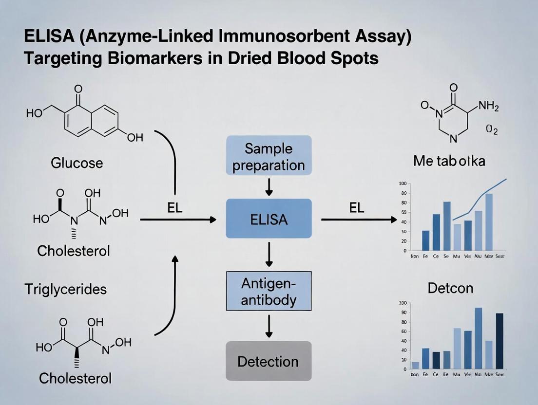
Dried Blood Spot ELISA: A Complete Guide to Metabolic Biomarker Analysis for Researchers
This article provides a comprehensive resource for researchers, scientists, and drug development professionals on the application of Enzyme-Linked Immunosorbent Assay (ELISA) for quantifying metabolic biomarkers in dried blood spot (DBS)...
Dried Blood Spot ELISA: A Complete Guide to Metabolic Biomarker Analysis for Researchers
Abstract
This article provides a comprehensive resource for researchers, scientists, and drug development professionals on the application of Enzyme-Linked Immunosorbent Assay (ELISA) for quantifying metabolic biomarkers in dried blood spot (DBS) samples. We cover the foundational principles of DBS sampling and its advantages over conventional methods. A detailed methodological section guides users through sample preparation, protocol adaptation, and data analysis. We address common troubleshooting challenges and optimization strategies to improve sensitivity and reproducibility. Finally, we examine the critical processes of assay validation, comparative performance against other analytical platforms, and regulatory considerations. This guide synthesizes current best practices to enable robust and reliable metabolic profiling from minimally invasive DBS specimens.
Why Dried Blood Spots? Understanding the Fundamentals of Metabolic Biomarker Sampling
History and Evolution
Dried Blood Spot (DBS) sampling, a microsampling technique, originated from Dr. Robert Guthrie’s work in the early 1960s for phenylketonuria (PKU) screening in newborns. The method involved collecting capillary blood from a heel or finger prick onto filter paper, which was then dried and analyzed. Over decades, its application expanded from neonatal screening to therapeutic drug monitoring, epidemiology, and biomarker research. The integration with advanced analytical techniques like LC-MS/MS and, more recently, ELISA, has cemented DBS as a cornerstone in modern biomedical research and drug development.
Principles of DBS Sampling
The core principle involves the application of a small volume of whole blood (typically 10-50 µL) onto a specially manufactured cellulose or polymer-coated filter paper card. The blood saturates the paper and is air-dried at ambient temperature, stabilizing many analytes. The dried spot is then punched, and the analyte is eluted from the paper matrix into a suitable buffer for downstream analysis, such as ELISA.
Key Advantages of DBS in Research
DBS sampling offers transformative advantages, particularly within biomarker research frameworks:
Table 1: Key Advantages of DBS Sampling
| Advantage | Description | Quantitative Impact |
|---|---|---|
| Minimally Invasive | Capillary blood from finger/heel prick vs. venipuncture. | Reduces sample volume to 10-50 µL vs. >1 mL for serum/plasma. |
| Enhanced Stability | Drying inactivates many degrading enzymes & pathogens. | Many analytes stable for weeks at ambient temp vs. hours for liquid blood. |
| Logistical Simplicity | Easy shipping & storage; no cold chain often required. | Shipping cost reduction up to ~90%; storage at -20°C vs. -80°C for some assays. |
| Biohazard Reduction | Pathogen inactivation upon drying lowers biosafety risk. | Reduced BSL requirements for many endemic pathogens. |
| Ethical & Practical | Enables remote, pediatric, & frequent sampling. | Enables high-frequency sampling in trials; critical for neonatal studies. |
Application in ELISA for Metabolic Biomarkers
Within the context of a thesis on ELISA for metabolic biomarkers, DBS serves as a powerful sample collection matrix. Metabolic biomarkers (e.g., hormones, lipids, inflammatory cytokines) can be quantitatively measured using sensitive ELISA protocols adapted for DBS eluates. Key considerations include:
- Elution Optimization: Maximizing analyte recovery from the paper matrix.
- Matrix Effects: Addressing interference from paper components or hemolysis.
- Correlation: Establishing robust correlation between DBS and plasma/serum values.
Experimental Protocol: ELISA for a Metabolic Biomarker from DBS
Protocol Title: Quantitative Analysis of Adiponectin in Human DBS Samples using a Commercial ELISA Kit.
Objective: To determine the concentration of the metabolic hormone adiponectin in human capillary blood collected via DBS.
I. Materials & Reagent Solutions (The Scientist's Toolkit) Table 2: Essential Research Reagent Solutions & Materials
| Item | Function & Specification |
|---|---|
| DBS Cards | Specially manufactured cellulose cards (e.g., Whatman 903). Provide consistent absorbency and purity. |
| Punch Tool/Hole Punch | Sterile, single-use 3-6 mm punch to obtain a uniform disc from the DBS center. |
| Elution Buffer | Assay-specific buffer (e.g., PBS with 0.1% Tween 20, BSA) to extract analyte from paper matrix. |
| Commercial Adiponectin ELISA Kit | Contains pre-coated plate, standards, detection antibodies, enzyme conjugate, and substrates. |
| Microplate Reader | For measuring absorbance at 450 nm (with 620 nm reference). |
| Humidity Indicator Cards | Packed with DBS cards during drying to ensure proper dryness (<20% humidity). |
| Desiccant Packs & Ziplock Bags | For long-term storage of dried DBS cards at -20°C. |
II. Step-by-Step Methodology
A. Sample Collection & Preparation
- Collection: Clean finger with alcohol. Use lancet for puncture. Touch capillary blood to filter paper circle. Fill completely.
- Drying: Air-dry cards horizontally for ≥3 hours at ambient temperature (15-22°C). Avoid direct sunlight or heat.
- Storage: Place dried card in a ziplock bag with desiccant and humidity card. Store at -20°C until analysis.
B. DBS Elution
- Punch a 3 mm disc from the center of the DBS using a sterile punch.
- Place the disc into a low-protein-binding microcentrifuge tube.
- Add 150 µL of the provided ELISA assay diluent (or optimized elution buffer).
- Seal tube and elute via gentle shaking (2 hours at room temperature) or overnight at 4°C.
- Centrifuge at 10,000 x g for 5 minutes to pellet paper debris. Carefully transfer supernatant (eluate) to a new tube.
C. ELISA Procedure (Adapted from Kit Protocol)
- Preparation: Reconstitute standards in provided diluent. Prepare all reagents.
- Loading: Add 100 µL of standards, DBS eluates (neat or diluted), and controls to appropriate wells of the antibody-coated microplate.
- Incubation: Cover plate. Incubate 2 hours at room temperature.
- Washing: Aspirate and wash each well 4x with 300 µL Wash Buffer.
- Detection Antibody: Add 100 µL of biotinylated detection antibody to each well. Incubate 1 hour. Wash as in step 4.
- Enzyme Conjugate: Add 100 µL of HRP-Streptavidin solution. Incubate 30 minutes. Wash as in step 4.
- Substrate & Stop: Add 100 µL of TMB Substrate. Incubate 15 minutes in the dark. Add 100 µL Stop Solution.
- Read: Measure absorbance at 450 nm within 30 minutes.
D. Data Analysis
- Generate a standard curve by plotting mean absorbance vs. standard concentration.
- Fit a 4- or 5-parameter logistic curve.
- Interpolate sample concentrations from the curve.
- Apply any necessary dilution factor. Report concentration in ng/mL of eluate.
- Optional: Correlate with hematocrit-corrected values or parallel plasma assays.
Visualizations
Diagram Title: DBS-ELISA Workflow for Metabolic Biomarkers
Diagram Title: Key Advantages of DBS vs. Venous Sampling
Application Notes: ELISA for Metabolic Biomarkers in Dried Blood Spot Research
Metabolic biomarkers, defined as measurable indicators of metabolic processes, pathways, or states, are crucial for understanding health, disease progression, and therapeutic response. Their quantification in dried blood spots (DBS) offers significant advantages in sample stability, logistics, and minimal invasiveness, making them ideal for large-scale epidemiological studies and therapeutic drug monitoring. Enzyme-Linked Immunosorbent Assay (ELISA) remains a cornerstone technique for the sensitive and specific quantification of proteinaceous metabolic biomarkers from DBS eluates. This note details protocols and considerations for applying ELISA in this context, framed within a thesis on advancing DBS-based metabolic profiling.
Key Biomarker Classes and Relevance: Table 1: Major Classes of Metabolic Biomarkers with Examples and Relevance
| Biomarker Class | Definition | Example Biomarkers | Primary Clinical/Research Relevance |
|---|---|---|---|
| Lipids & Lipoproteins | Molecules involved in fat metabolism and transport. | LDL-C, HDL-C, Apolipoprotein B, Triglycerides | Cardiovascular disease risk assessment, metabolic syndrome monitoring. |
| Carbohydrate Metabolism | Indicators of sugar metabolism and control. | Hemoglobin A1c (HbA1c), Insulin, C-peptide, Fructosamine | Diagnosis and management of diabetes mellitus, insulin resistance studies. |
| Amino Acids & Derivatives | Building blocks of proteins and their metabolites. | Homocysteine, Phenylalanine, Branched-Chain Amino Acids (BCAAs) | Nutritional status, inborn errors of metabolism (e.g., PKU), cardiovascular risk. |
| Inflammatory Cytokines | Signaling proteins mediating inflammation. | TNF-α, IL-6, CRP (C-reactive protein) | Tracking systemic inflammation, autoimmune diseases, cardiometabolic risk. |
| Oxidative Stress Markers | Molecules indicating redox imbalance. | Malondialdehyde (MDA), 8-OHdG, Nitrotyrosine | Research in aging, neurodegenerative disorders, and metabolic diseases. |
Quantitative Data from Recent DBS-ELISA Studies: Table 2: Representative Performance Metrics for ELISA of Metabolic Biomarkers in DBS
| Biomarker | Sample Volume (µL) | ELISA Kit (Example) | Reported Correlation (DBS vs. Plasma) | Key Advantage Demonstrated |
|---|---|---|---|---|
| C-reactive Protein (CRP) | ~3.2 mm punch | Human High Sensitivity CRP ELISA | r = 0.98 | Stability at room temp >7 days enables remote sampling. |
| Interleukin-6 (IL-6) | ~3.2 mm punch | Human IL-6 Quantikine ELISA | r = 0.95 | Suitable for pediatric and geriatric populations due to minimal blood draw. |
| Insulin | 6 µL whole spot | Human Insulin ELISA | r = 0.97 | Effective for large-scale diabetes screening programs. |
| Apolipoprotein A1 | 3.2 mm punch | Human ApoA1 ELISA | r = 0.93 | Reliable for cardiovascular risk stratification in field studies. |
Detailed Experimental Protocols
Protocol 1: DBS Sample Collection, Processing, and Elution for ELISA
Objective: To obtain a consistent, high-quality protein eluate from a DBS sample for subsequent ELISA analysis.
Materials (Research Reagent Solutions Toolkit):
- Whatman 903 Protein Saver Cards
- Disposable lancets & capillary tubes
- Desiccant packs & zip-lock bags
- Harris 3.2 mm Uni-Core punch
- Low-protein-binding microcentrifuge tubes
- ELISA Elution Buffer (e.g., 1% BSA, 0.1% Tween-20 in PBS, pH 7.4)
- Orbital shaker/rocker
- Centrifuge
Methodology:
- Collection: Apply a single drop of capillary blood from a finger prick to fill a pre-printed circle on the DBS card. Allow to dry completely for a minimum of 3 hours at ambient temperature in a clean, dust-free environment.
- Storage: Place dried cards in individual zip-lock bags with desiccant packs. Store at -20°C or below for long-term stability until analysis.
- Punching: Using a clean 3.2 mm punch, take a single disc from the center of the DBS. Transfer the disc to a labeled low-protein-binding microcentrifuge tube.
- Elution: Add 250 µL of ELISA Elution Buffer to the tube. Seal and incubate on an orbital shaker (gentle agitation) at 4°C for 2-3 hours, or overnight for optimal recovery.
- Clarification: Centrifuge the tube at 10,000 x g for 5 minutes at 4°C to pellet paper debris and cellular remnants.
- Analysis: Carefully transfer the clear supernatant (eluate) to a new tube. The eluate is now ready for direct analysis in the ELISA protocol. Note: The elution volume defines the theoretical sample concentration factor. A 3.2 mm punch from a 6 µL spot eluted into 250 µL gives an approximate 1:42 dilution.
Protocol 2: Direct Quantification of a Metabolic Biomarker via Sandwich ELISA
Objective: To quantify the concentration of a target protein biomarker (e.g., IL-6) in the DBS eluate.
Materials (Research Reagent Solutions Toolkit):
- Commercial Sandwich ELISA Kit (matched antibody pairs, standards, detection reagents)
- DBS eluates from Protocol 1
- Coated 96-well microplate (provided in kit)
- Plate washer or manual wash bottle
- Wash Buffer (PBS with 0.05% Tween-20)
- Microplate reader capable of measuring appropriate absorbance (e.g., 450 nm with 570 nm correction)
Methodology:
- Plate Preparation: Bring all reagents and samples to room temperature. Prepare serial dilutions of the protein standard according to the kit manual.
- Sample Loading: Add 100 µL of each standard, DBS eluate (undiluted or diluted as determined by pilot study), and blank (elution buffer) to appropriate wells in duplicate.
- Incubation: Cover plate and incubate at room temperature for 2 hours.
- Washing: Aspirate and wash each well 4 times with 300 µL Wash Buffer, ensuring complete removal of liquid after each wash.
- Detection Antibody Addition: Add 100 µL of biotinylated detection antibody to each well. Incubate for 1-2 hours at room temperature.
- Washing: Repeat wash step as in #4.
- Enzyme Conjugate Addition: Add 100 µL of Streptavidin-Horseradish Peroxidase (HRP) solution. Incubate for 30 minutes at room temperature, protected from light.
- Washing: Repeat wash step as in #4.
- Substrate Addition: Add 100 µL of TMB (3,3',5,5'-Tetramethylbenzidine) substrate solution. Incubate for 15-30 minutes at room temperature, protected from light, until color develops.
- Stop Reaction: Add 100 µL of Stop Solution (e.g., 1M H2SO4). The blue color will turn yellow.
- Reading & Analysis: Measure the absorbance at 450 nm within 30 minutes. Subtract the 570 nm reference reading. Generate a standard curve (4-parameter logistic fit recommended) and interpolate sample concentrations. Apply any necessary dilution factors to calculate the original concentration in the DBS.
Mandatory Visualizations
Diagram 1: DBS-ELISA Workflow for Metabolic Biomarkers
Diagram 2: Metabolic Inflammation Pathway & Biomarker Detection
The Scientist's Toolkit: Key Research Reagent Solutions for DBS-ELISA Table 3: Essential Materials for DBS-Based Metabolic Biomarker ELISA
| Item | Function & Rationale |
|---|---|
| Protein Saver DBS Cards | Cellulose-based filter paper treated to denature proteins and enhance stability during drying and storage. |
| BSA/Tween-20 Elution Buffer | Optimized buffer to efficiently elute proteins from cellulose matrix while preventing non-specific binding in subsequent ELISA. |
| Validated Sandwich ELISA Kit | Provides matched, affinity-purified antibody pairs, calibrated standards, and optimized buffers for specific, quantitative detection. |
| Low-Protein-Binding Tubes/Tips | Minimizes adsorptive loss of low-abundance target proteins during sample processing. |
| Certified DBS Punch | Provides a precise, consistent disc size (e.g., 3.2 mm) for volumetric or spot-area-based quantitative analysis. |
| Microplate Reader with Analysis Software | Enables accurate optical density measurement and curve-fitting for concentration interpolation. |
This application note details the integration of Enzyme-Linked Immunosorbent Assay (ELISA) with Dried Blood Spot (DBS) sampling, a synergistic approach offering significant logistical and analytical advantages for metabolic biomarker research. Framed within a thesis on ELISA for metabolic biomarkers in DBS, this document provides current data, protocols, and reagent toolkits for researchers and drug development professionals.
The confluence of DBS technology—enabling simplified collection, stabilization, and transport of blood samples—with the specificity and throughput of ELISA creates a powerful platform for metabolic biomarker quantification. This synergy addresses critical challenges in large-scale epidemiological studies, pediatric research, and decentralized clinical trials.
Table 1: Comparative Analysis of Sample Collection & Logistics
| Parameter | Conventional Venipuncture + Serum ELISA | DBS + ELISA | Advantage % |
|---|---|---|---|
| Sample Volume Required | 3-5 mL whole blood | 50-100 µL (spot) | ~98% reduction |
| Sample Stability (Ambient) | Hours (requires cold chain) | 2-4 weeks (for many analytes) | >700% improvement |
| Collection Cost (Est.) | $50-100 per draw | $5-15 per card | ~80% reduction |
| Transport & Storage Cost | High (cold chain) | Low (ambient, lightweight) | ~90% reduction |
| Required Personnel Phlebotomist | Patient/caregiver (self-sampling possible) | Enables remote sampling |
Table 2: Analytical Performance of ELISA on DBS Eluates (Example Biomarkers)
| Metabolic Biomarker | Correlation (r) vs. Plasma/Sera | Average Recovery from DBS | Key Pre-Analytical Factor |
|---|---|---|---|
| HbA1c | >0.95 | 95-102% | Hematocrit effect (critical) |
| C-Peptide | 0.90-0.94 | 88-95% | Spot homogeneity, punch location |
| 25-Hydroxy Vitamin D | 0.92-0.98 | 92-101% | Drying time, humidity control |
| CRP (hs) | 0.88-0.93 | 85-92% | Hematocrit, elution efficiency |
Detailed Experimental Protocols
Protocol 1: Standardized DBS Sample Collection & Preparation for Metabolic Biomarker ELISA
Objective: To obtain consistent, high-quality DBS eluates for downstream quantitative ELISA. Materials: FDA-approved filter paper cards, lancets, desiccant packs, zip-lock bags with humidity indicators, 3-4 mm DBS punch, rocking platform. Procedure:
- Collection: Fill pre-printed circle on filter paper card completely with a single, saturated blood spot (~50 µL). Allow to dry horizontally for a minimum of 3 hours at ambient temperature (15-22°C).
- Storage & Transport: Place dried card in a zip-lock bag with desiccant and humidity indicator. Store at ≤-20°C for long-term; transport at ambient temperature if stability data permits.
- Punching: Using a calibrated dis punch, remove a single 3 mm (or as validated) punch from the center of the DBS, avoiding uneven edges.
- Elution: Place punch into a low-protein-binding microcentrifuge tube. Add appropriate elution buffer (e.g., PBS with 0.1% Tween 20, 0.5% BSA) at a fixed ratio (e.g., 1 punch:200 µL). Elute on a rocking platform for 2 hours at 4°C.
- Clarification: Centrifuge at 10,000 x g for 5 minutes. Carefully transfer supernatant (the DBS eluate) to a new tube for immediate ELISA analysis or storage at ≤-80°C.
Protocol 2: Modified Sandwich ELISA for C-Peptide in DBS Eluates
Objective: To quantify C-Peptide, a key metabolic biomarker for beta-cell function, from DBS samples. Materials: Commercial human C-Peptide ELISA kit (validated for serum/plasma), DBS eluates, calibrators re-constituted in elution buffer matched for hematocrit. Modified Procedure:
- Calibrator/Control Preparation: Prepare kit calibrators in an artificial matrix mimicking the final composition of the DBS eluate (critical for accuracy).
- Assay Procedure: Follow kit insert with key modifications:
- Add 50 µL of DBS eluate or modified calibrator per well.
- Incubate plate overnight at 4°C (increases sensitivity for low-concentration analytes common in DBS).
- Proceed with washing, detection, and stop steps as per manufacturer.
- Data Analysis: Generate standard curve using modified calibrators. Apply correction factor determined from recovery experiments if necessary. Report concentration adjusted for elution volume and punch size.
Visualizing the Workflow & Context
Diagram Title: Integrated DBS-ELISA Workflow for Biomarker Analysis
The Scientist's Toolkit: Key Research Reagent Solutions
Table 3: Essential Materials for DBS-ELISA Integration
| Item | Function & Importance |
|---|---|
| Whatman 903 Protein Saver Card | Standardized cellulose matrix for consistent blood absorption and analyte stability. |
| Hematocrit-Adjusted Elution Buffer (PBS + 0.5% BSA + 0.1% Tween-20) | Maximizes protein recovery, minimizes nonspecific binding in ELISA wells. |
| Pre-Punched Calibrator/Control Spots | Lyophilized biomarker spotted on cards; provides process control from punch to plate. |
| Low-Binding Microcentrifuge Tubes & Tips | Prevents adsorption of low-abundance biomarkers onto plastic surfaces. |
| Validated, High-Sensitivity ELISA Kits | Kits with lower limits of detection accommodate sample dilution from elution step. |
| Ambient Humidity Indicator Cards | Critical for monitoring sample integrity during transport/storage before analysis. |
Application Notes
The application of Enzyme-Linked Immunosorbent Assay (ELISA) for metabolic biomarker analysis in dried blood spots (DBS) represents a pivotal methodological bridge between public health diagnostics and modern drug development. Within the thesis framework of "ELISA for Metabolic Biomarkers in Dried Blood Spot Research," this dual utility underscores a powerful convergence of simplicity, scalability, and analytical robustness.
1. Newborn Screening (NBS) for Inborn Errors of Metabolism (IEMs): Public health programs globally utilize DBS-ELISA for high-throughput, cost-effective screening of newborns. The method quantifies specific proteins or enzyme activities indicative of disorders like congenital hypothyroidism (via thyroxine, T4), congenital adrenal hyperplasia (via 17-hydroxyprogesterone), and lysosomal storage disorders (via enzymatic activity assays). The stability of analytes in DBS allows for efficient sample transport from remote collection sites to centralized laboratories, enabling equitable screening coverage.
2. Therapeutic Drug Monitoring (TDM) and Pharmacokinetic (PK) Studies: In drug development, DBS-ELISA provides a minimally invasive sampling method crucial for serial PK profiling. It allows for the quantification of therapeutic proteins, monoclonal antibodies, and biomarkers of drug response or toxicity from a single drop of blood. This facilitates dense sampling schedules in early-phase clinical trials, even in outpatient settings, improving data quality and patient compliance while reducing logistical burdens and biohazard risks associated with liquid plasma/serum.
3. Biomarker Validation in Clinical Research: DBS-ELISA serves as a tool for validating novel metabolic biomarkers in longitudinal cohort studies. Its ability to use archived DBS samples from biobanks enables retrospective analysis, linking biomarker levels to clinical outcomes. This application is critical for identifying prognostic or diagnostic signatures for complex metabolic diseases.
Table 1: Key Performance Metrics of DBS-ELISA in Selected Applications
| Application | Target Analyte | Typical Assay Range | Sensitivity (LLoQ) | Key Advantage (vs. Serum/Plasma) |
|---|---|---|---|---|
| Newborn Screening | 17-OH Progesterone | 5-100 nmol/L | ~2 nmol/L | High-throughput, stable transport |
| Newborn Screening | TSH (Thyroid) | 5-200 mIU/L | ~1 mIU/L | Enables centralized mass screening |
| PK Studies (mAb) | Anti-TNFα mAb | 0.5-50 µg/mL | ~0.2 µg/mL | Microsampling; sparse/serial sampling |
| Therapeutic Monitoring | Tacrolimus | 1-50 ng/mL | ~0.5 ng/mL | Patient self-collection potential |
| Biomarker Research | Cystatin C | 0.5-10 mg/L | ~0.1 mg/L | Stability for retrospective analysis |
Table 2: Comparison of Sample Requirements: DBS vs. Conventional Venipuncture
| Parameter | DBS Sample | Conventional Venous Draw | Advantage |
|---|---|---|---|
| Volume Required | ~10-20 µL per spot | 3-10 mL | >99% reduction |
| Collection Method | Finger/heel stick, home kit | Phlebotomist, clinic | Minimally invasive, decentralized |
| Transport/Storage | Ambient, low biohazard | Frozen, cold chain | Simplified logistics, lower cost |
| Stability (Typical) | Weeks to months at room temp | Hours to days at 4°C | Facilitates biobanking |
Experimental Protocols
Protocol 1: DBS-ELISA for a Novel Therapeutic Monoclonal Antibody (mAb) Pharmacokinetics
Objective: To quantify serum concentrations of a humanized IgG1 mAb (Drug-X) from DBS samples in a Phase I clinical trial.
Materials: DBS cards (Whatman 903), 3 mm DBS punch, calibration standards (Drug-X in whole blood), quality controls, anti-human IgG Fc-specific capture antibody, biotinylated anti-idiotype detection antibody, streptavidin-HRP, chemiluminescent substrate, assay buffer.
Procedure:
- Sample Collection & Preparation: Collect 20 µL of capillary whole blood via finger prick onto DBS card. Dry for ≥3 hours at room temperature. Store with desiccant at ≤-20°C until analysis.
- Punch Extraction: Punch a single 3 mm disc from the center of the DBS spot into a microtiter plate well. Add 100 µL of extraction buffer (PBS with 0.5% BSA, 0.1% Tween-20, 0.05% sodium azide). Seal plate and shake (800 rpm) for 2 hours at room temperature.
- Standard Curve: Prepare Drug-X in drug-free whole blood at 0.5, 1, 5, 20, 50 µg/mL. Spot 20 µL, dry, and process identically to subject samples.
- Sandwich ELISA: Transfer 50 µL of extract to a separate assay plate pre-coated with capture antibody. Incubate 1.5 hours. Wash 5x. Add 50 µL biotinylated detection antibody (1 µg/mL). Incubate 1 hour. Wash 5x. Add 50 µL streptavidin-HRP (1:10,000). Incubate 30 min. Wash 5x. Add 50 µL chemiluminescent substrate, read immediately.
- Data Analysis: Generate a 4-parameter logistic (4PL) standard curve. Apply hematocrit correction factor if necessary. Report concentrations in µg/mL.
Protocol 2: DBS-ELISA for Newborn Screening of 17-Hydroxyprogesterone (17-OHP)
Objective: To screen for congenital adrenal hyperplasia (CAH) by quantifying 17-OHP in newborn DBS.
Materials: Commercially available 17-OHP ELISA kit (validated for DBS), DBS calibrators/controls, 3 mm punch, microtiter plate shaker, plate washer, spectrophotometer.
Procedure:
- Punching: Punch one 3.2 mm disc from each unknown, calibrator, and control DBS into respective wells of the antibody-coated plate.
- Competitive ELISA: Add 100 µL of enzyme conjugate (17-OHP-HRP) to each well. Incubate with shaking (700 rpm) for 60 minutes at room temperature.
- Washing: Aspirate and wash wells 4 times with wash buffer.
- Detection: Add 100 µL of TMB substrate. Incubate for 20 minutes in the dark.
- Stop & Read: Add 100 µL of stop solution (1M H2SO4). Measure absorbance at 450 nm (reference 620-650 nm) within 15 minutes.
- Interpretation: Calculate 17-OHP concentration from the standard curve. Values > 30 nmol/L typically trigger reflex testing (e.g., LC-MS/MS confirmation).
Diagrams
DBS Workflow for Newborn Screening of CAH
DBS-ELISA Workflow for Pharmacokinetic Study
General ELISA Detection Pathway from DBS
The Scientist's Toolkit: Research Reagent Solutions
Table 3: Essential Materials for DBS-ELISA Research
| Item | Function & Specification | Rationale for DBS Application |
|---|---|---|
| Filter Paper Cards | High-quality cellulose (e.g., Whatman 903). Consistent porosity and blood spreading. | Ensures uniform spot formation, critical for accurate volumetric sampling via punch. |
| Pre-Punched Plates / Manual Punch | 3-6 mm diameter. Disposable punches or automated systems. | Enables transfer of a precise, reproducible volume of dried blood into the assay well. |
| Extraction Buffer | PBS or Tris-based with surfactant (Tween-20) and protein (BSA). | Efficiently elutes analytes from cellulose matrix while maintaining immuno-reactivity. |
| Hematocrit Correction Solution | Standardized buffers or bioinformatics algorithms. | Corrects for the variable volume of plasma in a fixed punch due to donor hematocrit effects. |
| Analyte-Specific ELISA Kit | Validated for DBS (matrix effects assessed). Includes DBS-specific calibrators. | Provides optimized antibody pairs, reagents, and a protocol adapted for the DBS matrix. |
| DBS Calibrators & Controls | Whole blood spiked with known analyte levels, spotted and dried identically to samples. | Crucial for constructing an accurate standard curve that mirrors the sample extraction efficiency. |
| Stabilizing Desiccant Packs | Silica gel. | Placed in bags with DBS cards to prevent humidity-related degradation during storage/transport. |
Key Challenges and Limitations of DBS for Immunoassays
1. Introduction Within the context of advancing enzyme-linked immunosorbent assay (ELISA) methodologies for metabolic biomarkers in dried blood spots (DBS), understanding the inherent limitations of the DBS matrix is crucial. While DBS sampling offers logistical advantages, its application in quantitative immunoassays presents distinct challenges that must be addressed to ensure data robustness for research and drug development.
2. Core Challenges & Quantitative Data Summary The primary challenges are categorized and summarized in the table below with supporting quantitative data from current literature.
Table 1: Key Quantitative Challenges in DBS Immunoassay
| Challenge Category | Specific Issue | Impact & Representative Data | Proposed Mitigation Strategy |
|---|---|---|---|
| Hematocrit (HCT) Effect | Variable spreading & analyte concentration. | ±30% bias in analyte quantitation across HCT range of 0.20-0.60 L/L. | Use pre-punched discs from calibrated volumetric DBS devices; HCT correction algorithms. |
| Sample Homogeneity | Non-uniform analyte distribution within spot. | >20% CV for punches from same DBS card vs. <5% CV for liquid duplicate. | Entire spot elution; automated, non-punch-based image analysis/elution. |
| Extraction Efficiency | Incomplete analyte recovery from cellulose matrix. | Recovery varies 70-120%, dependent on analyte size (e.g., antibodies vs. small peptides) and spot age. | Optimized buffer (e.g., surfactants, pH); prolonged shaking; sonication. |
| Matrix Effects | High background, non-specific binding, interference. | Can suppress or enhance signal by up to 25% vs. liquid serum/plasma controls. | Enhanced blocking (casein/BSA); selective immunoaffinity capture; sample dilution. |
| Stability & Storage | Analyte degradation over time. | Some labile metabolic biomarkers show >15% loss after 30 days at room temperature. | Controlled storage (-20°C or lower); use of desiccant and humidity indicator cards. |
| Volume/Spot Size | Inaccurate volumetric assumption. | Assumed 10-15 µL per 3 mm punch, but actual volume can vary by ±50% due to HCT/viscosity. | Volumetric absorbent papers; area-based normalization via hemoglobin assay. |
3. Detailed Experimental Protocol: Evaluating HCT Impact on DBS-ELISA This protocol is essential for validating any DBS-ELISA method for metabolic biomarkers.
Aim: To quantify the effect of hematocrit on the measured concentration of a target metabolic biomarker (e.g., adiponectin) in a DBS-based ELISA.
Materials & Reagents (The Scientist's Toolkit): Table 2: Essential Research Reagent Solutions
| Item | Function |
|---|---|
| Anti-Adiponectin ELISA Kit | Provides matched antibody pairs, standards, and optimized buffers for detection. |
| DBS Cards (Whatman 903) | Standardized cellulose-based collection paper. |
| Defibrinated Whole Blood | Allows for controlled adjustment of hematocrit without clotting. |
| Phosphate-Buffered Saline (PBS) | Used for adjusting hematocrit and as an elution buffer component. |
| BSA (1% in PBS) | Used as a blocking agent and as a stabilizing additive in elution buffers. |
| Tween-20 | Non-ionic surfactant added to elution buffer to improve protein recovery. |
| Hemoglobin Assay Kit | For normalizing eluted sample volume based on total blood content. |
| Hematocrit Centrifuge | To prepare and verify blood samples at specific HCT levels. |
| 3 mm DBS Punch | For consistent sub-sampling (if used). |
Procedure:
- Prepare HCT-Adjusted Blood: Using defibrinated blood and autologous plasma (or PBS), prepare aliquots at five distinct hematocrit levels (e.g., 0.20, 0.30, 0.40, 0.50, 0.60 L/L). Spike a known concentration of the purified target biomarker into each aliquot.
- DBS Spotting: Apply a precise volume (e.g., 20 µL) of each HCT-adjusted blood sample onto DBS cards in quintuplicate. Allow spots to dry for ≥3 hours at ambient temperature in a desiccated environment.
- Elution: Punch a single 3 mm disc from the center of each DBS (or use whole-spot elution). Elute in 150 µL of optimized elution buffer (PBS, 0.5% BSA, 0.1% Tween-20) with shaking (500 rpm, 2 hours, 4°C).
- Hemoglobin Quantification: Transfer 10 µL of each eluate to a separate plate for hemoglobin measurement via spectrophotometry (e.g., 415 nm) or a commercial kit. This serves as a volume/erythrocyte content normalizer.
- ELISA Analysis: Perform the standard ELISA protocol on the remaining eluate, using kit standards prepared in the same elution buffer to match the matrix.
- Data Analysis:
- Calculate biomarker concentration from the ELISA standard curve.
- Normalize concentrations using the corresponding hemoglobin reading.
- Plot measured biomarker concentration (normalized and non-normalized) against the HCT level. Calculate bias relative to the known spiked concentration at the median HCT (e.g., 0.40 L/L).
4. Visualization of Workflow & Challenges
Diagram 1: DBS-ELISA Workflow with Key Challenge Points
Diagram 2: Mitigation Strategy Decision Pathway
5. Conclusion Successful implementation of ELISA for metabolic biomarkers in DBS requires systematic investigation and mitigation of the challenges outlined. A rigorous validation protocol must include assessment of HCT effect, extraction efficiency, and stability under intended storage conditions. The strategies and detailed protocols provided here form a critical foundation for generating reliable, high-quality data in pharmaceutical research and development using DBS technology.
Step-by-Step Protocol: Adapting and Running ELISA on Dried Blood Spot Specimens
This application note details optimal pre-analytical protocols for dried blood spot (DBS) sampling, a cornerstone technique for metabolic biomarker analysis via ELISA in clinical and pharmaceutical research. The integrity of pre-analytical procedures directly impacts the accuracy, reproducibility, and clinical relevance of downstream ELISA quantification. This guide is framed within a broader thesis investigating the standardization of DBS methodologies for robust metabolic biomarker research in drug development.
Optimal Blood Collection Guidelines
Effective DBS analysis begins with meticulous blood collection. The choice of method influences hematocrit (Hct) effects, analyte stability, and spot homogeneity.
Key Considerations:
- Source: Capillary (finger/lancet) or venous blood.
- Anticoagulant: EDTA is preferred for most metabolic biomarkers (e.g., peptides, small molecules) to prevent clotting and ensure uniform spot formation. Heparin may interfere with some antibody-binding in ELISA.
- Hematocrit (Hct): The most significant pre-analytical variable. Hct affects blood viscosity, diffusion radius, and drying kinetics, leading to the "hematocrit effect" which can bias punch location and analyte recovery.
Quantitative Data Summary:
Table 1: Impact of Collection Variables on DBS Quality for Metabolic Biomarkers
| Variable | Optimal Condition | Effect of Deviation | Quantitative Impact (Typical Range) |
|---|---|---|---|
| Blood Source | Venous (controlled Hct) or Capillary (vol. limitation) | Capillary blood may have higher interstitial fluid. | Hct variation: Capillary ±7% vs. venous. |
| Anticoagulant | K2EDTA (1.5-2.2 mg/mL blood) | Heparin can interfere with some ELISAs; Clotting causes inhomogeneity. | EDTA recovery >95% vs. heparin for most protein targets. |
| Hematocrit (Hct) | Target range: 35-45% | High Hct: smaller spot, biased periphery. Low Hct: larger spot, biased center. | Spot diameter change: ~0.5 mm per 5% Hct change. Analyte bias up to 20-30%. |
| Collection Device | Capillary tube or syringe with precise volume control. | Inconsistent volume leads to spot size variability. | Target volume: 10-20 µL per spot (3-4 mm punch). CV >15% with poor volumetric control. |
Spotting & Drying Protocols
Detailed Protocol: Manual Spotting and Drying
- Objective: To apply uniform, reproducible DBS samples onto filter paper cards.
- Materials: Whatman 903 protein saver card or equivalent, calibrated pipette (10-50 µL), safety lancet, capillary tubes/EDTA tubes, drying rack.
- Procedure:
- Perform skin puncture or draw venous blood into EDTA tube.
- Gently mix blood tube by inversion 8-10 times. Do not vortex.
- Using a calibrated pipette, aspirate a precise volume (e.g., 15 µL).
- Hold the pipette tip perpendicular to the filter paper card, approximately 0.5 cm above the pre-printed circle.
- Dispense the blood in a single, steady action onto the center of the circle. Allow the blood to wick freely into the paper. Do not touch the tip to the paper.
- Repeat for required number of spots, using a fresh pipette tip for each sample.
- Immediately place the card horizontally on a drying rack in a biohazard laminar flow hood or a dedicated, clean drying area.
- Dry at ambient temperature (15-25°C) with moderate humidity (<60%) for a minimum of 3 hours. Ensure cards are protected from dust, insects, and direct sunlight.
- Validate complete drying by visual inspection (spots should be a consistent dark brown/matte, not glossy red).
- Quality Control: Record drying time, temperature, and humidity. Use visual QC for spot uniformity and complete saturation.
Quantitative Data Summary:
Table 2: Spotting and Drying Optimization Parameters
| Parameter | Optimal Protocol | Recommended Validation | Performance Metric |
|---|---|---|---|
| Spotting Volume | 15 µL for standard 3.2 mm punch | Validate for specific biomarker linearity. | Consistent spot diameter within ±1 mm. |
| Drying Time | ≥3 hours at 20-25°C, <60% RH. | Weigh card to constant mass. | Residual moisture <5% by weight. |
| Drying Environment | Clean, ambient, with air circulation. | Microbial culture of random spots. | No microbial growth after 48h incubation. |
| Card Type | Whatman 903 (cellulose) for most biomarkers. | Compare analyte recovery vs. other papers. | >90% recovery of spiked analyte post-drying. |
Storage and Stability Guidelines
Long-term storage stability is critical for batch analysis in longitudinal studies.
Detailed Protocol: Post-Drying Processing and Storage
- Objective: To preserve DBS integrity and analyte stability for long-term storage prior to ELISA.
- Materials: Low gas-permeable plastic bags with desiccant, oxygen scavengers, humidity indicator cards, -20°C or -80°C freezer, barcode labels.
- Procedure:
- After confirmed drying, place each card in a individual low-permeability zip-lock bag.
- Add 1-2 desiccant packets (e.g., silica gel) and one oxygen scavenger sachet per bag.
- Include a humidity indicator card. Target internal humidity <10%.
- Seal the bag completely, expelling as much air as possible.
- Affix a durable, barcoded label with sample ID and date.
- For long-term storage (>1 month), store bags at ≤-20°C. For maximum stability of labile metabolic biomarkers (e.g., certain phosphorylated peptides), store at -80°C.
- Avoid repeated freeze-thaw cycles. For analysis, remove the entire bag and allow it to equilibrate to room temperature while sealed (approx. 30 min) before opening to prevent condensation on spots.
- Stability Monitoring: Create stability QC pools from control blood. Store alongside samples and test at pre-defined intervals (e.g., 1, 3, 6, 12 months) using your ELISA protocol.
Quantitative Data Summary:
Table 3: DBS Storage Stability for Metabolic Biomarkers
| Storage Condition | Target Analytes | Demonstrated Stability (Literature Range) | Key Degradation Factor |
|---|---|---|---|
| Room Temp. (<25°C) with desiccant | Very stable small molecules (e.g., creatinine) | 7-30 days | Enzymatic activity, oxidation. |
| 4°C with desiccant | Most proteins, peptides, many metabolites | 1-6 months | Limited microbial growth, hydrolysis. |
| -20°C with desiccant/O2 scavenger | Broad range (proteins, hormones, lipids) | 6-24 months | Oxidation, residual enzyme activity. |
| -80°C with desiccant/O2 scavenger | Labile biomarkers (e.g., phosphorylated proteins, unstable metabolites) | 24+ months | Minimizes all chemical/enzymatic degradation. |
The Scientist's Toolkit: Research Reagent Solutions
Table 4: Essential Materials for DBS-Based Metabolic Biomarker Research
| Item | Function/Description | Example Product/Criteria |
|---|---|---|
| Filter Paper Cards | Porous cellulose matrix for blood absorption and storage. Must be consistent in thickness and purity. | Whatman 903 Protein Saver Card, PerkinElmer 226. |
| Punch Tool/Die Cutter | To excise a precise sub-punch from the DBS for elution. | 3.2 mm or 6.0 mm single-hole punch, semi-automated punch platforms. |
| Desiccant | Absorbs moisture within storage bags to prevent microbial growth and hydrolysis. | Silica gel packets (indicating or non-indicating). |
| Oxygen Scavengers | Removes residual O2 in storage bags to prevent oxidative degradation of analytes. | Mitsubishi Ageless ZPT sachets. |
| Humidity Indicator Card | Monitors internal humidity of the storage bag to ensure dry conditions. | Cards with cobalt chloride dots (blue = dry, pink = humid). |
| Low-Gas Permeability Bags | Provides a stable, sealed microenvironment for the DBS card. | Zip-lock bags with aluminum foil laminate or high-barrier plastic. |
| ELISA-Compatible Elution Buffer | Efficiently extracts the target biomarker from the DBS matrix without interfering with antibody binding. | PBS pH 7.4 with 0.1% Tween-20 and 0.5% BSA is common; may require optimization. |
| Whole Blood Quality Controls | Stabilized blood spiked with high/low concentrations of target analyte for process monitoring. | Commercial QC material or lab-prepared pools from donor blood. |
Visualized Workflows and Relationships
Title: DBS Pre-Analytical Workflow for ELISA Biomarker Analysis
Title: Pre-Analytical Factors Influencing DBS ELISA Outcomes
Within the broader thesis on ELISA for metabolic biomarkers in dried blood spot (DBS) research, the initial elution step is the critical foundation. Incomplete or inefficient recovery of analytes from the cellulose matrix directly compromises downstream quantification, leading to inaccurate biomarker profiles. This application note details current, optimized protocols for maximizing elution efficiency, enabling robust and reproducible ELISA results.
The Elution Challenge: Factors Influencing Recovery
Efficient elution must overcome analyte adsorption to the filter paper and cellular components. Key variables include the elution buffer composition, incubation parameters, and physical disruption methods. The optimal strategy is analyte-dependent, particularly for large proteins versus small molecule metabolites.
Quantitative Comparison of Elution Strategies
Table 1: Comparison of Elution Method Efficiencies for Different Biomarker Classes
| Elution Method | Buffer Typical Composition | Incubation Time/Temp | Reported Avg. Recovery (%) - Proteins | Reported Avg. Recovery (%) - Small Molecules | Key Advantage | Primary Limitation |
|---|---|---|---|---|---|---|
| Passive Soaking | PBS + 0.1% Tween-20, or ELISA Sample Diluent | 2-4h, RT or 4°C with shaking | 65-80% | 75-90% | Simple, preserves labile epitopes | Incomplete, long duration |
| Sonication-Assisted | PBS + 0.5% BSA | 15-30 min in ice-water bath | 85-95% | 90-98% | High efficiency, rapid | Potential heat/foam denaturation |
| Vortex-Mixing with Beads | Modified buffer with surfactants (e.g., CHAPS) | 3 x 5 min cycles, RT | 80-90% | 85-95% | Good disruption of cellular material | Increased risk of hemolysis |
| Acetonitrile/MeOH Precipitation | 70:30 ACN:H2O or Methanol | 1h, -20°C | Low (precipitated) | >95% (for metabolites) | Excellent for small molecules, deproteinizes | Denatures proteins, not for ELISA |
Detailed Experimental Protocols
Protocol 1: Sonication-Assisted Elution for Protein Biomarkers (e.g., HbA1c, Cytokines)
Principle: Ultrasonic waves agitate the punch, physically disrupting paper and cell matrices to liberate adsorbed proteins into a stabilizing buffer.
- Punching: Using a calibrated 3.2 mm or 6 mm punch, excise the DBS spot center into a low-protein-binding microcentrifuge tube.
- Buffer Addition: Add 150 µL (for 3.2 mm punch) or 300 µL (for 6 mm punch) of ice-cold elution buffer (PBS, pH 7.4, 0.5% w/v BSA, 0.1% Tween-20, 0.05% ProClin 300).
- Sonication: Place tubes in a microplate-horn sonicator equipped with a cooling ice-water bath. Sonicate at 40 kHz for 25 minutes, ensuring temperature remains below 15°C.
- Incubation & Separation: Transfer tubes to a thermo-shaker and incubate at 4°C with gentle shaking (500 rpm) for an additional 60 minutes. Centrifuge at 14,000 x g for 10 minutes at 4°C.
- Sample Collection: Carefully collect the supernatant for immediate ELISA analysis or storage at -80°C. Avoid the paper pellet and any insoluble debris.
Protocol 2: Organic Solvent Elution for Small Molecule Metabolites (e.g., Amino Acids, Steroids)
Principle: Organic solvents efficiently dissociate small molecules from the paper matrix while precipitating proteins, simplifying the sample matrix.
- Punching: Excise DBS punch into a microcentrifuge tube.
- Extraction: Add 200 µL of 70:30 v/v acetonitrile:water containing 0.1% formic acid and internal standards. Cap and vortex vigorously for 10 seconds.
- Agitation: Shake on a high-speed vortex mixer (with tube holder) for 30 minutes at room temperature.
- Precipitation & Recovery: Centrifuge at 16,000 x g for 15 minutes. Transfer the clear supernatant to a clean tube.
- Evaporation & Reconstitution: Dry the supernatant under a gentle stream of nitrogen or in a vacuum concentrator. Reconstitute the dry residue in 50-100 µL of ELISA sample diluent or a compatible aqueous buffer, vortexing thoroughly for 2 minutes prior to assay.
The Scientist's Toolkit: Research Reagent Solutions
Table 2: Essential Materials for DBS Elution Protocols
| Item / Reagent | Function / Rationale |
|---|---|
| Calibrated DBS Punch | Ensures consistent sample volume (linked to blood volume, not spot size). |
| Low-Protein-Bind Microtubes | Minimizes adsorptive loss of protein biomarkers during elution. |
| PBS with Tween-20 & Stabilizing Protein | Standard aqueous buffer; surfactant reduces adsorption, protein stabilizes analytes. |
| HPLC-Grade Acetonitrile/Methanol | High-purity solvents for efficient small molecule elution and protein precipitation. |
| Protease/Phosphatase Inhibitor Cocktail | Crucial for preserving phosphorylation states and preventing protein degradation during aqueous elution. |
| Ceramic or Stainless-Steel Beads | Used in bead-milling methods for physical homogenization of the DBS matrix. |
| Ultrasonic Bath with Cooling | Applies cavitation energy for efficient elution while cooling prevents analyte denaturation. |
| Internal Standards (Isotope-Labeled) | Added during elution to correct for recovery variability in quantitative assays. |
Visualizing Workflows and Strategies
Title: DBS Elution Strategy Decision Workflow
Title: Protein Biomarker Sonication Elution Protocol
Selecting and optimizing the elution protocol is the first decisive step in generating valid data for DBS-based ELISA. While sonication in a stabilizing buffer offers high recovery for protein biomarkers, organic solvent extraction is superior for small molecules. Integration of these protocols into a standardized workflow minimizes pre-analytical variability, ensuring that subsequent ELISA results accurately reflect the original metabolic biomarker profile within the dried blood sample.
Within the broader research on ELISA for metabolic biomarkers in dried blood spots (DBS), adapting existing commercial plasma/serum ELISA kits presents a significant opportunity to accelerate method development. This application note details the critical considerations and protocols for successful adaptation, focusing on overcoming the challenges of dilution factors, matrix effects, and elution buffer optimization to ensure accurate and reproducible quantification of analytes from DBS samples.
Core Challenges in DBS Adaptation
Matrix Effects and Sample Elution
DBS samples introduce a complex matrix of cellular components, hemoglobin, and paper extractables not present in liquid plasma or serum. This can lead to:
- High background signals: Interference in the antibody-antigen binding or colorimetric reaction.
- Signal suppression/enhancement: Altered assay sensitivity and dynamic range.
- Altered hook effect: Shifts in the prozone phenomenon at high analyte concentrations.
Determining the Correct Dilution Factor
The dilution factor (DF) must account for:
- The hematocrit (Hct) of the blood sample, which influences blood viscosity and spot homogeneity.
- The volume of elution buffer used per punch.
- The subsequent dilution required to bring the analyte concentration into the kit's standard curve range and to mitigate matrix interference.
Elution Buffer Optimization
The standard kit sample diluent is often insufficient for efficient elution and matrix neutralization. Optimization is required to maximize analyte recovery and minimize interference.
Table 1: Impact of Hematocrit on Effective Analyte Concentration in a 3.2 mm DBS Punch
| Hematocrit (%) | Approximate Serum Volume in Punch (µL)* | Implied Dilution Factor (for 200 µL elution) | Recommended Initial Test DF Range |
|---|---|---|---|
| 30 | ~0.83 | ~240x | 200x - 300x |
| 45 | ~0.71 | ~280x | 250x - 350x |
| 60 | ~0.58 | ~345x | 300x - 400x |
*Assumes a 3.2 mm punch from a DBS of 50 µL applied blood. Calculated based on the DBS area-to-volume relationship.
Table 2: Comparison of Elution Buffer Additives for Matrix Effect Mitigation
| Buffer Additive | Primary Function | Effect on Background (vs. Kit Diluent) | Typical % Recovery Improvement* |
|---|---|---|---|
| 1-2% BSA or 5% Casein | Blocks non-specific protein binding sites | Reduces by 15-30% | 10-20% |
| 0.05% Tween-20 or Triton X-100 | Surfactant for improved elution & wetting | May slightly increase | 5-15% |
| 0.5% Cholic Acid | Disrupts membranes, reduces lipoprotein binding | Reduces by 10-25% | 15-30% |
| Protease Inhibitor Cocktail | Prevents analyte degradation during elution | Neutral | 5-20% (stability-dependent) |
| Commercially Available DBS Elution Buffers | Proprietary formulations for broad-spectrum blocking | Reduces by 20-40% | 20-35% |
*Hypothetical data based on common findings in method adaptation literature; actual results are analyte and kit-dependent.
Experimental Protocols
Protocol 1: Initial Scouting for Dilution Factor and Matrix Effects
Objective: To determine the approximate necessary dilution factor and assess the magnitude of matrix interference.
Materials:
- Commercial ELISA Kit (designed for plasma/serum).
- DBS samples on filter paper (e.g., Whatman 903) and matched liquid plasma/serum controls.
- Precision punch (3.2 or 6 mm).
- Orbital shaker.
- Optimized elution buffer (start with kit diluent + 1% BSA).
- Multi-channel pipette and microplate reader.
Methodology:
- Prepare a standard curve in kit diluent as per manufacturer's instructions.
- Prepare "neat" DBS eluates: Punch a single disc from each DBS sample into a microcentrifuge tube. Add 200 µL of elution buffer. Seal and shake on an orbital shaker (800 rpm, 1 hour, RT). Centrifuge (10,000 x g, 5 min). Transfer supernatant.
- Prepare "spiked" DBS eluates: Add a known concentration of analyte standard to a DBS punch from a low-concentration sample before elution. Process as in Step 2.
- Prepare a "matrix-matched" standard curve: Use eluate from a blank DBS (analyte-free blood) as the diluent for the kit standards.
- Run the ELISA assay according to kit protocol, testing the neat eluates, spiked eluates, and matrix-matched standard curve alongside the regular standard curve.
- Calculations:
- % Recovery in Spike:
(Measured [spiked] - Measured [neat]) / Theoretical Spike Concentration * 100. - Matrix Effect: Compare the slopes of the standard curve in kit diluent vs. the matrix-matched curve. A significant difference indicates interference.
- Initial DF Estimate: Based on the measured concentration in the neat eluate and the kit's assay range.
- % Recovery in Spike:
Protocol 2: Systematic Elution Buffer Optimization
Objective: To identify the buffer composition that maximizes analyte recovery and minimizes background.
Materials:
- As in Protocol 1, plus buffer additives: BSA, casein, detergents (Tween-20, Triton X-100), cholic acid, protease inhibitors.
Methodology:
- Formulate a panel of 5-8 elution buffers:
- Buffer A: Kit diluent (control).
- Buffer B: Kit diluent + 1% BSA.
- Buffer C: Kit diluent + 5% Casein.
- Buffer D: PBS, pH 7.4 + 1% BSA + 0.05% Tween-20.
- Buffer E: PBS, pH 7.4 + 0.5% Cholic Acid + 0.1% BSA.
- Buffer F: Commercial DBS elution buffer.
- Using a pooled DBS sample with known (spiked) analyte concentration, perform elution in triplicate with each buffer (as per Protocol 1, Step 2).
- Run the ELISA, including a standard curve in the kit's recommended diluent.
- Calculate for each buffer: % Recovery, Intra-assay CV%, and Signal-to-Background Ratio (mean absorbance of low-concentration sample / mean absorbance of blank DBS eluate).
- Select the buffer yielding recovery closest to 100%, lowest CV, and highest S/B ratio for final validation.
Visualization of Workflows and Relationships
DBS ELISA Adaptation Decision Workflow
DBS Elution Buffer Component Interactions
The Scientist's Toolkit: Research Reagent Solutions
Table 3: Essential Materials for DBS-ELISA Adaptation
| Item | Function in DBS-ELISA Adaptation | Example/Note |
|---|---|---|
| Quality Filter Paper | Standardized cellulose matrix for consistent blood absorption, spotting, and punching. | Whatman 903 Protein Saver Card, FTA DMPK-C |
| Precision Punch | Obtains a reproducible disc of fixed diameter from the DBS for elution. | 3.2 mm (1/8") or 6.0 mm disposable biopsy punches; automated punchers for high throughput. |
| Elution Buffer Additives | Modify commercial kit diluent to overcome DBS-specific matrix effects. | BSA (1-2%), Casein (5%), Tween-20 (0.05-0.1%), Cholic Acid (0.5%), proprietary commercial blends. |
| Orbital Shaker | Provides consistent agitation for efficient and reproducible analyte elution from the paper matrix. | Bench-top model capable of 800-1000 rpm with a platform for microcentrifuge tubes or deep-well plates. |
| Hematocrit Measurement Tool | Critical for understanding and correcting for blood composition variability in DBS. | Hematocrit capillaries & reader, or results from full blood count analyzer of paired liquid sample. |
| Matrix-Matched Standards | Calibrants prepared in blank DBS eluate to correct for matrix-induced signal bias. | Essential for validation. Requires analyte-free blood (e.g., from antibody-stripped serum). |
| Commercial DBS Elution Buffer | Proprietary, pre-optimized solutions designed to elute a broad range of analytes while minimizing interference. | SeraCare DBS Elution Buffer, PerkinElmer DBS Elution Buffer. A good starting point for scouting. |
| Internal Standard (if applicable) | Corrects for variability in punch location, spot homogeneity, and elution efficiency. | Stable isotope-labeled version of the analyte (for MS assays); a different, co-detected biomarker is less common for ELISA. |
Application Notes: Procedural Timeline for DBS ELISA Workflow
This protocol details the integrated process from dried blood spot (DBS) sampling to final ELISA readout for metabolic biomarker quantification. The timeline is designed to maximize analyte stability, assay precision, and throughput for clinical research and drug development applications. Critical control points are established at each phase to mitigate pre-analytical variability inherent in DBS specimens.
Table 1: Quantitative Timeline Summary for DBS ELISA Workflow
| Phase | Step | Typical Duration | Key Parameters | Impact on CV% |
|---|---|---|---|---|
| Pre-Punch | DBS Collection & Drying | 3-4 hours | Temperature (15-25°C), Humidity (<60%) | Up to 15% if uncontrolled |
| Pre-Punch | Storage (Desiccated, -20°C) | Long-term | Desiccant, O2 scavenger | <5% degradation/year for most biomarkers |
| Punch | Punch Location Selection | <1 min | Avoid periphery, visual inspection | Major (Can introduce >20% bias) |
| Punch | Single-Punch Extraction | 2-3 hours | Solvent (e.g., 70:30 Methanol:Water), Agitation | Extraction efficiency 85-98% |
| Plate | Extract Handling & Loading | 30 min | Plate type (MSD/High-bind), Evaporation control | Pipetting CV should be <8% |
| Plate | ELISA Incubation & Wash | 5-8 hours | Temp Stability (±0.5°C), Wash buffer composition | Intra-assay CV target: <10% |
| Plate | Detection & Data Reduction | 1-2 hours | Reader calibration, 4-5PL curve fitting | Inter-assay CV target: <15% |
Experimental Protocols
Protocol: DBS Punching and Targeted Extraction
Objective: To obtain a consistent volumetric sample (typically equivalent to ~3.1 µL blood per 3.2 mm punch) and efficiently elute target metabolic biomarkers.
- Equipment/Software: DBS punch (manual or automated), calibrated punch head (e.g., 3.2 mm or 5 mm), 96-well polypropylene deep-well plate, plate shaker/rotator, centrifuge with plate adapters.
- Using a precision punch, excise a single disc from the central region of a DBS, avoiding visible hematocrit rings, clots, or saturation zones.
- Transfer the punch directly to the assigned well of a deep-well plate.
- Extraction Buffer: Add 150 µL of extraction buffer (e.g., 70:30 v/v methanol:water with 0.1% formic acid and stable isotope-labeled internal standards) to each well. Seal plate.
- Agitate on a orbital shaker (750 rpm) at 4°C for 2 hours.
- Centrifuge the plate at 2000 × g for 5 minutes at 4°C to pellet paper debris.
- Critical Step: Transfer 100-120 µL of the clarified supernatant to a fresh 96-well assay plate (compatible with downstream ELISA). Store at -80°C if not proceeding immediately.
Protocol: ELISA for Metabolic Biomarkers in DBS Extracts
Objective: Quantify specific metabolic biomarkers (e.g., amino acids, hormones, inflammatory cytokines) from DBS extracts using a validated sandwich or competitive ELISA.
- Coating: Coat high-binding 96-well plate with capture antibody (100 µL/well in carbonate-bicarbonate buffer, pH 9.6). Incubate overnight at 4°C. Wash 3x with PBS + 0.05% Tween-20 (PBST).
- Blocking: Add 200 µL/well blocking buffer (e.g., PBS with 1% BSA, 5% sucrose). Incubate 2 hours at RT. Wash 3x with PBST.
- Sample & Standard Addition: Reconstitute DBS extracts and prepare calibrators in a matching matrix (serum/plasma stripped of the analyte). Load 100 µL/well of standard, quality control (QC) (prepared from pooled DBS extracts), and unknown samples. Incubate 2 hours at RT with gentle shaking. Wash 5x with PBST.
- Detection Antibody Addition: Add 100 µL/well of biotinylated detection antibody. Incubate 1 hour at RT. Wash 5x with PBST.
- Streptavidin-Enzyme Conjugate: Add 100 µL/well of streptavidin-HRP (1:10,000 dilution). Incubate 30 minutes at RT in the dark. Wash 5x with PBST.
- Substrate & Stop: Add 100 µL/well of TMB substrate. Incubate for precisely 10-15 minutes. Stop reaction with 50 µL/well of 2M H2SO4.
- Readout: Measure absorbance at 450 nm (reference 570 nm or 620 nm) on a plate reader within 30 minutes.
- Analysis: Generate a standard curve using a 4- or 5-parameter logistic (4PL/5PL) model. Apply the curve fit to interpolate sample concentrations, applying dilution factors from extraction.
Visualizations
DBS to ELISA Workflow Timeline
Sandwich ELISA Detection Pathway
The Scientist's Toolkit: Key Research Reagent Solutions
Table 2: Essential Materials for DBS ELISA Workflows
| Item | Function & Critical Specification | Example Vendor/Product |
|---|---|---|
| DBS Collection Cards | Cellulose or polymer-based cards for standardized blood application. Must be pre-treated for analyte stability. | Whatman 903, PerkinElmer 226 |
| Precision Punch Tool | Provides consistent punch diameter (3.2mm standard) for volumetric sampling. Automated systems increase throughput. | BSD600 DBS Puncher, PerkinElmer |
| Internal Standards (IS) | Stable isotope-labeled analogs of target biomarkers. Corrects for extraction efficiency and matrix effects. | Cambridge Isotopes, Sigma-Isotec |
| ELISA Kit / Matched Antibody Pair | Validated for detection in the relevant matrix (e.g., serum, plasma, DBS extract). Low cross-reactivity. | R&D Systems, Meso Scale Discovery |
| MSD / High-Bind Plates | Plate type optimized for protein binding. MSD plates allow multiplexing and wider dynamic range. | Meso Scale Discovery, Nunc MaxiSorp |
| Blocking Buffer Additives | Proteins (BSA) and sugars (sucrose) reduce nonspecific binding and stabilize coated antibodies. | Rockland, Thermo Scientific |
| Streptavidin-HRP Conjugate | High-sensitivity conjugate for signal amplification. Consistent activity (U/mL) is critical for low CV%. | Vector Laboratories, Thermo Scientific |
| TMB Substrate | Stable, sensitive chromogenic substrate for HRP. Stop solution (acid) stabilizes endpoint signal. | KPL, Seracare |
| Plate Reader | Absorbance reader capable of 450 nm measurement with temperature control. | BioTek Synergy, Molecular Devices |
| Data Analysis Software | For 4/5PL curve fitting and concentration interpolation. | GraphPad Prism, SoftMax Pro |
Within the broader thesis on ELISA for metabolic biomarkers in dried blood spots (DBS) research, accurate quantification is paramount. A primary analytical challenge is the influence of hematocrit (HCT) on blood spot size, morphology, and analyte diffusion, which directly impacts the volume of blood sampled from a fixed-diameter punch. This, in turn, biases the calculated volumetric concentration (e.g., ng/mL). These Application Notes detail the protocols and calculations necessary to correct for HCT effects and convert measured spot analyte mass into accurate plasma or whole blood volumetric concentrations.
Theoretical Background and Key Equations
The Hematocrit Problem
Hematocrit, the volume percentage of red blood cells in whole blood, affects the viscosity and spreading characteristics of blood on filter paper. Low HCT blood spreads further, resulting in a larger, less dense spot for a given volume, while high HCT blood yields a smaller, more concentrated spot. Consequently, a standard 3 mm or 6 mm punch from spots of different HCT values contains unequal volumes of whole blood.
Core Calculation Framework
The fundamental conversion from a measured analyte mass in a DBS punch to a volumetric concentration requires an estimate of the blood volume in that punch.
Basic Conversion (Without HCT Correction):
Concentration (C) = [Analyte Mass in Punch (M_punch)] / [Blood Volume in Punch (V_punch)]
Where V_punch is often derived from a mean blood volume per unit area calibrated using blood of a standard HCT (e.g., 45%). This introduces error if the sample HCT deviates from the calibration standard.
HCT-Corrected Volume Calculation:
The volume of blood in a punch (V_punch) can be modeled as a function of hematocrit. A common empirical relationship is:
V_punch (µL) = k * A_punch / (1 - (α * HCT))
Where:
kis a paper-specific constant (µL/mm²) related to the volume absorbed per unit area at a reference HCT.A_punchis the area of the punch (mm²).αis a fitted parameter representing the HCT-dependent spreading factor.HCTis the fractional hematocrit (e.g., 0.45 for 45%).
A simplified, widely used form for a fixed punch diameter is:
V_punch (µL) = V_calib / (1 - β*(HCT_sample - HCT_calib))
Where:
V_calibis the calibrated blood volume for the punch at the referenceHCT_calib.βis the correction factor (typically between 1.5 and 2.5 for cellulose paper).HCT_sampleis the patient's hematocrit.
Final HCT-Corrected Concentration:
C_corrected (ng/mL) = (M_punch / V_punch) * Dilution Factor
Conversion to Plasma Concentration (for plasma-analytes):
Many biomarkers are reported in plasma equivalents. For a DBS from whole blood, the plasma concentration (C_plasma) is derived by accounting for the plasma fraction in the punch.
C_plasma (ng/mL) = C_corrected / (1 - HCT_sample)
Summarized Quantitative Data
Table 1: Impact of Hematocrit on Effective Blood Volume in a Standard 3.2 mm DBS Punch
| Hematocrit (%) | Uncorrected Assumed Volume (µL)* | HCT-Corrected Volume (µL) (β=2.0) | % Deviation from Calibrated Volume |
|---|---|---|---|
| 30 | 3.13 | 3.70 | +18.2% |
| 35 | 3.13 | 3.45 | +10.2% |
| 40 (Calib) | 3.13 | 3.13 | 0.0% |
| 45 | 3.13 | 2.88 | -8.0% |
| 50 | 3.13 | 2.68 | -14.4% |
| 55 | 3.13 | 2.50 | -20.1% |
Calibrated using a mean volume of 3.13 µL/punch at HCT=40%. Source: Derived from O’Mara et al. (2011) & other empirical studies.
Table 2: Resulting Error in Calculated Analyte Concentration Without HCT Correction
| Analyte (Example) | True Conc. at HCT=30% (ng/mL) | Calculated Conc. (Uncorrected) | Relative Error | Calculated Conc. (HCT-Corrected) | Relative Error |
|---|---|---|---|---|---|
| Biomarker A | 100.0 | 84.6 | -15.4% | 100.0 | 0.0% |
| Biomarker B | 250.0 | 211.5 | -15.4% | 250.0 | 0.0% |
Experimental Protocols
Protocol 1: Determination of the HCT Correction Factor (β) for a Specific Paper Type
Objective: To empirically determine the β parameter for use in the HCT-corrected volume equation.
Materials: See The Scientist's Toolkit below.
Methodology:
- Prepare whole blood samples with at least 5 different, precisely measured hematocrit values (e.g., 30%, 35%, 40%, 45%, 50%) using centrifugation and reconstitution with autologous plasma or saline.
- Precisely spot a known volume (e.g., 15 µL) of each HCT-adjusted blood sample onto the designated DBS paper in triplicate. Allow to dry overnight at ambient temperature.
- Using a calibrated punch, excise the center of each spot. For absolute volume determination, use a radioisotope (⁵¹Cr-tagged RBCs or ¹²⁵I-albumin) or a hemoglobin spectrophotometric method.
- Radioisotope Method: Spike blood with a known activity of tracer. Measure the activity in the spotted volume and subsequently in the excised punch using a gamma counter. Calculate the volume in the punch as:
(Activity_punch / Activity_spotted_volume) * Spotted_Volume. - Hemoglobin Method: Hemolyze the punch in a known volume of elution buffer. Measure absorbance of hemoglobin derivatives (e.g., at 415 nm or using the cyanmethemoglobin method) and compare to a standard curve from known volumes of the same blood.
- Radioisotope Method: Spike blood with a known activity of tracer. Measure the activity in the spotted volume and subsequently in the excised punch using a gamma counter. Calculate the volume in the punch as:
- Plot the measured blood volume per punch (
V_punch) against the hematocrit value. - Fit the data to the linearized model:
1/V_punch = m * HCT + c. - The correction factor
βis derived from the slope (m) and intercept (c) and the calibrated volume at reference HCT:β = m / (c * V_calib).
Protocol 2: Integrated Workflow for HCT-Corrected ELISA of Metabolic Biomarkers from DBS
Objective: To quantify a metabolic biomarker (e.g., Leptin, Adiponectin) in DBS with full HCT correction and reporting in plasma-equivalent concentrations.
Workflow:
- Sample Collection & Storage: Collect capillary or venous whole blood onto specified DBS cards. Dry for ≥3 hours. Store with desiccant at ≤-20°C.
- Hematocrit Determination:
- Method A (Direct): Measure patient HCT via standard lab methods (e.g., centrifugation, hematology analyzer) on a separate venous sample.
- Method B (Indirect from DBS): Use a punched segment from the same DBS for a dedicated HCT assay (e.g., potassium-based spectrophotometry, non-invasive reflectance scanning).
- DBS Punching: Excise a single, fixed-diameter punch (e.g., 3.2 mm) from the center of each DBS sample into a microtiter plate well.
- Analyte Elution: Add a fixed volume (e.g., 150 µL) of appropriate ELISA assay buffer/blocker solution. Seal plate and shake vigorously (700 rpm) for 1-2 hours at room temperature or overnight at 4°C.
- ELISA Analysis: Perform the standard ELISA protocol (capture, detection, substrate, stop) directly on the eluate or a diluted fraction. Include DBS-specific calibrators (spotted calibrators on the same paper) and controls.
- Data Calculation:
a. Determine analyte mass in the punch (
M_punch) from the ELISA standard curve. b. Calculate HCT-corrected blood volume in the punch:V_punch = V_calib / (1 - β*(HCT_sample - HCT_calib)). c. Calculate whole blood concentration:C_wb = M_punch / V_punch. d. Convert to plasma concentration:C_plasma = C_wb / (1 - HCT_sample). e. Apply any dilution factors from the elution/ELISA step.
Visualizations
Diagram 1: HCT Correction and Concentration Calculation Workflow
Diagram 2: Protocol for Empirical Determination of the β Factor
The Scientist's Toolkit: Essential Research Reagents & Materials
Table 3: Key Reagents and Materials for HCT-Corrected DBS Analysis
| Item | Function/Application in Protocol | Example Product/Criteria |
|---|---|---|
| Standardized DBS Cards | Consistent cellulose or modified paper matrix for controlled blood spreading and analyte stability. | Whatman 903, FTA DMPK-C, PerkinElmer 226. |
| Precision Punch | To excise fixed-area discs from DBS for reproducible volume sampling. | Harris 3.2 mm or 6.0 mm Micro-Punch. |
| HCT-Calibrated Blood | For generating calibration curves and determining V_calib and β. | Commercial whole blood standards or freshly prepared blood with HCT verified by hematology analyzer. |
| Radioisotope Tracer (e.g., ⁵¹Cr, ¹²⁵I-HSA) | Gold-standard method for direct, absolute measurement of blood volume in a DBS punch. | ⁵¹Cr for RBC tagging; ¹²⁵I-Human Serum Albumin for plasma volume. |
| Hemoglobin Assay Kit | Alternative, non-radioactive method for estimating blood volume via spectrophotometric hemoglobin quantification. | Drabkin's reagent/Cyanmethemoglobin assay kits. |
| HCT Assay Kits (DBS-based) | To determine sample HCT value directly from a companion DBS punch. | Potassium (flame photometry) or hemoglobin/hematocrit reflectance-based systems. |
| Biomarker-Specific ELISA Kits | To quantify the target metabolic analyte in the DBS eluate. | Kits validated or adaptable for DBS matrices (e.g., Mercodia, R&D Systems, ALPCO). |
| DBS Calibrators & Controls | Quality controls spotted on the same card matrix to monitor assay performance and extraction efficiency. | In-house prepared or commercial lyophilized/spotted controls. |
Solving Common Pitfalls: Optimization Strategies for Robust DBS-ELISA Results
Within the broader thesis investigating ELISA quantification of metabolic biomarkers (e.g., amino acids, hormones, enzyme activities) from dried blood spot (DBS) specimens, assay performance is paramount. Low sensitivity can obscure clinically relevant low-abundance analytes, while high background compromises specificity and dynamic range. This document details targeted reagent and incubation adjustments to rectify these issues, ensuring robust data for downstream pharmacokinetic or disease progression analyses.
Table 1: Common Reagent Adjustments and Expected Effects on Assay Parameters
| Adjustment Target | Specific Action | Primary Expected Effect | Typical Magnitude of Change (Quantitative Range) | Risk / Consideration |
|---|---|---|---|---|
| Capture Antibody | Increase coating concentration (1-10 µg/mL) | Increased signal & potentially sensitivity | Signal Increase: 20-150% | Can increase background; may reach plateau. |
| Optimize coating buffer (carbonate vs. PBS) | Improved antibody binding/ orientation | Sensitivity Gain: Up to 2-fold in EC₅₀ | Buffer pH and ionic strength critical. | |
| Blocking Agent | Increase blocking concentration (1-5% BSA/Casein) | Reduced non-specific binding (background) | Background Reduction: 30-70% | Over-concentration can mask epitopes. |
| Extend blocking time (1-2 hours to overnight) | Further background reduction | Additional 10-30% reduction | Diminishing returns; assay timeline impact. | |
| Add surfactants (e.g., 0.05% Tween-20) | Reduced hydrophobic interactions | Background Reduction: 20-50% | Can disrupt weak antibody-antigen bonds. | |
| Detection System | Increase conjugate dilution | Lower background | Background Reduction: 25-60% | Must balance with signal loss. |
| Switch enzyme substrate (e.g., TMB to Ultra-TMB) | Increased signal-to-noise ratio | Signal-to-Noise Increase: 1.5-3 fold | Cost and stability factors. | |
| Incubation Parameters | Increase sample/Ab incubation time | Enhanced sensitivity for low [analyte] | Signal Increase: Up to 100% for kinetically limited steps | Risk of analyte degradation or increased NSB. |
| Increase temperature (4°C to RT or 37°C) | Faster kinetics, potentially higher signal | Time Reduction: Up to 50% for same signal | Increased background and reagent instability. |
Table 2: Protocol for Systematic Optimization of Incubation Conditions
| Step | Variable | Test Range | Recommended Starting Point for DBS Eluates | Evaluation Metric |
|---|---|---|---|---|
| Coating | Time | 1 hr @ 37°C to O/N @ 4°C | O/N @ 4°C | Max Signal (S) - Background (B) |
| Blocking | Buffer | 1-5% BSA, Casein, or proprietary | 3% BSA in PBS | Background OD (<0.15 for TMB) |
| Sample/Analyte | Incubation Time | 1-3 hours | 2 hours | Signal at low QC vs. Background |
| Detection Antibody | Incubation Time | 1-2 hours | 1.5 hours | S/B Ratio |
| Enzyme Conjugate | Incubation Time | 30-90 min | 60 min | S/B Ratio |
| Substrate | Development Time | 5-30 min | 10-15 min (kinetic read if possible) | Linear rate of color change |
Experimental Protocols
Protocol 3.1: Method for Determining Optimal Capture Antibody Coating Concentration
Objective: To identify the antibody concentration that maximizes the signal-to-background (S/B) ratio.
- Prepare Coating Solutions: Dilute the capture antibody in 0.1 M carbonate-bicarbonate buffer (pH 9.6) to final concentrations of 0.5, 1, 2, 5, and 10 µg/mL.
- Coat Plate: Add 100 µL/well of each concentration to a 96-well microplate (in triplicate). Include wells with coating buffer only for background control. Seal and incubate overnight at 4°C.
- Wash: Aspirate and wash plates 3x with 300 µL/well of wash buffer (0.05% Tween-20 in PBS, PBST).
- Block: Add 200 µL/well of blocking buffer (3% BSA in PBS). Incubate for 2 hours at room temperature (RT). Wash 3x.
- Assay Run: Proceed with standard assay protocol using a mid-to-high level quality control (QC) DBS sample eluate and a zero calibrator (blank). Use standardized detection steps.
- Analysis: Plot mean signal (OD) for the QC sample and background (blank) against coating concentration. The optimal concentration is the lowest point that gives maximal QC signal with minimal increase in background.
Protocol 3.2: Method for Optimizing Blocking Conditions to Minimize Background
Objective: To evaluate blocking agents and durations for minimal non-specific binding in DBS eluates.
- Prepare Blocking Buffers: Create solutions of 1%, 3%, and 5% (w/v) Bovine Serum Albumin (BSA) in PBS. Also prepare 1% Casein and a commercial protein-free blocker.
- Coat & Block: Coat plate with optimal antibody concentration (from 3.1). After washing, apply 200 µL/well of each blocking buffer to separate wells (n=6 per blocker). Incubate for 1 hour at RT for half the wells, and overnight at 4°C for the other half.
- Simulate Assay Conditions: Do not add analyte. Instead, after blocking and washing (5x with PBST), add detection antibody and conjugate as per standard protocol, using the highest concentration/duration.
- Develop: Add substrate and stop solution. Read absorbance.
- Analysis: Compare background OD values across blockers and times. The condition yielding the lowest background while maintaining maximum signal in positive control wells (run in parallel) is optimal.
Visualizations
Title: ELISA Troubleshooting Decision Pathway
Title: Optimized DBS-ELISA Workflow for S/B Ratio
The Scientist's Toolkit: Key Research Reagent Solutions
Table 3: Essential Materials for Troubleshooting DBS-Based ELISAs
| Item / Reagent | Primary Function | Key Consideration for DBS/Metabolic Biomarkers |
|---|---|---|
| High-Affinity, Validated Antibody Pair | Specific capture and detection of target metabolic biomarker. | Must be validated for use with denatured or processed proteins/analytes from DBS eluates. |
| Protein-Based Blockers (BSA, Casein) | Saturate non-specific binding sites on the plate and sample matrix components. | Must be free of interfering agents (e.g., azide, target analyte). Casein can reduce background from phosphorylated epitopes. |
| Non-Ionic Detergent (Tween-20) | Reduces hydrophobic interactions and non-specific binding in wash and sample buffers. | Critical for DBS eluates containing hemoglobin and cellular debris. Optimal ~0.05%. |
| Enhanced Chemiluminescent (ECL) or Ultrasensitive Colorimetric Substrate (e.g., Ultra-TMB) | Amplifies signal from enzyme label, improving detection limit. | Essential for low-abundance metabolic biomarkers in sub-microliter blood volumes from DBS. |
| Stable, Reproducible DBS Calibrators & Controls | Provide matrix-matched standard curve and QC for validation. | Should be prepared from spiked whole blood, dried, and punched identically to patient samples. |
| Precision Microplate Washer | Ensures consistent and thorough removal of unbound reagents, lowering background. | Manual washing for DBS eluates is less reproducible and can increase well-to-well variability. |
| Kinetic/Multimode Plate Reader | Allows kinetic reads for substrate optimization and broader dynamic range. | Useful for identifying optimal linear signal development period, avoiding plateau-related inaccuracy. |
In the context of enzyme-linked immunosorbent assay (ELISA) for metabolic biomarkers in dried blood spots (DBS), hematocrit (HCT) bias represents a critical pre-analytical and analytical challenge. Hematocrit, the volume percentage of red blood cells in whole blood, significantly influences the physical properties of the DBS, including viscosity, spreading behavior, and drying kinetics. This leads to non-uniform analyte distribution within the spot, commonly termed the "hematocrit effect." For quantitative ELISA, this manifests as a variable relationship between the measured concentration in the DBS punch and the true concentration in the originating whole blood, independent of the biomarker's actual level. Failure to mitigate HCT bias compromises data accuracy, introduces variability, and can lead to erroneous conclusions in research and drug development, particularly in population studies where HCT ranges widely.
Assessment Methods for Hematocrit Bias
A systematic assessment is the first step toward mitigation. The following protocols outline standardized approaches.
Protocol 2.1: Experimental Design for HCT Bias Characterization
Objective: To quantify the relationship between hematocrit level and measured analyte concentration in DBS-ELISA. Materials: See "Scientist's Toolkit" below. Procedure:
- Blood Preparation: Using ethically sourced whole blood from a single donor, prepare serial dilutions with autologous plasma or packed red blood cells to generate at least 5 distinct HCT levels spanning the physiological range (e.g., 0.20-0.60 L/L).
- Analyte Spiking: Spike a known, fixed concentration of the target metabolic biomarker into each HCT-adjusted blood aliquot. Use a non-aqueous solvent if necessary to avoid altering HCT.
- DBS Creation: Apply a fixed volume (e.g., 20 µL) of each HCT-adjusted, spiked blood onto a specified DBS card. Use a calibrated pipette and consistent application technique. Allow spots to dry for ≥3 hours at ambient temperature in a humidity-controlled environment (<60% RH).
- Punching & Elution: Using a calibrated punch, excise a fixed-diameter disc (e.g., 6 mm) from the center of each DBS. Transfer the punch to a microplate well and elute using an optimized buffer (containing surfactants and blockers) for a defined period with shaking.
- ELISA Analysis: Perform the standard ELISA protocol on the eluates. Include calibrators prepared in the elution buffer (not in blood/DBS) to generate a standard curve.
- Data Analysis: Plot the measured analyte concentration (from ELISA) against the nominal HCT value. Fit linear or non-linear regression models. The slope indicates the magnitude of HCT bias.
Protocol 2.2: Non-Invasive HCT Estimation via Spot Image Analysis
Objective: To estimate HCT from DBS physical characteristics for use in correction algorithms. Procedure:
- Image Acquisition: After drying, scan or photograph DBS cards under standardized, diffuse lighting against a high-contrast background.
- Image Processing: Use software (e.g., ImageJ, Python OpenCV) to:
- Convert to grayscale.
- Apply thresholding to define spot boundary.
- Calculate metrics: Total spot area, perimeter, circularity, and mean pixel intensity of a defined inner region.
- Model Calibration: Create a calibration set of DBS with known HCT (as in Protocol 2.1). Correlate image metrics with known HCT using multiple linear regression or machine learning (e.g., random forest).
- Validation: Apply the derived model to predict HCT in independent DBS samples.
Table 1: Summary of HCT Bias Assessment Methods
| Method | Principle | Key Measured Output | Advantages | Disadvantages |
|---|---|---|---|---|
| Direct Experimental Correlation (Protocol 2.1) | Measures ELISA output from DBS of known, manipulated HCT. | Slope of [Analyte] vs. HCT plot. | Directly quantifies bias for specific assay. Gold standard. | Destructive, time-consuming, requires blood manipulation. |
| Spot Image Analysis (Protocol 2.2) | Correlates DBS physical appearance with HCT. | Predictive model for HCT from area, intensity, etc. | Non-destructive, rapid, potential for automation. | Model is card/application specific; requires calibration. |
| Co-measurement of Reference Analyte | Measures a second, HCT-sensitive analyte (e.g., hemoglobin) in eluate. | Ratio of target analyte to reference signal. | Corrects for punch volume and extraction efficiency. | Adds assay complexity; reference analyte must be stable. |
Correction Algorithms
Once characterized, HCT bias can be mathematically corrected. The choice of algorithm depends on the assessed bias pattern.
Algorithm 3.1: Linear Model Correction
Apply when bias shows a linear relationship with HCT.
Corrected [Analyte] = Measured [Analyte] / (1 + β * (HCT - HCT_ref))
Where β is the slope from Protocol 2.1, and HCT_ref is the reference HCT (e.g., population mean).
Algorithm 3.2: Polynomial or Non-linear Correction
Apply for curvilinear relationships.
Corrected [Analyte] = Measured [Analyte] / f(HCT)
Where f(HCT) is a polynomial or power function derived from fitting the bias characterization data.
Algorithm 3.3: Normalization to Hemoglobin (Hb)
Uses endogenous Hb as an internal standard for blood volume in the punch.
- Measure Hb concentration in the DBS eluate via a validated colorimetric assay (e.g., Drabkin's method).
- Calculate corrected value:
Corrected [Analyte] = Measured [Analyte] * (Mean Population Hb / Individual Sample Hb)
Table 2: Comparison of HCT Correction Algorithms
| Algorithm | Required Inputs | Complexity | Best For | Limitations |
|---|---|---|---|---|
| Linear Model | Measured [Analyte], Sample HCT, β (slope). | Low | Assays with a consistent, linear HCT effect. | Fails if relationship is non-linear. |
| Non-linear Model | Measured [Analyte], Sample HCT, fitted f(HCT). | Medium to High | Assays with demonstrable curvilinear HCT bias. | Overfitting risk; requires extensive calibration data. |
| Hb Normalization | Measured [Analyte], Sample Hb (from eluate). | Medium (adds assay) | Broad applicability; corrects for punch volume and extraction. | Assumes Hb stability and uniform distribution; adds cost/time. |
The Scientist's Toolkit: Key Research Reagent Solutions
| Item | Function in HCT Bias Mitigation |
|---|---|
| Quality DBS Cards (e.g., Whatman 903, FTA DMPK) | Standardized cellulose matrix for consistent blood absorption and drying. Critical for image-based HCT estimation. |
| Calibrated Precision Punches (e.g., 3-6 mm) | Ensure consistent punch area, reducing one source of volume variability linked to HCT. |
| Enhanced ELISA Elution Buffers | Buffers containing surfactants (e.g., Tween-20, CHAPS) and proteins (BSA) to improve elution efficiency of analytes from high-HCT, viscous spots. |
| Synthetic Blood/Plasma Matrices | For preparing calibration standards that mimic blood composition, allowing separate characterization of HCT vs. matrix effects. |
| Hemoglobin Quantification Kit (Colorimetric, e.g., Drabkin's) | Essential for implementing Hb-normalization correction algorithms. |
| Image Analysis Software (e.g., ImageJ, Proprietary DBS scanners) | To implement non-invasive HCT estimation via spot morphology analysis. |
Diagrams
Title: DBS-ELISA Workflow with HCT Bias Mitigation
Title: Decision Flow for HCT Correction Algorithms
Within ELISA-based quantification of metabolic biomarkers from dried blood spots (DBS), major sources of analytical variability stem from the DBS sample itself. This application note details protocols to mitigate variability from spot homogeneity, punch location, and punch size—critical for ensuring reliable data in metabolic phenotyping and therapeutic drug monitoring research.
Table 1: Comparative Impact of Pre-Analytical Variables on DBS-ELISA CV%
| Variability Source | Typical CV% Increase (Uncontrolled) | Controlled CV% Target | Key Mitigation Strategy |
|---|---|---|---|
| Spot Homogeneity | 15-25% | <8% | Standardized Drying & Hematocrit Management |
| Punch Location (Edge vs. Center) | 20-35% | <5% | Central Sub-Punch Protocol |
| Punch Size (3 mm vs. 6 mm) | 10-30%* | <3% | Precision Punch Tools & Weight Normalization |
| Overall DBS-ELISA Assay (with controls) | N/A | 10-12% | Combined Implementation of Below Protocols |
*Variability is biomarker-concentration dependent.
Experimental Protocols
Protocol 1: Assessing and Ensuring Spot Homogeneity
Objective: To evaluate and standardize spotting for uniform biomarker distribution. Materials: Capillary blood (adjusted for hematocrit), filter paper cards (Whatman 903), humidity-controlled chamber, calibrated pipette.
- Hematocrit Adjustment: Adjust test blood samples to 45% Hct using autologous plasma. Note: High Hct (>55%) significantly increases drying time and edge effects.
- Standardized Spotting: Apply 50 µL of blood in a single, steady deposition onto the pre-marked center of the card circle. Do not layer multiple drops.
- Controlled Drying: Place cards horizontally in a sealed container with desiccant and humidity indicator. Dry at 15-25°C, 20-40% relative humidity for ≥4 hours.
- Homogeneity Validation: Visually inspect for uniform color. For quantitative assessment, use a non-destructive hemoglobin reflectometer to scan multiple zones of the spot.
Protocol 2: Central Sub-Punching for Location Consistency
Objective: To obtain a representative sample by avoiding the non-uniform hematocrit-driven "ring effect" at the spot periphery. Materials: Dried blood spot cards, precision punch (e.g., 3 mm or 6 mm), precision mat with alignment guide.
- Spot Alignment: Place the DBS card on the alignment mat. Center the target spot under the guide template.
- Central Marking: Using a template, mark a secondary, smaller circle (e.g., 8 mm diameter) concentrically within the original blood spot.
- Sub-Punch Extraction: Using a disinfected precision punch, extract a punch from the exact center of this inner circle. For a 6 mm total punch, ensure the outer 1-2 mm of the spot periphery is excluded.
- Elution: Transfer the punch directly to the ELISA plate well or a microcentrifuge tube containing the specified elution buffer (e.g., PBS with 0.1% Tween 20 and protein stabilizers). Elute with shaking for 60-90 minutes.
Protocol 3: Normalization for Punch Size Variability
Objective: To correct for analyte amount differences due to variable punch area or blood volume. Materials: Precision punches (3, 4.8, 6 mm), analytical microbalance (±0.01 mg), elution buffer. A. Volume-Based Normalization (Recommended):
- Punch Weighing: Weigh each individual DBS punch on a microbalance. Record the mass (M_punch).
- Calculate Volume: Estimate the blood volume (V) in the punch using the formula:
V (µL) = M_punch (mg) / [Density (mg/µL) * (1 - Hct/100)]. Assume blood density ~1.055 mg/µL for calculation. - Concentration Correction: Adjust the final ELISA-derived concentration (Craw) using:
C_corrected = C_raw * (V_std / V), where Vstd is the average blood volume in punches from a standardized control spot. B. Analyte-Specific Normalization: For hemoglobin-related metabolites, elute the punch and measure hemoglobin content via a Drabkin’s assay or a validated spectrophotometric method. Use the Hb value as a normalizing divisor for the biomarker concentration.
Visualizations
Title: DBS Spot Homogeneity Control Workflow
Title: Decision Logic for DBS Punch Normalization
The Scientist's Toolkit: Research Reagent Solutions
Table 2: Essential Materials for High-Precision DBS-ELISA
| Item | Function & Rationale |
|---|---|
| Whatman 903 Protein Saver Cards | Standardized cellulose matrix for consistent blood absorption and biomolecule stability. |
| Precision Hollow Punch (e.g., 3.0 mm, 6.0 mm) | Disposable or sterilizable steel punches for exact, consistent punch area extraction. |
| Humidity-Controlled Drying Chamber | Ensures uniform, controlled drying to prevent cracking and ring formation. |
| Analytical Microbalance (±0.01 mg) | Enables punch weight measurement for volume-based normalization calculations. |
| DBS Elution Buffer (PBS + 0.5% BSA + 0.1% Tween 20) | Efficiently elutes proteins/metabolites while preserving antigen integrity for ELISA. |
| Hemoglobin Quantification Kit (Drabkin’s) | Provides an independent, precise measure of total blood in a punch for normalization. |
| Calibrated Digital Pipette (10-100 µL) | Critical for applying consistent, accurate blood volumes during spot creation. |
| Desiccant Packs & Humidity Indicator Cards | Maintains low moisture in stored DBS cards to prevent analyte degradation. |
1. Introduction and Thesis Context Within the broader thesis on developing robust ELISA protocols for quantifying metabolic biomarkers (e.g., phenylalanine, succinyl-acetone) from dried blood spots (DBS), the elution step is a critical pre-analytical variable. Efficient and reproducible recovery of analytes from the cellulose matrix is paramount for assay accuracy and precision. This document details systematic optimization of elution parameters—buffer composition, volume, time, and temperature—to maximize biomarker recovery while minimizing matrix interference for downstream immunoassay detection.
2. Research Reagent Solutions Toolkit Table 1: Essential Materials for DBS Elution Optimization
| Item | Function/Benefit |
|---|---|
| Punch Tool (3-6 mm) | Provides standardized DBS disc size for consistent sample area. |
| Low-Binding Microplates/Tubes | Minimizes nonspecific adsorption of low-abundance biomarkers. |
| Phosphate-Buffered Saline (PBS) | Common isotonic eluent; baseline for comparison. |
| PBS with 0.1% Tween-20 | Adds non-ionic detergent to improve protein/analyte solubility. |
| Tris-Buffered Saline (TBS) | Alternative buffer system; may offer better pH stability. |
| Extraction Buffer with BSA (0.5-1%) | Protein additive (e.g., BSA) blocks binding sites, improving recovery. |
| Organic/Aqueous Mix (e.g., MeOH:PBS) | Can enhance elution of small molecules; requires compatibility with ELISA. |
| Plate Sealer & Plate Shaker | Ensures consistent incubation and agitation during elution. |
| Temperature-Controlled Incubator | For precise temperature optimization studies. |
3. Optimized Elution Parameters: Data Summary Table 2: Summary of Optimized Elution Conditions for Metabolic Biomarkers from DBS
| Parameter | Tested Range | Optimal Condition | Key Finding |
|---|---|---|---|
| Buffer Composition | PBS, PBS+0.1%Tween, TBS, TBS+1%BSA | TBS + 0.5% BSA | 0.5% BSA increased recovery of protein-bound biomarkers by ~25% vs. plain PBS, reducing surface adsorption. |
| Elution Volume | 50 µL - 200 µL per 3.2 mm disc | 100 µL | Optimal balance between sufficient analyte concentration for detection (avoiding dilution) and complete disc immersion. Volumes <100 µL led to incomplete elution. |
| Elution Time | 30 min - 24 hours | 2 hours | >90% recovery achieved within 2 hours with agitation; overnight elution offered <5% additional gain. |
| Elution Temperature | 4°C, 22°C (RT), 37°C | Room Temperature (22°C) | 37°C showed slight increase (<8%) but risked analyte degradation for some biomarkers. RT offered robust and stable recovery. |
| Agitation | Static, Orbital Shaking | Orbital Shaking (800 rpm) | Agitation improved elution kinetics, reducing required time by 50% compared to static incubation. |
4. Detailed Experimental Protocols
Protocol 1: Systematic Screening of Elution Buffers Objective: To identify the buffer yielding the highest immuno-reactive recovery of target biomarkers. Materials: Pre-spotted and calibrated DBS cards for target biomarker, 3.2 mm punch, low-binding 96-well plate, candidate buffers (see Table 1), plate sealer, orbital shaker. Procedure:
- Punch a single 3.2 mm DBS disc from each calibrated spot into individual wells of a low-binding microplate.
- Add 100 µL of each candidate elution buffer to respective wells (n=6 per buffer).
- Seal the plate and incubate with orbital shaking (800 rpm) at room temperature for 2 hours.
- Carefully pipette the eluate from each well into a fresh tube, avoiding transfer of the disc.
- Proceed with standardized ELISA protocol. Calculate recovery relative to a calibrator solution of known concentration.
Protocol 2: Optimization of Elution Time and Temperature Objective: To determine the kinetic profile and thermal stability of the elution process. Materials: Optimized buffer from Protocol 1, DBS discs, low-binding plates, temperature-controlled incubators/shakers. Procedure:
- Aliquot DBS discs into multiple low-binding plates as in Protocol 1.
- Add optimized elution buffer (100 µL).
- Incubate separate plates at 4°C, RT, and 37°C.
- At each time point (30 min, 1h, 2h, 4h, 24h), remove one plate from each temperature condition and transfer eluate.
- Analyze all eluates in the same ELISA run. Plot recovery vs. time for each temperature.
5. Visualization of Experimental Workflow and Impact
Title: DBS ELISA Workflow with Elution Optimization
Title: Elution Quality Directly Influences ELISA Data Reliability
Managing Sample Matrix Interference and Ensuring Assay Linearity
Within the broader research on ELISA for metabolic biomarkers in dried blood spots (DBS), two paramount analytical challenges are sample matrix interference and non-linearity. DBS samples present a complex matrix of hematocrit-dependent cellular components, hemoglobin, and paper-derived leachates that can significantly interfere with antigen-antibody binding. Simultaneously, ensuring the assay’s response is linear across the expected physiological and pathological range is critical for accurate quantification. This document details application notes and protocols to address these issues, enabling robust and reliable data generation for drug development and clinical research.
Matrix effects in DBS analyses arise from both the blood composition and the sample collection medium.
- Hematocrit Effect: Variations in hematocrit affect blood viscosity, spot size, and diffusion characteristics, leading to uneven analyte distribution and altered extraction efficiency.
- Hemoglobin & Cellular Lysates: High concentrations of hemoglobin and other proteins can cause non-specific binding or quench assay signals.
- DBS Card Components: Chemicals in filter paper (e.g., surfactants, sizing agents) can leach during extraction, interfering with the immunoassay.
- Sample Processing: Incomplete elution of the analyte from the punch or evaporation effects can introduce bias.
Experimental Protocols for Interference Assessment and Mitigation
Protocol 3.1: Assessment of Matrix Effects via Standard Addition
Objective: To quantify and correct for matrix-induced suppression or enhancement of the assay signal.
Materials:
- Blank DBS matrix (stripped blood spotted on the intended card type).
- Calibration standards of the target metabolic biomarker.
- Test DBS samples.
- Standard ELISA reagents (coat. antibody, detection antibody, streptavidin-HRP, substrate, stop solution).
Procedure:
- Prepare a high-concentration stock of the analyte in the appropriate buffer.
- Spike the stock into aliquots of a homogenized blank DBS eluate to create a matrix-matched calibration curve.
- In parallel, prepare the same concentrations of the analyte in pure assay buffer/diluent (neat curve).
- Run both sets of calibrators through the full ELISA protocol in duplicate.
- Plot the absorbance vs. concentration for both curves.
- Calculation: Calculate the % recovery at each point:
(Slope of matrix-matched curve / Slope of neat buffer curve) * 100. Consistent recovery of 80-120% indicates minimal interference. A significant divergence indicates matrix effects.
Data Interpretation: Suppression (<80% recovery) suggests the presence of interfering substances binding to the analyte or antibodies. Enhancement (>120%) may indicate cross-reactivity.
Protocol 3.2: Verification of Assay Linearity and Hook Effect
Objective: To confirm the assay's linear dynamic range and identify potential high-dose hook effects.
Materials:
- DBS samples or pooled DBS eluate.
- High-purity analyte standard.
Procedure:
- Prepare a serial dilution of the analyte standard in blank DBS eluate, covering a range that exceeds the expected physiological maximum by at least 10-fold.
- Analyze all dilutions in the same ELISA run.
- Plot the measured signal (absorbance) against the expected concentration.
- Perform linear regression analysis on the central, apparently linear portion of the curve.
- Assess linearity by examining residual plots and calculating the coefficient of determination (R²). Acceptable linearity is typically R² ≥ 0.99.
- Hook Effect Identification: Observe the curve at the highest concentrations. A plateau followed by a decrease in signal indicates a prozone (hook) effect, where excess analyte saturates both capture and detection antibodies, preventing sandwich formation.
Protocol 3.3: Mitigation via Sample Dilution and Protein Buffering
Objective: To reduce matrix interference by optimizing the sample dilution factor and adding blocking agents.
Procedure:
- Perform a dilution linearity test. Prepare a high-interference sample (e.g., low hematocrit DBS) and serially dilute it with assay diluent.
- Assay each dilution. The measured concentration, when corrected for dilution, should remain constant. The dilution factor where consistency is achieved is the minimum required dilution (MRD).
- Enhance Diluent: Formulate the assay diluent with high concentrations of inert protein (e.g., 1-2% BSA, 5% normal animal serum) and non-ionic detergents (e.g., 0.05% Tween-20) to minimize non-specific binding.
- Incorporate heterophilic blocking reagents or species-specific IgG into the diluent to prevent interference from human anti-animal antibodies.
Table 1: Assessment of Matrix Effects for Biomarker X in DBS
| Spiked Concentration (ng/mL) | Mean Signal (Neat Buffer) | Mean Signal (DBS Eluate) | % Recovery | Conclusion |
|---|---|---|---|---|
| 1.0 | 0.125 | 0.098 | 78.4% | Suppression |
| 5.0 | 0.580 | 0.520 | 89.7% | Acceptable |
| 25.0 | 1.950 | 1.920 | 98.5% | Acceptable |
| 100.0 | 3.200 | 3.250 | 101.6% | Acceptable |
Summary: Significant matrix suppression observed at the low end of the range. An MRD of 1:2 or higher is recommended.
Table 2: Linearity and Hook Effect Assessment for Biomarker Y
| Theoretical Conc. (µg/mL) | Observed Signal (OD) | Measured Conc. (µg/mL) | Deviation from Linearity |
|---|---|---|---|
| 0.5 | 0.210 | 0.48 | -4.0% |
| 5.0 | 1.550 | 5.10 | +2.0% |
| 50.0 | 2.980 | 52.50 | +5.0% |
| 250.0 | 3.050 (Plateau) | 245.00 | -2.0% |
| 500.0 | 2.750 (Decrease) | 380.00 | -24.0% (Hook Effect) |
Summary: Assay is linear from 0.5-50 µg/mL (R² = 0.998). A clear hook effect is observed at 500 µg/mL.
Diagrams
DBS ELISA Interference Mitigation Workflow
Sources of DBS Matrix Interference
The Scientist's Toolkit: Essential Research Reagents & Materials
| Item | Function & Rationale |
|---|---|
| Blank DBS Matrix (Stripped Blood) | Serves as an interference-free base for preparing matrix-matched calibration standards and for spike-and-recovery experiments. |
| Hematocrit-Adjusted Whole Blood | Donor blood adjusted to low, normal, and high hematocrit levels (e.g., 30%, 45%, 60%). Critical for validating assay performance across physiological ranges. |
| Commercial Heterophilic Blocking Reagent (HBR) | A cocktail of immunoglobulins and inert proteins that neutralizes interfering human antibodies, reducing false results. |
| Assay Diluent with High Protein (e.g., 2% BSA, 5% Serum) | Minimizes non-specific binding of biomolecules to the plate and antibodies, improving signal-to-noise ratio. |
| DBS Punches (Standardized Size, e.g., 3.2 mm) | Ensures consistent sample volume. Automatic punches reduce variability. |
| Optimized Extraction Buffer | Contains buffers (Tris, PBS), detergents (Tween-20), and protease inhibitors to efficiently elute the analyte while stabilizing it and reducing interference. |
| Hook Effect Control (High [Analyte] Sample) | A sample with a concentration near the top of the assay's range. Used in each run to monitor for any hook effect occurrence. |
Ensuring Reliability: Validation Parameters and Comparative Analysis of DBS-ELISA
Within the thesis on developing ELISA for metabolic biomarkers (e.g., phenylalanine for PKU, G6PD, or lysosomal enzymes) from dried blood spots (DBS), robust method validation is the cornerstone of generating credible, reproducible research data fit for clinical or pharmaceutical decision-making. DBS sampling introduces unique matrix effects and pre-analytical variables distinct from plasma or serum. Therefore, validating the DBS-ELISA assay requires a specific focus on parameters that confirm the method's reliability despite these challenges. This application note details the essential validation parameters—Precision, Accuracy, Lower Limit of Quantification (LLOQ), and Stability—providing protocols and data interpretation frameworks.
Essential Validation Parameters: Protocols & Data
Precision (Repeatability & Intermediate Precision)
Precision measures the closeness of agreement between independent test results under stipulated conditions. For DBS-ELISA, it is assessed at multiple levels.
Experimental Protocol:
- Sample Preparation: Prepare DBS cards from venous blood spiked with the target biomarker at Low, Medium, and High concentrations (covering the assay range). Use a minimum of 5 different blood sources (donors) to assess donor-to-donor variability.
- Punching & Elution: For within-run precision, punch six 3.2 mm discs from a single DBS per concentration level. Elute in 100-150 µL of specified extraction buffer (e.g., PBS with 0.1% Tween-20 and 0.1% BSA) for 60-120 minutes with shaking.
- ELISA Analysis: Analyze all eluates in a single ELISA run.
- Intermediate Precision: Repeat the entire process (new punches from same cards, new reagent preparations) on three different days, by two different analysts.
- Calculation: Calculate the mean, standard deviation (SD), and coefficient of variation (%CV) for each concentration level, both within-run and between-run.
Data Presentation: Table 1: Precision Data for a Hypothetical DBS Phenylalanine ELISA
Concentration Level (µg/mL) Within-Run (n=6) %CV Intermediate Precision (n=18, 3 days) %CV Acceptance Criteria LLOQ (2 µg/mL) 8.5% 12.3% ≤20% / ≤25% Low QC (6 µg/mL) 5.2% 8.7% ≤15% / ≤20% Medium QC (30 µg/mL) 4.1% 6.5% ≤15% / ≤20% High QC (80 µg/mL) 3.8% 7.1% ≤15% / ≤20%
Accuracy (Bias) and Recovery
Accuracy reflects the closeness of agreement between the measured value and the true value. For DBS, it is confounded by extraction efficiency, making recovery a critical component.
Experimental Protocol:
- Spiking Methods:
- Whole Blood Spiking (Pre-spot): Spike biomarker into liquid whole blood at known concentrations (Low, Med, High). Spot and dry.
- DBS Spot Spiking (Post-spot): Apply known volumes of biomarker solution directly onto blank DBS cards and dry. This assesses the impact of the spotting process.
- Reference Sample: Prepare matching quality control (QC) samples in the extraction buffer matrix (bypassing DBS) at identical concentrations.
- Analysis: Analyze all DBS and reference QC samples in the same ELISA run (n=6 per level).
- Calculation:
- Accuracy (% Bias): (Mean Measured Conc. / Nominal Conc.) x 100%.
- Extraction Recovery (%): (Mean DBS Result / Mean Reference QC Result) x 100%.
- Spiking Methods:
Data Presentation: Table 2: Accuracy and Recovery for DBS-ELISA Validation
Nominal Conc. (µg/mL) Mean Measured (µg/mL) Accuracy (% Bias) Recovery (%) Acceptance Criteria 2 (LLOQ) 1.92 -4.0% 85% ±20% Bias; 80-120% Rec. 6 (Low) 5.89 -1.8% 92% ±15% Bias; 85-115% Rec. 30 (Med) 30.9 +3.0% 96% ±15% Bias; 85-115% Rec. 80 (High) 81.6 +2.0% 94% ±15% Bias; 85-115% Rec.
Lower Limit of Quantification (LLOQ)
The LLOQ is the lowest analyte concentration that can be quantitatively determined with acceptable precision (≤20% CV) and accuracy (80-120% bias).
- Experimental Protocol:
- Prepare DBS samples at 5-6 concentrations near the expected limit, descending from the low QC.
- Analyze a minimum of 6 replicates per concentration in at least 3 separate runs.
- Calculate the %CV and %Bias for each concentration level.
- The LLOQ is the lowest concentration where both precision and accuracy meet acceptance criteria, and the signal is at least 5 times the signal of a blank DBS (from biomarker-free blood).
Stability
DBS stability is multifaceted and must be assessed under conditions mimicking storage and handling.
Experimental Protocol:
- Bench-Top Stability: Store prepared DBS cards at room temperature (e.g., 25°C) and protected from light for 24h, 72h, and 1 week. Compare to baseline (analyzed immediately after drying).
- Long-Term Storage Stability: Store DBS cards in low-permeability bags with desiccant at the intended storage temperature (e.g., -20°C or -80°C). Test at 1, 3, 6, and 12 months.
- Post-Extraction Stability: Assess stability of the analyte in the eluate when stored at the ELISA plate preparation temperature (e.g., 4°C for 24h).
- Freeze-Thaw Stability: Subject DBS cards or eluates to multiple freeze-thaw cycles (e.g., 3 cycles).
- Analysis: For each condition, analyze replicates (n=3-6) at Low and High QC levels against freshly prepared calibrators.
- Calculation: Stability is confirmed if mean results at each stability timepoint are within ±15% of the baseline mean.
Data Presentation: Table 3: Stability Profile of a Hypothetical DBS Biomarker
Stability Condition Low QC (% of Baseline) High QC (% of Baseline) Conclusion RT, 72h 98.5% 101.2% Stable -20°C, 6 months 94.8% 96.3% Stable 3 Freeze-Thaw Cycles (DBS) 102.1% 97.5% Stable Post-Extraction, 4°C, 24h 88.2% 91.0% Not Stable
The Scientist's Toolkit: Research Reagent Solutions
Table 4: Essential Materials for DBS-ELISA Validation
| Item | Function in DBS-ELISA Validation |
|---|---|
| Filter Paper Cards (e.g., Whatman 903) | Standardized cellulose matrix for consistent blood absorption, drying, and storage. Critical for inter-lot comparisons. |
| Precision Punch (3.2 mm or 6 mm) | Ensures consistent blood volume sampled from the DBS, directly impacting reproducibility and concentration calculations. |
| Extraction Buffer (PBS-based + Additives) | Elutes the biomarker from the paper matrix. Additives (detergents, proteins, inhibitors) maximize recovery and stabilize the analyte. |
| Biomarker-Specific ELISA Kit/Reagents | Validated antibody pairs (capture/detection) and calibrators specific to the target metabolic biomarker. |
| Whole Blood from Multiple Donors | Assesses the impact of hematocrit and inter-individual matrix effects on accuracy and recovery. |
| Humidity-Controlled Desiccant Packs & Barrier Bags | For stable long-term DBS storage, preventing moisture-induced degradation. |
| DBS Spotting Simulator/ Automated Puncher | Improves precision in sample volume application and punching, reducing manual error in validation studies. |
Workflow and Relationship Diagrams
Diagram 1: DBS-ELISA Method Validation Workflow
Diagram 2: Relationship of Core Validation Parameters
The quantification of metabolic biomarkers (e.g., insulin, leptin, adiponectin, C-peptide) is central to metabolic disease research and drug development. Enzyme-Linked Immunosorbent Assay (ELISA) remains the gold standard for high-sensitivity quantification. However, matrix choice critically impacts results. This application note, framed within a thesis on DBS-based ELISA, details protocols and correlation studies for comparing biomarker measurements across matched venous whole blood, plasma, serum, and derived DBS samples. Establishing robust correlations is essential for validating DBS as a minimally invasive, scalable alternative for large-scale studies and therapeutic monitoring.
Table 1: Correlation Coefficients (Pearson's r) for Common Metabolic Biomarkers Across Matrices
| Biomarker | Plasma vs. Serum | Venous Whole Blood vs. Plasma | DBS Eluate vs. Plasma | Key Note |
|---|---|---|---|---|
| Insulin | 0.98 - 0.99 | 0.92 - 0.96 | 0.87 - 0.93 | Hemolysis can elevate plasma values. DBS requires hematocrit correction. |
| C-Peptide | 0.97 - 0.99 | 0.94 - 0.98 | 0.89 - 0.95 | More stable than insulin; excellent correlation for DBS. |
| Adiponectin | 0.99 | 0.91 - 0.95 | 0.85 - 0.90 | High molecular weight forms may show matrix-dependent recovery. |
| Leptin | 0.98 - 0.99 | 0.90 - 0.94 | 0.82 - 0.88 | Concentration-dependent correlation; lower in DBS may need adjustment. |
| GLP-1 (active) | 0.90 - 0.95 | 0.85 - 0.90 | 0.75 - 0.85 | Rapid degradation necessitates immediate stabilization for all matrices. |
Table 2: Typical Recovery and CV% Across Matrices in Validation Studies
| Matrix | Typical Biomarker Recovery (%) | Intra-assay CV (%) | Inter-assay CV (%) | Primary Pre-Analytical Factor |
|---|---|---|---|---|
| Serum | 95-105 | < 8 | < 12 | Clot time/temperature. |
| Plasma (EDTA) | 95-105 | < 8 | < 12 | Time to centrifugation, hemolysis. |
| Venous Whole Blood | (Reference) | < 10 | < 15 | Homogeneity, anticoagulant mixing. |
| DBS (3.2 mm punch) | 80-95* | < 15 | < 20 | Hematocrit, spot volume/drying time, punch location. |
*Recovery is highly hematocrit-dependent and requires model-based correction.
Experimental Protocols
Protocol 1: Parallel Sample Collection and Processing for Correlation Studies
Objective: To collect matched samples for method comparison from a single venipuncture. Materials: See "Scientist's Toolkit" (Section 5). Procedure:
- Venipuncture: Draw venous blood into a serum separator tube (SST) and a K2EDTA tube.
- Whole Blood Aliquot: From the EDTA tube, immediately pipette 50 µL of freshly mixed whole blood onto each circle of a pre-marked DBS card. Dry horizontally for 3 hours at room temperature.
- Plasma Processing: Centrifuge the EDTA tube at 1500-2000 x g for 10 minutes at 4°C within 30 minutes of draw. Aliquot plasma.
- Serum Processing: Allow SST to clot for 30 minutes at room temperature. Centrifuge at 1500-2000 x g for 10 minutes. Aliquot serum.
- Storage: Store plasma/serum at -80°C. Store dried DBS cards with desiccant at ≤ -20°C.
Protocol 2: DBS Sample Elution for Downstream ELISA
Objective: To efficiently elute metabolic biomarkers from DBS punches. Procedure:
- Punching: Using a calibrated punch, take a 3.2 mm or 6 mm punch from the center of a DBS, avoiding saturated edges.
- Elution: Place the punch in a low-protein-binding microcentrifuge tube. Add 100-150 µL of appropriate elution buffer (e.g., PBS with 0.1% BSA, 0.05% Tween-20, and protease inhibitors). The buffer volume should be optimized for the target analyte and hematocrit range.
- Incubation: Seal the tube and incubate on a plate shaker at 4°C for 2-4 hours, or overnight for maximum recovery.
- Clarification: Centrifuge the tube at >10,000 x g for 5 minutes. Carefully transfer the supernatant (eluent) to a fresh tube for immediate ELISA analysis or storage at -80°C.
Protocol 3: Modified Sandwich ELISA Protocol for Plasma/Serum and DBS Eluates
Note: This is a generalized protocol. Follow specific kit instructions with modifications. Procedure:
- Plate Preparation: Coat a 96-well microplate with capture antibody in coating buffer. Incubate overnight at 4°C. Wash 3x.
- Blocking: Block with 1-5% BSA or proprietary blocker for 1-2 hours. Wash 3x.
- Sample & Standard Addition:
- Prepare standards in a matrix that mimics the sample (e.g., analyte-free serum/plasma for liquid samples, elution buffer for DBS).
- Run plasma/serum samples at recommended dilutions (e.g., 1:2 to 1:10).
- Run DBS eluates neat and/or at a low dilution (e.g., 1:2).
- Incubate for 2 hours at room temperature or specified time. Wash 3-6x.
- Detection Antibody Addition: Add detection antibody conjugated to biotin or HRP. Incubate 1-2 hours. Wash 3-6x.
- Signal Development: Add Streptavidin-HRP (if biotinylated) followed by TMB substrate, or directly add HRP substrate. Incubate for 10-30 minutes.
- Stop & Read: Add stop solution. Measure absorbance at 450 nm (reference 570-650 nm).
- Data Correction for DBS: Apply hematocrit- and volume-based correction factors to the calculated DBS eluate concentrations.
Visualizations
The Scientist's Toolkit: Essential Research Reagent Solutions
Table 3: Key Materials for Correlation Studies
| Item | Function & Specification | Example/Catalog Consideration |
|---|---|---|
| High-Sensitivity ELISA Kits | Quantify low-abundance metabolic biomarkers. Must be validated for multiple matrices (plasma, serum, DBS). | Mercodia, R&D Systems, or Millipore kits with proven DBS applications. |
| Qualitative DBS Cards | Standardized cellulose matrix for consistent blood absorption and drying. | Whatman 903 Protein Saver Cards, PerkinElmer 226 Cards. |
| Precision DBS Punchers | Ensure accurate, reproducible disc removal from DBS cards. | PerkinElmer DBS Puncher, BSD Robotics Dual Punch. |
| Hematocrit Measurement Device | Critical for correcting DBS results, as hematocrit affects spot viscosity and diffusion. | Radiometer ABL90 FLEX, or micro-hematocrit centrifuge. |
| Stabilized ELISA Wash Buffer | Ensure consistent, low-background washing steps. Pre-mixed, surfactant-based. | Commercial 20-25x concentrates (e.g., Thermo Fisher). |
| ELISA Coating/Blocking Buffers | Optimize antibody binding and minimize non-specific background. | Carbonate/Bicarbonate Buffer (pH 9.6), PBS with 1-5% BSA. |
| DBS Elution Buffer | Efficiently releases analytes from cellulose matrix while preserving immunoactivity. | PBS with 0.1-1% BSA, 0.05-0.5% Tween-20, protease inhibitors. |
| Matrix-Matched Calibrators | Standards prepared in analyte-free serum/plasma or elution buffer to correct for matrix interference. | Commercial calibrators or in-house prepared from stripped serum. |
| Low-Binding Microplates/Tubes | Minimize analyte loss due to adsorption during elution and assay steps. | Polypropylene tubes/plates from Eppendorf, Nunc. |
The analysis of metabolic biomarkers in dried blood spots (DBS) is a cornerstone of modern clinical and translational research, offering advantages in sample stability, logistics, and minimal invasiveness. A core thesis in this field posits that while high-performance liquid chromatography-tandem mass spectrometry (LC-MS/MS) is the definitive method for novel biomarker discovery and absolute quantification, enzyme-linked immunosorbent assay (ELISA) provides a rapid, high-throughput, and cost-effective platform for targeted validation and routine monitoring. This application note details protocols and data to benchmark ELISA against LC-MS/MS, outlining their complementary roles in a cohesive DBS research workflow.
Comparative Performance Data: Immunoassay vs. LC-MS/MS
The following tables summarize key performance characteristics for the two analytical platforms when applied to the quantification of metabolic biomarkers (e.g., amino acids, hormones, therapeutic drugs) from DBS samples.
Table 1: Platform Characteristics & Suitability
| Parameter | ELISA/Immunoassay | LC-MS/MS |
|---|---|---|
| Throughput | High (96/384-well plates) | Moderate (batch processing) |
| Development Time | Short (commercial kits) | Long (method development) |
| Sample Volume (DBS) | ~3.2 mm disc (~3.5 µL blood) | ~3.2 mm disc (~3.5 µL blood) |
| Multiplexing Capacity | Limited (typically 1-10 plex) | High (100+ analytes) |
| Analytical Specificity | Subject to cross-reactivity | High (chromatographic separation + MRM) |
| Dynamic Range | 2-3 orders of magnitude | 4-6 orders of magnitude |
| Absolute Quantification | Relative (requires calibrators) | Absolute (with stable isotope standards) |
| Primary Application | Targeted screening, validation | Discovery, reference method, complex panels |
Table 2: Benchmarking Data for Representative Biomarkers (Theoretical Data)
| Biomarker (in DBS) | Platform | LOQ | Precision (%CV) | Correlation (R²) to LC-MS/MS | Turnaround (96 samples) |
|---|---|---|---|---|---|
| Phenylalanine | LC-MS/MS | 2 µM | 4.5% | 1.00 | ~24 hours |
| ELISA | 5 µM | 8.2% | 0.987 | ~4 hours | |
| 25-OH Vitamin D3 | LC-MS/MS | 2 ng/mL | 6.0% | 1.00 | ~24 hours |
| ELISA | 5 ng/mL | 10.5% | 0.975 | ~4 hours | |
| Testosterone | LC-MS/MS | 0.1 ng/mL | 5.5% | 1.00 | ~24 hours |
| ELISA | 0.25 ng/mL | 12.0% | 0.962 | ~4 hours |
Experimental Protocols
Protocol 3.1: DBS Sample Preparation for Comparative Analysis
Objective: To prepare DBS punches for parallel analysis by ELISA and LC-MS/MS. Materials: DBS cards (Whatman 903), manual punch (3.2 mm), deep-well plates, shaking incubator.
- Punching: Using a calibrated 3.2 mm punch, obtain a single disc from the center of each DBS spot. Transfer one disc per well into two separate plates: one for ELISA, one for LC-MS/MS extraction.
- Extraction for ELISA: To the plate, add 150 µL of the kit-specific commercial extraction/dilution buffer. Seal. Shake at 600 rpm, 37°C for 60 minutes.
- Extraction for LC-MS/MS: To the second plate, add 150 µL of methanol:water (80:20, v/v) containing stable isotope-labeled internal standards for all target analytes. Seal. Shake at 600 rpm, 20°C for 60 minutes.
- Supernatant Transfer: For both plates, centrifuge at 4°C, 3000 x g for 10 minutes. Carefully transfer 100-120 µL of supernatant to fresh plates for analysis. LC-MS/MS extracts may require dilution with water prior to injection.
Protocol 3.2: ELISA Protocol for Metabolic Biomarkers from DBS Extracts
Objective: Quantify a specific biomarker using a commercial sandwich or competitive ELISA. Materials: Commercial ELISA kit, DBS extracts, microplate reader.
- Preparation: Reconstitute all standards and prepare reagents as per kit instructions.
- Loading: Load 50 µL of standards, controls, and DBS extracts into appropriate wells. Add 50 µL of detection antibody/conjugate. Incubate per kit protocol (typically 1-2 hours, RT with shaking).
- Washing: Aspirate and wash wells 3-5 times with 300 µL wash buffer.
- Detection: Add 100 µL of substrate (e.g., TMB). Incubate for precisely 15-30 minutes in the dark.
- Stop & Read: Add 100 µL stop solution. Read absorbance at 450 nm (with 570 nm or 620 nm correction) within 30 minutes.
- Analysis: Generate a 4- or 5-parameter logistic (4PL/5PL) standard curve. Report sample concentrations after applying the dilution factor from DBS extraction.
Protocol 3.3: LC-MS/MS Quantification of Biomarkers from DBS Extracts
Objective: Perform absolute quantification using a validated LC-MS/MS method. Materials: LC-MS/MS system (triple quadrupole), C18 column, mobile phases.
- Chromatography: Inject 5-10 µL of extract. Use a reversed-phase C18 column (2.1 x 50 mm, 1.7 µm). Employ a gradient of water and acetonitrile, both with 0.1% formic acid, over 5-7 minutes at 0.4 mL/min.
- Mass Spectrometry: Operate in positive/negative electrospray ionization (ESI) mode with multiple reaction monitoring (MRM). Use optimized compound-specific transitions (Q1 > Q3).
- Quantification: Integrate peak areas for analyte and internal standard. Calculate response ratios (analyte IS area / internal standard area). Use a linear (weighted 1/x²) calibration curve from spiked matrix standards to determine concentration.
Visualizations
Diagram 1: Complementary Workflow for DBS Biomarker Analysis
(Title: DBS Biomarker Analysis Decision Workflow)
Diagram 2: Specificity Comparison: Immunoassay vs. LC-MS/MS
(Title: Analytical Specificity Comparison)
The Scientist's Toolkit: Research Reagent Solutions
Table 3: Essential Materials for DBS Biomarker Assay Benchmarking
| Item | Function & Importance |
|---|---|
| Whatman 903 Protein Saver Cards | Standardized cellulose matrix for consistent DBS collection, punchability, and analyte recovery. |
| 3.2 mm Precision Punch | Ensures accurate and reproducible volumetric sampling from a DBS. |
| Stable Isotope-Labeled Internal Standards (e.g., 13C, 15N) | Critical for LC-MS/MS to correct for extraction efficiency, matrix effects, and ionization variability. |
| Methanol (LC-MS Grade) | High-purity organic solvent for protein precipitation and efficient analyte extraction from DBS matrix. |
| Commercial ELISA Kit (Validated for Serum/Plasma) | Requires verification for DBS matrix. Provides pre-optimized antibodies, standards, and buffers for rapid deployment. |
| Anti-Adhesive, Deep-Well Microplates | Prevents loss of DBS punches and extraction supernatants during vigorous shaking. |
| MRM Transition Library | Curated database of precursor > product ion pairs for LC-MS/MS method development for metabolic biomarkers. |
| Multi-Analyte Immunoassay Buffer | A universal buffer that minimizes nonspecific binding and matrix interference in DBS extracts for ELISA. |
Considerations for Regulatory Compliance (FDA/EMA) in Bioanalysis Using DBS
This document, framed within a thesis on ELISA for metabolic biomarkers in Dried Blood Spot (DBS) research, outlines critical regulatory considerations and provides detailed protocols for compliance. DBS sampling offers advantages in microsampling, but its implementation in regulated bioanalysis for drug development requires meticulous attention to guidelines from the U.S. Food and Drug Administration (FDA) and the European Medicines Agency (EMA).
Key Regulatory Considerations for DBS Analysis
Regulatory guidances, including FDA’s Bioanalytical Method Validation and EMA’s Guideline on bioanalytical method validation, apply to DBS. Specific challenges must be addressed.
Table 1: Core Regulatory Challenges and Mitigation Strategies for DBS Bioanalysis
| Regulatory Challenge | FDA/EMA Expectation | Recommended Mitigation Strategy |
|---|---|---|
| Hematocrit (Hct) Effect | Accuracy and precision must be demonstrated across the expected Hct range of the target population. | - Validate method over Hct range (e.g., 20-55%).- Use volumetric microsampling devices (e.g., Mitra).- Employ paired sample analysis (DBS vs. plasma). |
| Spot Homogeneity & Sub-punching | Demonstrate that a sub-punch is representative of the whole spot. | - Validate homogeneity across the spot.- Use whole spot analysis where feasible.- Document punch location precisely. |
| Sample Stability | Establish stability under conditions of collection, shipment, storage, and processing. | - Conduct stability studies on dried cards (ambient, refrigerator, freezer).- Assess stability after desiccant removal.- Evaluate freeze-thaw stability of extracts. |
| Correlation to Plasma Concentrations | Justify the use of DBS concentration vs. traditional plasma PK. | - Conduct a robust clinical correlation study (DBS vs. plasma).- Establish a conversion factor if needed and justify its use. |
| Method Validation Parameters | All standard validation parameters apply (precision, accuracy, selectivity, sensitivity, etc.). | - Include Hct and spot volume as variables in validation.- Use quality controls (QCs) prepared at different Hct levels. |
Application Notes: ELISA for Metabolic Biomarkers in DBS
Sample Preparation Protocol for DBS ELISA
Title: DBS Punch Elution and Sample Preparation for ELISA
Principle: Quantitative extraction of the target metabolic biomarker from a defined DBS punch into a buffered solution compatible with downstream ELISA procedures.
Materials & Reagents:
- DBS cards (Whatman 903 or equivalent validated paper).
- Standard hole punch (e.g., 3 mm or 6 mm diameter).
- Deep-well polypropylene plates.
- Plate shaker.
- Elution Buffer: PBS (pH 7.4) containing 0.1% Tween-20 and 1% BSA (for protein biomarkers). Optimization may require surfactants or specific salts.
- Sealing mats for plates.
Procedure:
- Punching: Using a calibrated punch, obtain a single disc from the center of the DBS. Transfer the disc to a well of a deep-well plate.
- Elution: Add 200 µL of chilled elution buffer to each well. Seal the plate with a mat.
- Incubation: Shake the plate on an orbital plate shaker at ~800 rpm for 2 hours at 4°C to prevent degradation.
- Clarification: Centrifuge the plate at 3000 × g for 10 minutes at 4°C to sediment paper debris and cellular material.
- Supernatant Transfer: Carefully transfer 150 µL of the clear supernatant to a fresh polypropylene plate. This is the sample extract for ELISA analysis.
- Analysis: Proceed with the validated ELISA protocol. Include DBS-specific QCs and calibrators prepared on card and processed identically.
Critical Experiment: Hematocrit Impact Assessment
Title: Protocol for Evaluating Hematocrit Effect on DBS ELISA Recovery
Objective: To quantitatively assess the impact of hematocrit on the measured concentration of a metabolic biomarker in a DBS ELISA.
Protocol:
- Preparation of Blood at Varying Hct:
- Obtain fresh blood with normal Hct.
- Prepare adjusted blood samples at five Hct levels (e.g., 20%, 30%, 40%, 50%, 60%) by adding/removing plasma.
- Spike a fixed concentration of the biomarker standard into each Hct-adjusted blood pool.
- DBS Spot Creation:
- Spot 20 µL (using a calibrated pipette) of each spiked blood sample onto DBS cards (n=6 per Hct level).
- Dry cards horizontally for a minimum of 3 hours at ambient temperature.
- Sample Analysis:
- Process all DBS samples and corresponding unspotted "neat" spiked plasma samples using the DBS ELISA protocol (Section 2.1) and the standard plasma ELISA protocol, respectively.
- Data Analysis:
- Calculate % Recovery for each Hct level: (Measured DBS conc. / Measured plasma conc.) × 100.
- Statistically analyze (e.g., ANOVA) recovery across Hct levels. Acceptance criterion: Recovery within ±15% of nominal (plasma value) across all Hct levels, or a defined correlation established.
Table 2: Example Data Output from Hematocrit Effect Experiment
| Target Hct (%) | Mean DBS Conc. (ng/mL) | Mean Plasma Conc. (ng/mL) | % Recovery | %CV (DBS) |
|---|---|---|---|---|
| 20 | 9.5 | 10.1 | 94.1 | 5.2 |
| 30 | 9.9 | 10.0 | 99.0 | 4.8 |
| 40 | 10.2 | 10.2 | 100.0 | 3.7 |
| 50 | 8.8 | 10.1 | 87.1 | 6.1 |
| 60 | 8.1 | 9.9 | 81.8 | 7.5 |
The Scientist's Toolkit: Key Research Reagent Solutions
Table 3: Essential Materials for Regulated DBS-ELISA Workflows
| Item | Function in DBS-ELISA | Example/Note |
|---|---|---|
| Validated DBS Cards | Cellulose or polymer-based cards for consistent blood absorption and analyte stability. | Whatman 903 Protein Saver Card, FTA DMPK-C. |
| Volumetric Microsampler | Provides accurate blood volume independent of Hct, mitigating the primary DBS challenge. | Mitra device (VAMS technology), Capitainer qDBS. |
| Calibrated Punch | Obtains a precise, reproducible sub-punch from DBS for analysis. | Harris Micro-Punch, BSD600 DBS Puncher. |
| ELISA Kit for Biomarker | Validated immunoassay for specific, sensitive detection of the target metabolic biomarker. | Must be validated for use with DBS eluates (matrix effects). |
| Hct-Adjusted QC Blood | Quality control materials covering the physiological Hct range for method validation and routine runs. | Prepared in-house from donated blood; stability must be established. |
| Stable Isotope-Labeled Internal Standard (if moving to LC-MS) | For biomarker assays transitioning to hybrid ELISA/LC-MS workflows, corrects for extraction variability. | Critical for quantitative LC-MS bioanalysis. |
| Card Desiccant | Maintains low humidity during card storage to ensure analyte stability. | Indicated silica gel packets. |
| Bar Code Labels & Scanner | Ensures complete chain of custody and sample tracking from collection to analysis (ALCOA+ principles). | Integral to regulatory compliance. |
Visualized Workflows and Relationships
Title: Regulatory Pathway for DBS Method Validation
Title: DBS Sample Processing Workflow for ELISA
Application Note AN-MD-001: Validation of Phenylalanine Quantification via ELISA for PKU Monitoring from Dried Blood Spots
Within the broader thesis on ELISA for metabolic biomarkers in dried blood spots (DBS), this application note details the successful validation of a competitive ELISA for the quantification of phenylalanine (Phe) from DBS. This method is critical for the monitoring of Phenylketonuria (PKU) patients, enabling routine therapeutic drug monitoring (TDM) and dietary adjustment.
The assay validation was performed according to CLSI guidelines C62-A and ICH Q2(R2). Key performance metrics are summarized below.
Table 1: Validation Parameters for Phe ELISA from DBS
| Parameter | Result | Acceptance Criteria |
|---|---|---|
| Linear Range | 50 - 2000 µM | R² > 0.990 |
| Lower Limit of Quantitation (LLOQ) | 50 µM | CV <20%, Bias ±20% |
| Intra-assay Precision (CV%) | 4.2% (n=20) | <15% |
| Inter-assay Precision (CV%) | 7.8% (n=5 days) | <15% |
| Accuracy (% Recovery) | 98.5% | 85-115% |
| DBS Punch Size | 3.2 mm | N/A |
| Elution Efficiency | 95% | >90% |
| Correlation with LC-MS/MS (R²) | 0.987 | >0.950 |
Table 2: Clinical Sample Analysis (PKU Patients vs. Controls)
| Cohort | n | Mean Plasma Phe (µM) | Mean DBS Phe (µM) | Correlation (R) |
|---|---|---|---|---|
| PKU Patients (On Diet) | 25 | 452 ± 210 | 438 ± 198 | 0.981 |
| Healthy Controls | 25 | 62 ± 15 | 58 ± 12 | 0.972 |
Protocol 1: DBS Sample Preparation & Phe Elution
Title: Preparation of Dried Blood Spot Eluates for Phenylalanine ELISA
Principle: Phenylalanine is quantitatively eluted from a defined punch of a dried blood spot filter card using a mild acidic buffer, making it available for subsequent competitive ELISA.
Materials:
- DBS cards (Whatman 903 protein saver cards)
- Disposable punch (3.2 mm)
- Deep-well, 96-well elution plate
- Phe Elution Buffer: 50 mM phosphate buffer, pH 2.5, with 0.1% Tween-20
- Neutralization Buffer: 1 M Tris-HCl, pH 9.0
- Orbital plate shaker
- Centrifuge with plate adaptors
Procedure:
- Punching: Using a calibrated disposable punch, obtain a single 3.2 mm disc from the center of each DBS sample. Transfer each punch to an individual well of the elution plate.
- Elution: Add 150 µL of chilled Phe Elution Buffer to each well. Seal the plate.
- Incubation: Shake the plate on an orbital shaker at 800 rpm for 60 minutes at 4°C.
- Neutralization: Add 25 µL of Neutralization Buffer to each well and mix briefly by shaking.
- Clarification: Centrifuge the plate at 2000 x g for 5 minutes at 4°C to sediment paper debris.
- Supernatant Transfer: Carefully transfer 100 µL of the clear supernatant to a new plate for immediate ELISA analysis or store at -80°C.
Protocol 2: Competitive ELISA for Phe Quantification
Title: Competitive ELISA for Quantifying Phenylalanine in DBS Eluates
Principle: Native Phe in the sample competes with a fixed amount of biotinylated Phe analog for binding sites on a Phe-specific monoclonal antibody coated on the plate. Signal is inversely proportional to Phe concentration.
Materials:
- Coated Plate: 96-well plate coated with anti-Phe mAb (clone Phe-12B)
- Assay Diluent: PBS, 1% BSA, 0.05% Proclin-300
- Phe-Biotin Conjugate
- Streptavidin-Horseradish Peroxidase (SA-HRP) conjugate
- TMB Substrate Solution
- Stop Solution (1 M H₂SO₄)
- Plate washer and microplate reader.
Procedure:
- Plate Setup: Pipette 25 µL of calibrators (0, 50, 100, 250, 500, 1000, 2000 µM Phe), controls, and prepared DBS eluates into designated wells in duplicate.
- Add Conjugate: Add 100 µL of Phe-Biotin Conjugate (diluted 1:5000 in Assay Diluent) to each well.
- Incubate: Seal plate and incubate for 90 minutes at room temperature (25°C) on a shaker.
- Wash: Aspirate and wash wells 4 times with 300 µL of PBS-T wash buffer.
- Add Detection: Add 100 µL of SA-HRP (diluted 1:10,000) to each well. Incubate for 30 minutes at RT on shaker.
- Wash: Repeat wash step 4.
- Develop: Add 100 µL of TMB substrate. Incubate for exactly 15 minutes at RT in the dark.
- Stop: Add 100 µL of Stop Solution.
- Read: Measure absorbance at 450 nm (reference 620 nm) within 30 minutes.
- Analysis: Generate a 4-parameter logistic (4PL) standard curve. Calculate sample concentrations from the curve, applying the dilution factor from elution.
Visualizations
Title: DBS Phe ELISA Workflow
Title: PKU Metabolic Pathway & Assay Target
The Scientist's Toolkit
Table 3: Key Research Reagent Solutions for DBS Biomarker ELISA
| Item | Function in Application | Key Consideration |
|---|---|---|
| DBS Filter Cards (Whatman 903) | Uniform cellulose matrix for consistent blood absorption and stable biomarker storage. | Must be pre-treated for specific analytes; lot homogeneity is critical. |
| Calibrators & Controls in Whole Blood | Provide matrix-matched references for accurate quantification across the assay range. | Should mimic patient hematocrit range; prepared from spiked, defibrinated blood. |
| Anti-Phe Monoclonal Antibody (Clone Phe-12B) | High-affinity, specific capture reagent at the core of the competitive ELISA. | Specificity against tyrosine and other analogs must be validated (cross-reactivity <0.1%). |
| Phe-Biotin Conjugate | Labeled analog that competes with native Phe for antibody binding, enabling detection. | Conjugation must not alter epitope recognition; linker length is optimized. |
| Specialized Elution Buffer (pH 2.5) | Efficiently elutes amino acid biomarkers from DBS matrix without degrading them. | Low pH disrupts protein binding; must be compatible with subsequent ELISA step. |
| HRP-Streptavidin Conjugate | High-sensitivity detection reagent that binds to the biotinylated conjugate. | High specific activity and low non-specific binding to DBS eluate components are required. |
Application Note AN-TDM-001: Therapeutic Drug Monitoring of Metformin via DBS in Diabetes Management
This case study, framed within the thesis on DBS-based ELISA, presents a validated method for monitoring metformin levels from DBS. This supports TDM in Type 2 Diabetes Mellitus, particularly for adherence monitoring and renal function adjustment.
The sandwich ELISA for metformin was validated for specificity against common antidiabetic drugs.
Table 4: Validation of Metformin DBS ELISA for TDM
| Parameter | Result | Acceptance Criteria |
|---|---|---|
| Linear Range | 25 - 2500 ng/mL | R² > 0.990 |
| LLOQ | 25 ng/mL | CV <20% |
| Functional Sensitivity | 50 ng/mL | CV <10% |
| Intra-assay Precision (CV%) | 5.1% | <15% |
| Inter-assay Precision (CV%) | 8.5% | <15% |
| Cross-reactivity (Sitagliptin) | <0.01% | <1% |
| Cross-reactivity (Metabolite) | 2.5% | Documented |
| DBS Stability (-20°C) | 30 days | >80% recovery |
Table 5: Clinical TDM Data Correlation (n=40 Patients)
| Sample Type | Mean Concentration (ng/mL) | Passing-Bablok Slope (vs. Plasma LC-MS) | 95% CI |
|---|---|---|---|
| Plasma (LC-MS) | 845 ± 620 | 1.00 (Reference) | N/A |
| DBS (ELISA) | 798 ± 605 | 0.96 | [0.92, 1.01] |
| Capillary Whole Blood | 815 ± 590 | N/A | N/A |
Protocol 3: Metformin Extraction from DBS for TDM ELISA
Title: Organic Solvent Extraction of Metformin from DBS for Sensitive ELISA
Principle: Metformin is efficiently extracted from a DBS punch using a methanol-based solvent, which precipitates proteins and elutes the drug into a clean supernatant.
Materials:
- DBS cards
- Disposable punch (6.0 mm)
- Extraction Solvent: 80:20 Methanol:Water with 0.1% Formic Acid
- Evaporation system (Nitrogen or centrifugal vacuum)
- Reconstitution Buffer: PBS, pH 7.4
Procedure:
- Punch: Obtain a 6.0 mm punch from each DBS and transfer to a microcentrifuge tube.
- Extract: Add 500 µL of cold Extraction Solvent. Vortex vigorously for 1 minute.
- Shake & Centrifuge: Shake tubes on a platform shaker for 30 minutes at RT. Centrifuge at 12,000 x g for 10 minutes.
- Transfer Supernatant: Transfer 400 µL of the clear supernatant to a new glass tube.
- Dry Down: Evaporate the solvent to complete dryness under a gentle stream of nitrogen or in a centrifugal vacuum concentrator (~45-60 mins).
- Reconstitute: Reconstitute the dry residue in 200 µL of Reconstitution Buffer by vortexing for 2 minutes.
- Analyze: Proceed to the sandwich ELISA protocol. The reconstituted extract can be used directly.
Visualizations
Title: DBS Metformin TDM ELISA Workflow
Title: TDM Logic for Metformin Using DBS
Conclusion
ELISA adapted for dried blood spot analysis presents a powerful, minimally invasive tool for metabolic biomarker quantification, offering significant logistical advantages for large-scale and remote studies. Success hinges on a thorough understanding of DBS-specific pre-analytical variables, careful protocol optimization to overcome matrix and hematocrit effects, and rigorous method validation. While challenges in sensitivity and precision remain compared to traditional serum assays, ongoing advancements in elution techniques, normalization strategies, and kit design are steadily closing this gap. For researchers and drug developers, DBS-ELISA enables more accessible longitudinal monitoring and population screening. The future of this field lies in the development of standardized, automated platforms and multiplexed panels, further solidifying DBS as a cornerstone specimen type in precision medicine and decentralized clinical trials.