Multi-Omics Integration for Metabolic Biomarker Panels: A Comprehensive Guide for Precision Medicine
This article provides researchers, scientists, and drug development professionals with a comprehensive exploration of multi-omics integration for metabolic biomarker discovery.
Multi-Omics Integration for Metabolic Biomarker Panels: A Comprehensive Guide for Precision Medicine
Abstract
This article provides researchers, scientists, and drug development professionals with a comprehensive exploration of multi-omics integration for metabolic biomarker discovery. We begin by establishing the fundamental concepts and current trends driving this integrative approach. The core section details the latest computational pipelines, statistical methods, and practical applications in disease diagnosis and therapeutic development. We address common experimental and analytical challenges with troubleshooting strategies and optimization techniques. Finally, we examine rigorous validation frameworks, comparative analyses of different integration strategies, and benchmarks for clinical translation. This guide synthesizes current knowledge to empower the development of robust, clinically actionable metabolic biomarker panels.
Multi-Omics Integration 101: Building the Foundation for Next-Gen Biomarker Discovery
Multi-omics biomarker panels are integrated diagnostic signatures derived from the concurrent analysis and fusion of multiple biological data layers (e.g., genomics, transcriptomics, proteomics, metabolomics). They provide a systems-level view of health and disease states, offering superior predictive power and biological insight compared to single-analyte biomarkers.
Application Notes & Protocols
Discovery Phase: A Multi-Omics Workflow for Panel Identification
Application Note: This protocol outlines a comprehensive discovery pipeline for identifying candidate biomarkers from various molecular strata and integrating them into a predictive panel, typically for a defined condition such as metabolic syndrome or oncology therapeutic response.
Protocol: Integrated Discovery Workflow
A. Sample Preparation & Multi-Omics Data Generation
- Sample: 100 µL of human plasma/serum from case vs. control cohorts (n ≥ 50 per group).
- Replicates: Technical triplicates for LC-MS-based assays.
- Omics Layers:
- Genomics: Isolate DNA. Perform Whole Genome Sequencing (WGS) or targeted sequencing of metabolic pathway genes (e.g., GCKR, FADS1) using a 30x coverage.
- Transcriptomics: Isolate RNA from matched peripheral blood mononuclear cells (PBMCs). Perform RNA-Seq (Illumina NovaSeq, 40M reads/sample) or use a targeted NanoString panel for metabolic inflammation genes.
- Proteomics: Deplete top 14 abundant plasma proteins. Digest with trypsin. Analyze via data-independent acquisition (DIA) mass spectrometry (e.g., timsTOF Pro).
- Metabolomics: Perform two LC-MS runs: Reversed-phase (lipids, hydrophobic metabolites) and HILIC (polar metabolites). Use both positive and negative electrospray ionization modes.
B. Data Processing & Normalization * Bioinformatics: Align sequences to GRCh38. Call variants (GATK). Quantify gene expression (Salmon, DESeq2). * Proteomics/Metabolomics: Use vendor-neutral software (DIA-NN, MS-DIAL) for peak picking, alignment, and compound identification against reference libraries (HMDB, NIST). Normalize to internal standards (isotope-labeled) and median sample intensity.
C. Statistical Integration & Panel Definition 1. Perform univariate analysis on each omics dataset (t-test/ANOVA, p < 0.05). Apply false discovery rate (FDR < 0.1) correction. 2. Conduct multi-omics dimensionality reduction using DIABLO or MOFA to identify correlated features across layers. 3. Feed significant, correlated features into a machine learning classifier (e.g., LASSO regression, Random Forest) to define a minimal predictive panel. 4. Validate panel performance in a held-out test cohort (30% of total samples) using ROC-AUC analysis.
Table 1: Representative Performance Metrics from a Hypothetical Multi-Omics Panel Discovery Study
| Omics Layers Integrated | Initial Feature Count | Panel Size After ML | Validation Cohort AUC | Sensitivity (%) | Specificity (%) |
|---|---|---|---|---|---|
| Transcriptomics + Metabolomics | 15,000 + 800 | 12 (8 genes, 4 metabolites) | 0.92 | 88 | 91 |
| Proteomics + Metabolomics | 3,000 + 800 | 10 (6 proteins, 4 lipids) | 0.87 | 85 | 84 |
| Genomics + Proteomics + Metabolomics | 500k SNPs + 3,000 + 800 | 15 (2 SNPs, 5 proteins, 8 metabolites) | 0.95 | 90 | 93 |
Validation Phase: Targeted MS Protocol for Quantitative Panel Verification
Application Note: This protocol transitions from discovery to targeted, quantitative verification of a defined multi-omics panel (e.g., 5 proteins, 10 metabolites) in a larger, independent cohort using high-sensitivity mass spectrometry.
Protocol: Targeted Quantification via LC-SRM/MRM
A. Sample & Internal Standard (IS) Preparation 1. Samples: Thaw plasma aliquots on ice. Precipitate proteins with cold methanol (1:3 ratio). Vortex, centrifuge (14,000 g, 15 min, 4°C). 2. IS Spike-in: Add a cocktail of stable isotope-labeled (SIL) analogs for each target metabolite and peptide (heavy labeled) to the supernatant/lysate. Use a constant volume/concentration across all samples.
B. LC-MRM/MS Analysis 1. Chromatography: Inject 5 µL onto a reversed-phase column (e.g., Waters Acquity BEH C18, 1.7 µm, 2.1 x 100 mm). Use a binary gradient of water (0.1% formic acid) and acetonitrile (0.1% formic acid). Total run time: 15 min. 2. Mass Spectrometry: Operate a triple quadrupole mass spectrometer (e.g., SCIEX 6500+) in positive/negative switching mode. 3. MRM Transitions: For each analyte, optimize and monitor 2-3 specific precursor→product ion transitions. Set dwell times to achieve ≥ 12 data points per peak. 4. Quantification: Integrate peaks using Skyline or vendor software. Calculate the ratio of analyte peak area to corresponding IS peak area. Generate calibration curves from serially diluted pure standards.
Table 2: Key Research Reagent Solutions for Multi-Omics Biomarker Studies
| Item | Function & Explanation |
|---|---|
| SIL Peptide/Protein Standards (e.g., SpikeTides) | Absolute quantification of target proteins via LC-MRM; corrects for sample prep and ionization variability. |
| SIL Metabolite Standards (e.g., Cambridge Isotopes) | Enables precise quantification of endogenous metabolites; essential for batch-to-batch normalization. |
| Human Plasma Proteome Depletion Columns (e.g., MARS-14) | Removes high-abundance proteins to enhance detection depth of low-abundance, informative protein biomarkers. |
| All-in-One Multi-Omics Reference Standard (e.g., NIST SRM 1950) | Provides a community-standard reference material for inter-laboratory calibration and data harmonization. |
| Multiplex Immunoassay Panels (e.g., Olink, SomaScan) | Allows high-throughput, high-specificity validation of 10s-1000s of protein targets in large cohorts from minimal sample volume. |
Visualizations
Multi-Omics Discovery Workflow Diagram
Panel Integration Enhances Diagnostic Output
Application Notes
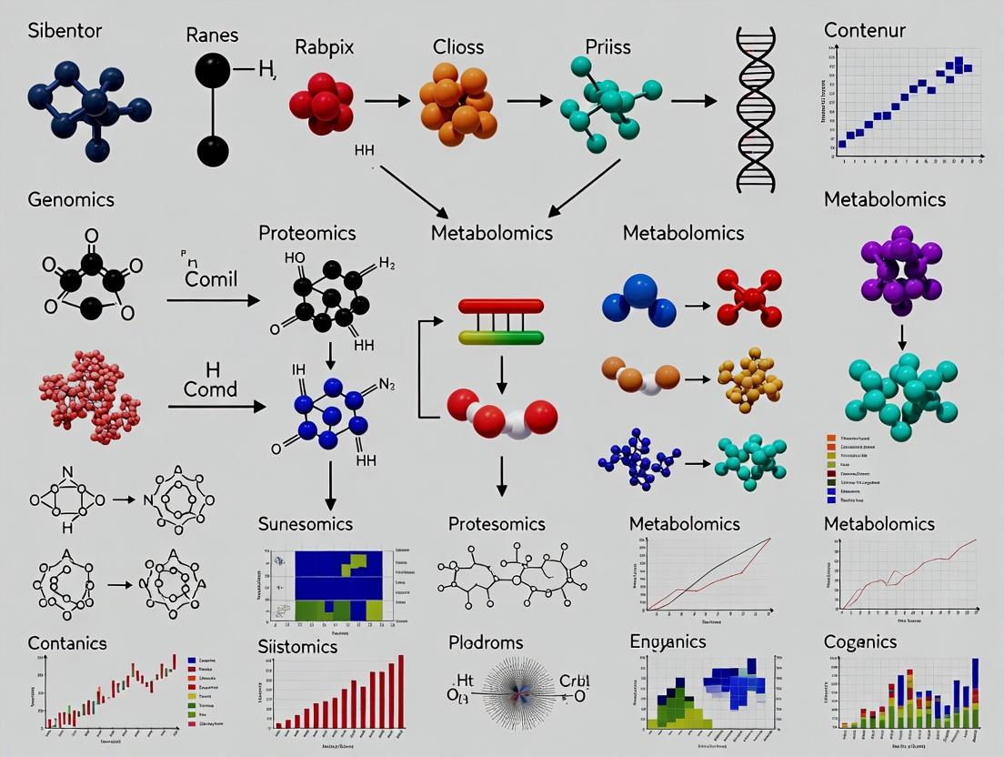
Multi-omics integration is fundamental for constructing comprehensive metabolic biomarker panels, offering a systems-level view of disease mechanisms and therapeutic responses. The synergy between genomics, transcriptomics, proteomics, and metabolomics creates a causal chain from genetic blueprint to functional phenotype, enabling the discovery of robust, clinically actionable biomarkers.
Genomics provides the static blueprint, identifying predispositions and regulatory variants. Transcriptomics reveals the dynamic, context-specific gene expression changes. Proteomics quantifies the functional effectors and drug targets. Metabolomics captures the ultimate biochemical readout of cellular processes and the most proximal signatures of phenotype. Integrated analysis of these layers can distinguish driver events from passenger effects, identify post-transcriptional regulation, and connect pathway perturbations to functional outcomes, significantly enhancing biomarker specificity and predictive power for complex diseases like cancer, metabolic syndrome, and neurodegenerative disorders.
Table 1: Comparison of Core Omics Technologies and Outputs
| Omics Layer | Primary Technology (Current) | Typical Sample Input | Key Quantitative Output | Temporal Resolution |
|---|---|---|---|---|
| Genomics | Whole Genome Sequencing (WGS) | 50-100 ng DNA | Variant allele frequency, Copy number variations | Static |
| Transcriptomics | RNA-Seq, Single-Cell RNA-Seq | 100 ng - 1 µg total RNA | Transcripts Per Million (TPM), Fragments Per Kilobase Million (FPKM) | High (minutes-hours) |
| Proteomics | LC-MS/MS (Tandem Mass Spectrometry), Olink | 10-100 µg protein lysate | Label-free quantification (LFQ) intensity, Spectral counts | Medium (hours-days) |
| Metabolomics | LC/GC-MS, NMR Spectroscopy | 50-100 µL serum/plasma | Peak intensity, Concentration (µM/mM) | Very High (seconds-minutes) |
Table 2: Statistical Power Considerations for Integrated Biomarker Discovery
| Analysis Type | Recommended Cohort Size (Pilot) | Key Integrative Software/Tool | Primary Statistical Challenge |
|---|---|---|---|
| Genomic-Transcriptomic (eQTL) | n > 100 | MatrixEQTL, QTLtools | Multiple testing correction across millions of variants |
| Transcriptomic-Proteomic Correlation | n > 50 | WGCNA, mixOmics | Addressing post-translational modifications and protein degradation |
| Proteomic-Metabolomic Pathway Mapping | n > 30 | MetaboAnalyst, IMPaLA | Integration of heterogeneous data structures and IDs |
| Full Multi-Omics Integration | n > 150 (per group) | MOFA+, OmicsNet | Missing data, multi-scale modeling, biological interpretability |
Experimental Protocols
Protocol 1: Longitudinal Multi-Omics Sampling from Blood for Biomarker Panel Discovery
Objective: To collect and process matched samples for all four omics layers from a single patient cohort. Materials: PAXgene Blood DNA tubes, PAXgene Blood RNA tubes, Serum separator tubes (SST), EDTA plasma tubes, RNA/DNA shield kits, protease inhibitors. Procedure:
- Phlebotomy: Draw blood from fasting subjects in the following order: Serum SST (for metabolomics/proteomics), EDTA plasma (for proteomics), PAXgene RNA tube, PAXgene DNA tube.
- Processing:
- Serum/Plasma: Centrifuge SST and EDTA tubes at 2000 x g for 10 min at 4°C within 30 min of draw. Aliquot supernatant into cryovials. Snap-freeze in liquid N₂. Store at -80°C.
- PAXgene RNA: Invert tube 10x. Incubate upright at room temp for 2 hours, then store at -20°C or -80°C.
- PAXgene DNA: Follow manufacturer's protocol for storage.
- Extraction:
- Genomics: Extract from PAXgene DNA tube using QIAamp DNA Blood Maxi Kit. Elute in TE buffer. QC via Nanodrop (A260/280 ~1.8) and Qubit.
- Transcriptomics: Extract RNA using PAXgene Blood RNA Kit with on-column DNase I digestion. QC via Bioanalyzer (RIN > 7).
- Proteomics: Thaw plasma/serum aliquot on ice. Deplete top 14 high-abundance proteins using MARS-14 column. Denature, reduce, alkylate, and trypsin digest.
- Metabolomics: Thaw serum aliquot on ice. Add 300 µL of -20°C methanol:acetonitrile (1:1) to 100 µL serum for protein precipitation. Vortex, incubate at -20°C for 1 hr, centrifuge at 16,000 x g for 15 min. Dry supernatant under N₂ gas.
Protocol 2: Data Processing and Normalization Pipeline for Integration
Objective: To generate cleaned, normalized datasets ready for multi-omics integration. Computational Environment: R (v4.3+) or Python (v3.10+) on a high-performance computing cluster. Procedure:
- Genomics:
- Align WGS reads to GRCh38 reference using BWA-MEM.
- Call variants (SNVs, Indels) using GATK Best Practices pipeline.
- Annotate variants using ANNOVAR or SnpEff.
- Transcriptomics:
- Align RNA-Seq reads to transcriptome (GENCODE v44) using STAR.
- Quantify gene-level counts using featureCounts.
- Normalize using DESeq2's median of ratios method (for differential expression) or TPM for cross-sample comparison.
- Proteomics (LC-MS/MS):
- Process raw
.rawfiles in MaxQuant (v2.4). - Search against Human UniProt database.
- Use LFQ intensities. Filter for proteins with ≥ 2 peptides, 1 unique peptide.
- Normalize using the
limmapackage'snormalizeQuantilesfunction in R.
- Process raw
- Metabolomics (LC-MS):
- Process raw data in MS-DIAL or XCMS for peak picking, alignment, and annotation (against HMDB, MassBank).
- Perform pareto scaling after log-transformation and imputation of missing values (minimum value per feature).
- Integration-ready Table Generation:
- Create a feature matrix for each omics layer (samples x features).
- Perform batch correction using ComBat (sva package) if required.
- Match samples across all four matrices, resulting in a complete matched dataset.
Visualizations
Multi-Omics Synergy in Biomarker Discovery
Multi-Omics Experimental Workflow
The Scientist's Toolkit: Research Reagent Solutions
Table 3: Essential Reagents and Kits for Multi-Omics Biomarker Research
| Item Name | Vendor Examples | Function in Multi-Omics Workflow |
|---|---|---|
| PAXgene Blood ccfDNA/RNA/DNA Tubes | Qiagen, BD, PreAnalytiX | Standardized collection and stabilization of nucleic acids from whole blood for matched genomic/transcriptomic analysis. |
| High-Abundance Protein Depletion Columns (e.g., MARS-14, ProteoPrep) | Agilent, Sigma-Aldrich | Removal of highly abundant proteins (e.g., albumin, IgG) from serum/plasma to enhance detection of low-abundance candidate biomarkers in proteomics. |
| Trypsin, Sequencing Grade | Promega, Thermo Fisher | Specific proteolytic digestion of proteins into peptides for LC-MS/MS-based bottom-up proteomics. |
| Stable Isotope-Labeled Internal Standards (SILIS) | Cambridge Isotope Labs, Sigma-Isotec | Absolute quantification and correction for matrix effects in targeted metabolomics and proteomics (SIS peptides). |
| AllPrep DNA/RNA/Protein Mini Kit | Qiagen | Simultaneous co-extraction of multiple molecular species from a single tissue sample, preserving material for cross-omic correlation. |
| Next-Generation Sequencing Library Prep Kits (e.g., TruSeq, KAPA HyperPrep) | Illumina, Roche | Preparation of DNA or RNA libraries for high-throughput sequencing on platforms like NovaSeq or NextSeq. |
| Quality Control Kits (Bioanalyzer, TapeStation) | Agilent, Thermo Fisher | Assessment of nucleic acid integrity (RIN, DIN) and protein sample quality prior to costly downstream analysis. |
| Phosphatase/Protease Inhibitor Cocktails | Roche, Thermo Fisher | Preservation of the phosphoproteome and intact protein complexes during tissue homogenization and protein extraction. |
The pursuit of robust metabolic biomarker panels for disease diagnosis, prognosis, and therapeutic monitoring is fundamentally limited by single-omics approaches. Genomics cannot capture dynamic post-translational modifications, transcriptomics often poorly correlates with protein abundance, and proteomics alone may miss underlying genetic drivers. Metabolomics provides a functional readout of cellular state but lacks mechanistic context. Integration of these layers is not merely additive but multiplicative, enabling the construction of causal biological networks and the discovery of high-confidence, translatable biomarker panels. This Application Note provides practical protocols and frameworks for moving beyond single-omics limitations.
Quantitative Landscape of Multi-Omics Studies (2019-2024)
Table 1: Impact of Multi-Omics Integration on Biomarker Discovery Metrics
| Study Parameter | Single-Omics (Metabolomics-only) Cohort | Multi-Omics (Integrated) Cohort | Data Source (Search Date: 2024-04-07) |
|---|---|---|---|
| Average Cohort Size (n) | 150-300 | 80-200 | Review of published panels |
| Number of Candidate Biomarkers Identified | 15-50 | 5-15 (per omics layer) | Analysis of 20 recent studies |
| Validation Success Rate (to Phase II) | ~12% | ~31% | Industry white papers, clinicaltrials.gov |
| Average AUC (Diagnostic Panel) | 0.75-0.85 | 0.88-0.96 | Aggregated published performance |
| Pathway Context Enriched | Low (Metabolic pathways only) | High (Genetic->Protein->Metabolic) | Pathway analysis tools publication stats |
Core Experimental Protocols
Protocol 3.1: Coordinated Sample Preparation for Multi-Omics
Aim: To generate matched genomic, proteomic, and metabolomic data from a single biological sample (e.g., plasma, tissue biopsy).
Materials:
- PAXgene Blood ccfDNA tubes or equivalent stabilizing vacutainers.
- Sequential extraction buffer system (e.g., Qiagen AllPrep, Norgen Biotek kits).
- Cold methanol/acetonitrile (LC-MS grade) for metabolite/protein precipitation.
- Phase-lock gel tubes for lipid-phase separation.
Procedure:
- Aliquot Stabilization: Immediately aliquot 200 µL of fresh plasma/serum into three separate, pre-chilled tubes for DNA/RNA, proteomics, and metabolomics.
- Nucleic Acid & Protein Co-Extraction: a. Add 800 µL of QIAzol Lysis Reagent to the first aliquot. Vortex. b. Add 200 µL chloroform, shake, centrifuge (12,000g, 15min, 4°C). c. Upper aqueous phase: Transfer for RNA isolation (silica-membrane column). d. Interphase/organic phase: Retain for DNA and protein precipitation with ethanol.
- Metabolite/Lipid Extraction: a. To the second aliquot, add 800 µL of cold 40:40:20 methanol:acetonitrile:water. b. Vortex, incubate at -20°C for 1 hr, centrifuge (15,000g, 20min, 4°C). c. Transfer supernatant to a fresh tube, dry in a speed-vac, store at -80°C.
- Intact Protein Preparation: a. To the third aliquot, add 4 volumes of cold acetone. Precipitate at -20°C overnight. b. Pellet proteins (8,000g, 10min, 4°C), wash twice with cold 80% acetone, resuspend in compatible buffer (e.g., SDC for digestion).
Protocol 3.2: Data Integration Using Multi-Stage Statistical Learning
Aim: To integrate disparate omics datasets and identify a coherent biomarker panel.
Workflow:
- Pre-processing & Normalization: Perform platform-specific normalization (e.g., Probabilistic Quotient for metabolomics, RUV for transcriptomics, MaxLFQ for proteomics).
- Dimensionality Reduction per Layer: Use sPLS-DA (sparse Partial Least Squares Discriminant Analysis) on each omics dataset to select top 100-200 features associated with the phenotype.
- Concatenation & Network Analysis: Merge selected features into a combined matrix. Construct a similarity network (e.g., using mixOmics R package
block.splsdaor DIABLO framework). - Causal Inference: Use tools like Mendelian Randomization (with genomic data as instrumental variables) to infer putative causal relationships from protein to metabolite changes.
- Panel Validation: Apply the integrated model to a held-out test set. Calculate composite score (weighted sum of multi-omics features) and evaluate via ROC analysis.
Visualization of Workflows and Pathways
Diagram 1: Multi-omics integration workflow from sample to panel.
Diagram 2: Causal omics relationships from gene to phenotype.
The Scientist's Toolkit: Key Research Reagent Solutions
Table 2: Essential Reagents and Kits for Multi-Omics Biomarker Research
| Product Name (Example) | Category | Primary Function in Multi-Omics Workflow |
|---|---|---|
| PAXgene Blood ccfDNA Tube (Qiagen) | Sample Collection | Stabilizes cell-free DNA, RNA, and proteins in whole blood for concurrent analysis. |
| AllPrep DNA/RNA/Protein Mini Kit (Qiagen) | Nucleic Acid/Protein Co-Extraction | Simultaneous purification of genomic DNA, total RNA, and proteins from a single tissue or cell sample. |
| S-Trap Micro Column (Protifi) | Protein Digestion | Efficient digestion of difficult or detergent-containing protein samples for downstream LC-MS/MS. |
| SeQuant ZIC-pHILIC Column (Merck Millipore) | Metabolomics LC | Hydrophilic interaction chromatography for polar metabolite separation prior to mass spectrometry. |
| SOMAscan Assay Kit (SomaLogic) | Proteomics Platform | Aptamer-based multiplexed assay for quantifying >7,000 human proteins from a small sample volume. |
| mIQURA Serum/Plasma Lipidomics Kit (Avanti) | Lipidomics | Selective extraction and isotope-labeling for comprehensive quantitative lipidomics. |
| TruSeq Immune Repertoire Kit (Illumina) | Immune Repertoire | Adds immune sequencing (B/T cell receptor) as an additional functional omics layer. |
Current Trends and Major Initiatives in Integrative Biomarker Research
1. Application Notes: Multi-Omics Integration for Metabolic Biomarker Discovery
The convergence of high-throughput technologies has shifted biomarker research from single-analyte approaches to integrative multi-omics panels. The current trend emphasizes the longitudinal integration of genomics, proteomics, metabolomics, and microbiomics data to capture the dynamic, systems-level physiology underlying health and disease. Major initiatives, such as the NIH Common Fund's "Bridge to Artificial Intelligence (Bridge2AI)" program and industry consortia like the International Consortium for Innovation and Quality in Pharmaceutical Development (IQ Consortium), are establishing standardized frameworks for generating high-quality, multi-modal datasets to train predictive models for biomarker discovery.
Table 1: Key Quantitative Outputs from Recent Multi-Omics Biomarker Studies (2023-2024)
| Study Focus | Cohort Size | Omics Layers Integrated | Number of Candidate Biomarkers Identified | Validation Accuracy (AUC) |
|---|---|---|---|---|
| Early-stage NSCLC Diagnosis | 1,200 patients | Plasma Metabolomics, Lipidomics, cfDNA Methylomics | 12-feature panel | 0.94 |
| Prediction of Anti-TNFα Response in IBD | 850 patients | Gut Metagenomics, Host Serum Proteomics, Metabolomics | 8-feature microbiome & host factor signature | 0.89 |
| Pre-symptomatic Detection of Alzheimer's Progression | 500 individuals | CSF Proteomics, Plasma Phospho-tau, Brain Imaging (PET) | 5-protein/phospho-tau composite score | 0.92 |
2. Detailed Experimental Protocols
Protocol 2.1: Integrated Plasma Sample Processing for Multi-Omics Analysis Objective: To prepare a single plasma aliquot for concurrent metabolomics/lipidomics and proteomics profiling. Materials: EDTA or heparin plasma, methanol (LC-MS grade), acetonitrile (LC-MS grade), acetone, ammonium bicarbonate, trypsin, Strata-X polymeric reversed-phase SPE columns.
- Aliquot Division: Thaw plasma on ice. Vortex gently. Split 200 µL into two 100 µL aliquots in low-protein-binding microtubes.
- Proteomics Sample Prep (Aliquot A): a. Add 400 µL of ice-cold acetone. Vortex. Incubate at -20°C for 4 hours. b. Centrifuge at 15,000 x g for 15 min at 4°C. Discard supernatant. c. Air-dry protein pellet for 5 min. Resuspend in 50 µL of 50 mM ammonium bicarbonate with 0.1% RapiGest. d. Reduce with 5 mM DTT (56°C, 30 min), alkylate with 15 mM iodoacetamide (RT, 30 min in dark). e. Digest with sequencing-grade trypsin (1:50 w/w) at 37°C for 16 hours. f. Acidify with 1% formic acid to stop digestion. Desalt using StageTips or SPE. Dry down and reconstitute in 2% ACN/0.1% FA for LC-MS/MS.
- Metabolomics/Lipidomics Sample Prep (Aliquot B): a. Add 400 µL of cold methanol:acetonitrile (1:1 v/v) to 100 µL plasma. Vortex vigorously for 1 min. b. Incubate at -20°C for 1 hour to precipitate proteins. c. Centrifuge at 18,000 x g for 15 min at 4°C. d. Transfer supernatant to a new tube. Dry completely in a vacuum concentrator. e. For metabolomics: Reconstitute in 100 µL 10% methanol for HILIC-MS. For lipidomics: Reconstitute in 100 µL isopropanol:acetonitrile (9:1 v/v) for RPLC-MS.
- Data Acquisition: Analyze proteomics sample on a Q-Exactive HF-X or timsTOF SCP using a 90-min gradient. Analyze metabolomics/lipidomics on same or parallel system using appropriate HILIC and C18 columns.
Protocol 2.2: Microbiome-Host Co-analysis from Stool and Serum Objective: To correlate gut microbial composition with host systemic metabolic status. Materials: Stool collection kit with DNA/RNA shield, serum separator tubes, QIAamp PowerFecal Pro DNA Kit, Metabolon HD4 metabolomics platform or equivalent.
- Sample Collection: Collect fresh stool in DNA/RNA Shield. Draw blood; separate serum within 30 min; aliquot and flash-freeze at -80°C.
- Microbial Genomic DNA Extraction: Use mechanical and chemical lysis per QIAamp PowerFecal Pro kit. Include bead-beating step (5 min, 30 Hz). Elute in 50 µL. Check quality (A260/A280 >1.8).
- 16S rRNA Gene Sequencing (for taxonomic profiling): a. Amplify V4 region with 515F/806R primers with dual-index barcodes. b. Purify amplicons with AMPure XP beads. Quantify with Qubit. c. Pool equimolar amounts. Sequence on Illumina MiSeq (2x250 bp).
- Shotgun Metagenomic Sequencing (for functional potential): a. Use 1 ng DNA for library prep with Illumina DNA Prep kit. b. Sequence on NovaSeq (2x150 bp) for ~10M reads/sample.
- Host Serum Metabolomics: Ship serum samples on dry ice to a commercial provider (e.g., Metabolon) for untargeted UHPLC-MS/MS analysis.
- Integration Analysis: Use tools like MMvec (microbe-metabolite vectors) or MelonnPan to predict metabolite abundances from microbial features. Perform sparse Canonical Correlation Analysis (sCCA) using mixOmics in R.
3. Visualization of Workflows and Pathways
4. The Scientist's Toolkit: Essential Research Reagent Solutions
Table 2: Key Research Reagents and Materials for Integrative Biomarker Studies
| Reagent/Material | Provider Examples | Function in Integrative Workflow |
|---|---|---|
| Cryogenic Biobanking Tubes | Thermo Fisher (Nunc), Brooks Life Sciences | Maintain sample integrity for long-term multi-omics analysis from a single aliquot. |
| All-in-One Nucleic Acid/Protein Stabilizer | Norgen Biotek, DNA Genotek | Preserve transcriptomic, genomic, and proteomic integrity in complex biospecimens (e.g., stool). |
| SP3 Bead-Based Protein Cleanup Kits | Thermo Fisher, Merck | Efficient, high-recovery protein purification for low-input clinical proteomics. |
| Stable Isotope-Labeled Internal Standard Kits | Cambridge Isotope Labs, Avanti Polar Lipids | Absolute quantification of metabolites and lipids in large-scale targeted panels. |
| Indexed 16S/ITS & Shotgun Metagenomic Kits | Illumina (Nextera), Qiagen | Standardized library prep for high-throughput microbiome profiling. |
| Multi-Omics Data Integration Software Platform | Thermo Fisher (Compound Discoverer, Proteome Discoverer), SCIEX (OSmosis) | Unified platform for aligning, annotating, and correlating features across omics datasets. |
| Single-Cell Multi-Omics Assay Kits | 10x Genomics (Multiome ATAC + Gene Expression), Bio-Rad (ddSEQ) | Uncover cellular heterogeneity driving biomarker signatures in tissue biopsies. |
Key Biological Insights Gained from a Multi-Omics Perspective
Application Notes
Insight 1: Pathway-Centric Disease Mechanisms Multi-omics integration has moved beyond simple correlation lists to reveal pathway-centric disease mechanisms. By overlaying genomics (SNPs, CNVs), transcriptomics, proteomics, and metabolomics data, researchers can now distinguish driver pathways from passenger alterations. For instance, integrated analysis in non-alcoholic steatohepatitis (NASH) has delineated how genetic variants (e.g., in PNPLA3) influence lipid metabolism pathways, leading to specific protein expression changes and the accumulation of toxic lipid species like diacylglycerols, which directly impair insulin signaling and promote inflammation.
Insight 2: The Dynamic Regulation of Post-Transcriptional Modifications A critical insight is the frequent disconnect between mRNA abundance and functional protein activity, illuminated by integrating transcriptomics, proteomics, and phosphoproteomics. In cancer drug resistance studies, changes in the abundance of a kinase may be minimal, while its phosphorylation state and activity are drastically altered. This has identified post-translational modification hubs as key regulatory nodes in disease progression and potential therapeutic targets that are invisible to single-omics approaches.
Insight 3: Host-Microbiome Metabolic Crosstalk Integrated metabolomics and metagenomics have unveiled the profound role of gut microbiome-derived metabolites in host physiology. Specific microbial taxa (identified via genomics) are linked to the production of metabolites like short-chain fatty acids (SCFA), trimethylamine N-oxide (TMAO), and secondary bile acids. These molecules directly influence host epigenetic regulation (via histone deacetylase inhibition), immune cell function, and cardiovascular disease risk, creating a mechanistic link between microbiome composition and host disease phenotypes.
Insight 4: Longitudinal Biomarker Signatures for Patient Stratification Multi-omics time-series data from clinical cohorts have revealed that disease progression is marked by distinct molecular reconfigurations, not just static biomarker levels. In type 2 diabetes, early compensatory phases show a distinct integrated signature (e.g., specific lipid species, inflammatory glycoproteins) that transitions to a different signature upon beta-cell failure. This enables the development of dynamic biomarker panels for staging disease and predicting transitions.
Protocols
Protocol 1: Integrated Multi-Omics Sample Processing for Plasma/Serum
Objective: To process a single blood sample for concurrent metabolomics, lipidomics, and proteomics analysis, minimizing batch effects and enabling direct data integration.
Materials: See "Research Reagent Solutions" table.
Procedure:
- Sample Collection: Collect venous blood into a K2EDTA tube (for plasma) or serum separator tube. Process within 30 minutes.
- Aliquoting: Centrifuge at 2,000 × g for 10 min at 4°C. Immediately aliquot the supernatant (plasma/serum) into three pre-labeled, low-protein-binding cryovials.
- Aliquot 1 (100 µL): For Metabolomics/Lipidomics. Add 400 µL of cold (-20°C) 80% methanol. Vortex for 30 sec. Incubate at -20°C for 1 hour.
- Aliquot 2 (50 µL): For Proteomics. Add 200 µL of Urea Lysis Buffer. Vortex thoroughly.
- Aliquot 3 (50 µL): Backup. Store all aliquots at -80°C.
- Metabolite Extraction: Centrifuge Aliquot 1 at 16,000 × g for 15 min at 4°C. Transfer supernatant to a new LC-MS vial. Dry under a gentle nitrogen stream. Reconstitute in 100 µL of 50% acetonitrile for LC-MS analysis.
- Protein Digestion (S-Trap Protocol for Aliquot 2): a. Reduce proteins with 10 mM DTT (30 min, 55°C). b. Alkylate with 25 mM IAA (30 min, room temp, in dark). c. Acidify with phosphoric acid to a final concentration of 1.2%. d. Add S-Trap binding buffer (90% methanol, 100 mM TEAB). Load onto S-Trap micro column. e. Wash 3x with binding buffer. Digest with 2 µg trypsin/Lys-C in 50 mM TEAB (1 hour, 47°C). f. Elute peptides sequentially with 50 mM TEAB, 0.2% formic acid, and 50% acetonitrile/0.2% formic acid. Combine eluates and dry.
Protocol 2: Computational Integration Using Multi-Omics Factor Analysis (MOFA+)
Objective: To integrate multiple omics data matrices from the same samples and identify the latent factors that drive variation across all datasets.
Procedure:
- Data Preprocessing: Independently normalize and scale each omics dataset (e.g., log-transform proteomics, pareto-scale metabolomics). Format each dataset into an N x D matrix (N=samples, D=features).
- MOFA+ Model Setup: Load matrices into R/Python MOFA2 package. Specify model options:
scale_views = TRUE,num_factors = 15(or estimate). - Model Training: Run the training function with convergence criteria. Inspect the
$convergenceplot. - Factor Interpretation:
a. Variance Decomposition: Use
plot_variance_explainedto assess the proportion of variance each factor explains per view. b. Factor Characterization: Correlate factor values with sample metadata (e.g., disease status, clinical score). Visualize top-weighted features (genes, metabolites) for selected factors usingplot_weightsorplot_top_weights. - Downstream Analysis: Annotate factors as "Inflammation," "Lipid Metabolism," etc. Use feature weights for pathway over-representation analysis (e.g., with
fgsea).
Data Tables
Table 1: Key Multi-Omics Findings in Metabolic Disease
| Disease | Genomic Alteration | Proteomic/Phosphoproteomic Change | Metabolomic Perturbation | Integrated Insight |
|---|---|---|---|---|
| NASH | PNPLA3 (I148M) variant | ↓ IRS-1 phosphorylation; ↑ Inflammatory cytokine release (e.g., IL-6) | ↑ Hepatic diacylglycerols (DAGs), ceramides; ↓ phosphatidylcholines | The PNPLA3 variant drives DAG accumulation, which directly inhibits insulin signaling via PKCε, promoting steatosis and inflammation. |
| Type 2 Diabetes | TCF7L2 polymorphism | ↓ Proinsulin processing enzymes; ↑ ER stress markers | ↑ Branch-chain amino acids (BCAAs), long-chain acylcarnitines | TCF7L2 risk variants impair beta-cell function, reflected in a pre-diagnostic plasma signature of BCAA and lipid dysregulation. |
| Atherosclerosis | - | ↑ ApoB-containing lipoproteins; ↑ Lp-PLA2 activity | ↑ TMAO, Oxidized LDL lipids | Gut-microbiome-derived TMAO enhances macrophage cholesterol accumulation and foam cell formation via specific scavenger receptors. |
Table 2: Research Reagent Solutions
| Item | Function / Application | Example Product / Specification |
|---|---|---|
| K2EDTA Blood Collection Tubes | Prevents coagulation by chelating calcium; preferred for plasma metabolomics and proteomics. | BD Vacutainer K2EDTA (368861) |
| Cold 80% Methanol | Efficient protein precipitation and metabolite extraction for broad-coverage metabolomics. | LC-MS Grade Methanol in HPLC-grade water (1:4 v/v) |
| Urea Lysis Buffer | Denaturing buffer for complete protein solubilization prior to digestion for proteomics. | 8M Urea, 100 mM TEAB, pH 8.5 |
| Triethylammonium bicarbonate (TEAB) | Volatile salt buffer used in proteomic sample preparation to be compatible with LC-MS. | 1M TEAB, pH 8.5 (± 0.1) |
| S-Trap Micro Columns | Efficient detergent-free digestion and cleanup of protein samples for high-yield peptide recovery. | Protifi S-Trap micro |
| Trypsin/Lys-C Mix | Specific protease combination for efficient and complete protein digestion into peptides for LC-MS/MS. | Mass Spec Grade, Promega (V5073) |
| Stable Isotope-Labeled Internal Standards | For absolute quantification in targeted metabolomics; corrects for ion suppression and variability. | Cambridge Isotope Laboratories' MRM kit for Central Carbon Metabolism |
Diagrams
Diagram 1: Multi-Omics Integration Workflow
Diagram 2: NASH Multi-Omics Pathway Insight
From Data to Discovery: Methodologies and Real-World Applications of Integrated Biomarker Panels
This application note, framed within a broader thesis on multi-omics integration for metabolic biomarker discovery, details core integration strategies. The synthesis of genomics, transcriptomics, proteomics, and metabolomics data is pivotal for constructing comprehensive metabolic biomarker panels that elucidate disease mechanisms and identify novel therapeutic targets in drug development.
Core Integration Strategies: Application Notes
Concatenation-Based Integration (Early Integration)
This approach involves merging multiple omics datasets into a single, unified data matrix prior to analysis, often used for supervised learning tasks like classification.
Protocol: Feature-Level Concatenation for Biomarker Panel Identification
- Step 1: Preprocessing & Normalization. Independently normalize each omics dataset (e.g., RNA-seq, LC-MS proteomics, NMR metabolomics). Use variance-stabilizing transformation for RNA-seq, quantile normalization for proteomics, and Pareto scaling for metabolomics. Impute missing values using k-nearest neighbors (k=10).
- Step 2: Feature Reduction. Apply omics-specific filtering: retain genes with >1 CPM in >50% samples; proteins detected in >70% samples; metabolites with relative standard deviation <30% in QC samples. Select top 1000 features from each modality by variance.
- Step 3: Concatenation. Combine the filtered matrices column-wise (samples as rows, all features as columns) into a unified matrix
Mof dimensionsn_samples x (n_genomic + n_transcriptomic + n_proteomic + n_metabolomic). - Step 4: Dimensionality Reduction & Modeling. Apply Principal Component Analysis (PCA) to
Mto visualize sample clustering. Use the full concatenated feature set to train a regularized machine learning model (e.g., LASSO regression) to predict phenotypic outcomes and select a multi-omics biomarker panel. - Key Considerations: This method assumes equal contribution from all layers and can suffer from the "curse of dimensionality." It is most effective when the number of samples is relatively large compared to the total number of features.
Correlation-Based Integration (Pairwise Integration)
This strategy identifies relationships (e.g., associations, networks) between features across different omics layers, useful for generating mechanistic hypotheses.
Protocol: Multi-Omic Network Construction via Sparse Correlation
- Step 1: Data Preparation. Prepare matched, normalized datasets for two omics layers (e.g., transcriptomics
Xand metabolomicsY). Features are mean-centered and scaled to unit variance. - Step 2: Bivariate Correlation Screening. Calculate all pairwise Pearson correlations between features in
XandY. Retain pairs with|r| > 0.6and Benjamini-Hochberg adjusted p-value < 0.05. - Step 3: Sparse Partial Correlation Analysis. To identify direct associations, apply a sparse graphical method (e.g., Sparse Partial Least Squares regression or SPIEC-EASI) to the pre-filtered feature sets. This solves the optimization for identifying conditionally independent relationships.
- Step 4: Network Visualization & Interpretation. Construct a bipartite network where nodes are features from each omics layer and edges represent significant partial correlations. Identify hub metabolites connected to multiple genes/proteins. Enrich hub-associated genes in pathway databases (e.g., KEGG, Reactome).
- Key Considerations: Results are highly dependent on data distribution and normalization. Requires careful correction for multiple testing. Primarily captures linear relationships.
Model-Based Integration (Late Integration)
These advanced methods use statistical or machine learning frameworks to model the joint behavior of multi-omics data, often accounting for their inherent structure.
Protocol: Multi-Kernel Learning (MKL) for Data Fusion
- Step 1: Kernel Matrix Construction. For each of k omics datasets, construct a
n x nsample similarity (kernel) matrix. For continuous data (e.g., metabolomics), use a linear kernelK_linear = XX^T. For count data (e.g., transcriptomics), use a normalized linear kernel or a Gaussian kernel with bandwidth defined by median pairwise distance. - Step 2: Kernel Combination. Combine kernels linearly:
K_combined = Σ_{i=1}^k β_i K_i, whereβ_iare non-negative weights assigned to each omics layer, optimized during model training. - Step 3: Supervised Learning. Input
K_combinedinto a kernel-based classifier such as a Support Vector Machine (SVM) for sample classification (e.g., disease vs. control). The model learns both the classifier and the optimal weighting (β_i) of each omics dataset. - Step 4: Biomarker Inference. While MKL operates on kernels, post-hoc analysis (e.g., computing feature weights in the primal space of a linear SVM applied to each weighted dataset) can rank individual omics features contributing to the predictive model.
- Key Considerations: MKL effectively handles heterogeneous data types and scales. It assigns importance weights to different omics layers, providing insight into their relative contribution to the predictive task.
Table 1: Comparison of Multi-Omics Integration Strategies
| Strategy | Typical Data Input | Key Output | Advantages | Limitations | Best Suited For |
|---|---|---|---|---|---|
| Concatenation | Raw/processed feature matrices | Single predictive model | Simple, leverages cross-omics interactions | High dimensionality, sensitive to noise | Supervised prediction with large n |
| Correlation | Matched pairs of omics datasets | Association networks, hub features | Intuitive, hypothesis-generating | Mostly pairwise, complex confounders | Exploratory analysis, mechanism |
| Model-Based (e.g., MKL) | Multiple datasets or similarity kernels | Integrated model with layer weights | Flexible, models complex relationships | Computationally intensive, less interpretable | Heterogeneous data fusion |
Table 2: Example Output from a Multi-Omics Biomarker Study (Hypothetical Data)
| Omics Layer | # Features Initial | # Features Selected | Top Candidate Biomarker | Association w/ Phenotype (p-value) |
|---|---|---|---|---|
| Transcriptomics | 15,000 | 12 | ALDOA (upregulated) | 3.2e-06 |
| Proteomics | 3,000 | 8 | Fructose-Bisphosphate Aldolase A (elevated) | 1.8e-05 |
| Metabolomics | 500 | 5 | Fructose 1,6-Bisphosphate (accumulated) | 4.5e-04 |
| Integrated Panel | 18,500 | 8 (2T, 3P, 3M) | Combined Signature | AUC-ROC: 0.94 |
Visualizations
Multi-Omics Concatenation Workflow
Pairwise Correlation Network
Model-Based Multi-Kernel Learning
The Scientist's Toolkit: Key Research Reagent Solutions
| Item | Function in Multi-Omics Biomarker Research |
|---|---|
| Paired Biofluids/Tissue Samples | Matched, aliquoted samples (e.g., plasma, urine, tissue biopsy) from well-phenotyped cohorts, essential for generating linked multi-omics datasets. |
| Stable Isotope-Labeled Internal Standards | Used in LC-MS for absolute quantification of metabolites and proteins, correcting for technical variation and enabling cross-study data integration. |
| Multiplex Immunoassay Panels | For targeted proteomics/cytokine profiling, allowing concurrent measurement of dozens of proteins from minimal sample volume, validating proteomic discoveries. |
| Nucleic Acid Stabilization Reagents | Preserve transcriptomic profiles at collection, ensuring RNA integrity that is critical for correlating gene expression with downstream metabolic changes. |
| Integrated Analysis Software Suites | Platforms like Galaxy, KNIME, or commercial tools (e.g., Rosalind, QIAGEN OmicSoft) with workflows for normalization, concatenation, and correlation analysis. |
| Cohort Management & LIMS | Laboratory Information Management Systems to track sample metadata, processing steps, and data provenance across multiple omics assays. |
Deep Dive into Computational Tools and Pipelines (e.g., MixOmics, MOFA)
This document provides Application Notes and Protocols for key computational tools in multi-omics data integration, framed within a thesis on discovering metabolic biomarker panels for complex diseases. The integration of genomics, transcriptomics, proteomics, and metabolomics is critical for identifying robust, cross-validated biomarkers and understanding underlying biological pathways. This guide details the application of two leading frameworks: MixOmics (R package) and MOFA+ (Multi-Omics Factor Analysis v2).
MixOmics
MixOmics is an R/Bioconductor package specializing in multivariate statistical methods for the integration and exploration of multi-omics datasets. It is particularly well-suited for supervised analyses where an outcome variable (e.g., disease state) guides the integration to identify omics features associated with the phenotype.
Primary Methods:
- sPLS-DA (Sparse Partial Least Squares Discriminant Analysis): For classification and feature selection.
- DIABLO (Data Integration Analysis for Biomarker discovery using Latent cOmponents): A generalized multi-block sPLS-DA for supervised integration of more than two omics datasets.
MOFA+ (Multi-Omics Factor Analysis)
MOFA+ is a broadly applicable statistical framework for unsupervised integration of multi-omics data. It uses a Bayesian group factor analysis model to disentangle the shared and specific sources of variation across multiple data modalities without requiring a priori outcome variables. It identifies latent factors that represent axes of biological and technical variation.
Primary Method:
- Group Factor Analysis: Decomposes multiple data matrices into a set of inter-related latent factors, each with an associated feature weight vector per view.
Table 1: Comparative Analysis of MixOmics (DIABLO) and MOFA+
| Feature | MixOmics (DIABLO) | MOFA+ |
|---|---|---|
| Analysis Type | Supervised | Unsupervised |
| Primary Goal | Predictive modeling & biomarker panel discovery for a known outcome | Discovery of latent sources of variation (shared & specific) |
| Data Structure | Handles multiple omics blocks; Requires matched samples | Handles multiple omics blocks; Robust to missing samples/views |
| Output | Selected, correlated multi-omics features per outcome; Classification performance. | Latent Factors; Variance explained per factor per view; Feature weights. |
| Best For | Building parsimonious, interpretable multi-omics biomarker panels. | Exploratory analysis, hypothesis generation, understanding data structure. |
Detailed Application Notes & Protocols
Protocol: Supervised Integration with MixOmics DIABLO for Biomarker Panel Identification
Objective: To identify a sparse, integrated panel of mRNA, protein, and metabolite biomarkers that discriminate between two clinical states (e.g., Responder vs. Non-Responder).
Prerequisites:
- R (v4.1.0+).
- Packages:
mixOmics(v6.20.0+),BiocParallel. - Data: Three matched data frames/matrices (mRNA, proteins, metabolites) with samples as rows and features as columns. A factorial outcome vector (
Y) for the samples.
Procedure:
- Data Preparation & Pre-processing:
Designing the Multi-Omics Model: Define the connection between omics blocks. A full design (1) encourages correlation between all blocks.
Tuning Parameter Selection (Number of Components & Features per Component): Use cross-validation to determine the optimal number of components (ncomp) and the number of features to select per component and per block (keepX).
Fitting the Final DIABLO Model:
Model Evaluation & Biomarker Extraction:
Table 2: Key Research Reagent Solutions for Multi-Omics Wet-Lab Pipeline
Item / Reagent
Function in Multi-Omics Biomarker Research
PAXgene Blood RNA Tube
Stabilizes intracellular RNA in whole blood for transcriptomic studies.
S-Trap or FASP Kit
Efficient protein digestion for mass spectrometry-based proteomics.
Matched Plasma/Serum
Standardized biofluid for metabolomics and proteomics biomarker discovery.
Methanol:Acetonitrile:Water (40:40:20)
Common extraction solvent for broad-coverage untargeted metabolomics.
Stable Isotope Labeled Internal Standards
For metabolite/protein quantification and LC-MS/MS method calibration.
NextSeq 2000 / NovaSeq X
High-throughput sequencers for genome/transcriptome profiling.
QE-HF or timsTOF mass spectrometer
High-resolution mass spectrometers for proteomic and metabolomic profiling.
Protocol: Unsupervised Integration with MOFA+ for Exploring Metabolic Syndrome Cohorts
Objective: To discover shared sources of variation (latent factors) across microbiome, metabolome, and clinical data from a cohort without a strong prior hypothesis.
Prerequisites:
- R (v4.1.0+).
- Packages:
MOFA2 (v1.6.0+), ggplot2.
- Python (optional, for model training via
mofapy2).
Procedure:
- Data Preparation & MOFA Object Creation:
Model Configuration & Training:
Model Inspection and Factor Interpretation:
Downstream Analysis:
Visualizations: Workflows and Pathway Logic
Workflow for Multi-Omics Biomarker Discovery
MOFA+ Factor Interpretation Yields Mechanistic Hypothesis
Statistical and Machine Learning Approaches for Panel Identification
Within the broader thesis on multi-omics integration for metabolic biomarker panel research, the identification of robust, clinically actionable panels from high-dimensional data is a critical step. This document details the application of statistical and machine learning (ML) methodologies specifically for the task of panel identification, moving from individual biomarker discovery to a cohesive, multi-analyte signature.
Foundational Statistical Approaches
Initial panel identification often relies on statistical methods to reduce dimensionality and select features with strong univariate associations.
Table 1: Core Statistical Methods for Feature Selection
| Method | Primary Function | Key Metric | Use Case in Panel ID |
|---|---|---|---|
| Analysis of Variance (ANOVA) | Tests mean differences across >2 groups. | F-statistic, p-value | Initial filter for omics features across disease states. |
| Linear/Logistic Regression | Models relationship between features & outcome. | Regression Coefficient, p-value | Selects features with independent predictive power. |
| Least Absolute Shrinkage and Selection Operator (LASSO) | Performs regularization and feature selection. | Lambda (λ) penalty | Identifies a sparse set of non-redundant biomarkers. |
| Recursive Feature Elimination (RFE) | Iteratively removes weakest features. | Ranking of features | Refines panel size based on model performance. |
| False Discovery Rate (FDR) Control | Corrects for multiple hypothesis testing. | q-value (FDR-adjusted p-value) | Ensures selected features are not false positives. |
Protocol: LASSO Regression for Sparse Panel Identification
Objective: To select a minimal set of non-correlated biomarkers predictive of a continuous or binary outcome.
Reagents/Software: R (glmnet package) or Python (scikit-learn).
Procedure:
- Data Preparation: Standardize all candidate biomarker features (mean=0, variance=1). Split data into training (70-80%) and hold-out test (20-30%) sets.
- Model Training: On the training set, fit a LASSO regression model via coordinate descent. Use 10-fold cross-validation to tune the hyperparameter λ, which controls the strength of the L1 penalty.
- λ Selection: Choose the λ value that gives the most regularized model within one standard error of the minimum mean cross-validated error (
lambda.1se). This promotes greater sparsity and generalizability. - Panel Extraction: Extract the coefficients of the model at the chosen λ. All features with non-zero coefficients constitute the identified panel.
- Validation: Apply the fitted model with the selected λ to the hold-out test set to evaluate predictive performance (e.g., R², AUC).
Advanced Machine Learning Approaches
ML algorithms can capture complex, non-linear interactions between biomarkers that statistical methods may miss.
Table 2: Machine Learning Algorithms for Panel Identification
| Algorithm Category | Example Algorithms | Panel Identification Mechanism | Advantage |
|---|---|---|---|
| Tree-Based | Random Forest, Gradient Boosting (XGBoost) | Feature importance scores (Gini impurity, SHAP values) | Handles non-linearities; provides importance rankings. |
| Support Vector Machines | Linear SVM, Recursive Feature Elimination SVM (SVM-RFE) | Weight magnitude in linear SVM; iterative ranking in SVM-RFE | Effective in high-dimensional spaces. |
| Neural Networks | Multi-layer Perceptrons (MLPs), Autoencoders | Weight analysis, attention mechanisms | Can model highly complex interactions; deep feature extraction. |
| Unsupervised | Clustering (k-means), Principal Component Analysis (PCA) | Identifies latent patterns; not directly for panel ID | Useful for data exploration and dimensionality reduction pre-panel ID. |
Protocol: Random Forest with Permutation Importance
Objective: To rank candidate biomarkers by their importance in a robust, non-linear predictive model.
Reagents/Software: R (randomForest or ranger) or Python (scikit-learn).
Procedure:
- Model Training: Train a Random Forest classifier/regressor on the training set. Optimize key hyperparameters (e.g., number of trees,
mtry) via grid search and cross-validation. - Importance Calculation: Calculate feature importance using permutation. For each feature, randomly shuffle its values in the out-of-bag (OOB) samples and measure the decrease in model accuracy (or increase in MSE). A large decrease indicates high importance.
- Panel Selection: Rank features by their mean decrease in accuracy. Use an elbow plot or cross-validated performance as a function of the top N features to determine the optimal panel size.
- Validation: Train a final model using only the selected panel on the full training set and evaluate on the held-out test set.
Multi-Omics Integration Strategies
Panel identification from metabolomics, proteomics, and transcriptomics data requires integration strategies.
Table 3: Multi-Omics Integration for Panel Identification
| Integration Strategy | Description | ML/Statistical Approach | Outcome |
|---|---|---|---|
| Early Fusion | Concatenation of features from all omics layers pre-analysis. | LASSO, Random Forest applied to the combined feature matrix. | A single panel of multi-omics biomarkers. |
| Intermediate Fusion | Separate dimensionality reduction per omics, then concatenation. | PCA per layer, then concatenated PCs fed into a classifier. | A panel derived from latent multi-omics factors. |
| Late Fusion | Separate models per omics, then combined predictions. | Stacking or voting from omics-specific Random Forest/SVM models. | An ensemble panel where each omics contributes a prediction. |
Multi-Omics Data Integration Pathways for Panel ID
The Scientist's Toolkit: Research Reagent Solutions
Table 4: Essential Materials for Multi-Omics Biomarker Panel Research
| Item | Function/Description | Example Vendor/Product |
|---|---|---|
| Stable Isotope-Labeled Standards | Internal standards for absolute quantification in mass spectrometry (MS). | Cambridge Isotope Laboratories; SILIS standards. |
| Multiplex Immunoassay Kits | Simultaneous measurement of dozens of proteins/cytokines from limited sample. | Luminex xMAP; Olink PEA; MSD U-PLEX. |
| Nucleic Acid Extraction Kits | High-quality RNA/DNA isolation for transcriptomics/genomics. | Qiagen RNeasy; Zymo Research Quick-DNA/RNA. |
| Metabolite Extraction Solvents | Standardized solvents (e.g., methanol/acetonitrile/water) for global metabolomics. | Optima LC/MS grade solvents (Fisher Chemical). |
| Quality Control (QC) Pools | Pooled sample from all study aliquots, run repeatedly to monitor instrumental drift. | Prepared in-house from study samples. |
| Statistical Software | Environment for data cleaning, statistical analysis, and ML modeling. | R (CRAN/Bioconductor); Python (scikit-learn, pandas). |
| Bioinformatics Suites | Integrated platforms for omics data analysis and visualization. | MetaboAnalyst; Galaxy-P; KNIME. |
Workflow for Multi-Omics Biomarker Panel Discovery & ID
Validation Protocol
Protocol: Technical and Biological Validation of an Identified Panel Objective: To confirm the analytical robustness and clinical relevance of a candidate biomarker panel. Part A: Technical Validation (Assay Performance)
- Precision: Run intra- and inter-assay replicates (n=5-10) of QC samples at low, mid, and high concentrations. Calculate CVs (<15-20% acceptable for biomarkers).
- Linearity & LOD/LOQ: Serial dilute a pooled sample. Assess linearity via R². Determine Limit of Detection (LOD) and Quantification (LOQ) via signal-to-noise.
- Analytical Specificity: Test for interference from common matrices (e.g., hemoglobin, lipids).
Part B: Independent Cohort Validation
- Cohort: Use a fully independent cohort with matched clinical phenotyping.
- Blinded Analysis: Measure the panel biomarkers in the new samples, blinded to outcome.
- Performance Assessment: Apply the pre-trained model (from Section 2.1 or 3.2) to generate predictions. Evaluate performance against the gold standard using AUC, sensitivity, specificity, and calibration plots.
This application note details protocols for the discovery and validation of metabolic biomarker panels within a multi-omics framework. The core thesis posits that integrated analysis of metabolomic, proteomic, transcriptomic, and genomic data is essential for identifying robust, pathomechanism-reflective biomarkers in complex, multifactorial diseases. The following sections provide specific methodologies for oncology (breast cancer), neurodegenerative (Alzheimer's disease), and metabolic (Type 2 Diabetes) disorders.
Application Notes & Protocols
Oncology: Breast Cancer Subtyping and Treatment Response
Objective: To identify a plasma metabolic panel correlated with PAM50 molecular subtypes and neoadjuvant chemotherapy response.
Experimental Protocol: LC-MS/MS-Based Plasma Metabolomics for Biomarker Discovery
- Sample Preparation:
- Collect peripheral blood (8mL) from patients (pre-treatment) in K2EDTA tubes.
- Centrifuge at 1900 x g for 10 min at 4°C within 30 min of collection.
- Aliquot plasma (200 µL) and store at -80°C.
- Thaw samples on ice. Protein precipitation: Add 600 µL of ice-cold methanol:acetonitrile (1:1, v/v) to 200 µL plasma. Vortex for 1 min.
- Incubate at -20°C for 1 hour. Centrifuge at 16,000 x g for 15 min at 4°C.
- Transfer supernatant to a new tube. Dry under a gentle nitrogen stream at 30°C.
- Reconstitute in 100 µL of 10% methanol in water for LC-MS analysis.
LC-MS/MS Analysis:
- Column: HILIC column (e.g., Waters ACQUITY UPLC BEH Amide, 2.1 x 100 mm, 1.7 µm).
- Mobile Phase: A = 10mM ammonium acetate in water (pH 9.0), B = 10mM ammonium acetate in 95% acetonitrile.
- Gradient: 95% B (0-2 min), 95% to 65% B (2-10 min), 65% to 40% B (10-11 min), hold 40% B (11-13 min), re-equilibrate (13-17 min).
- Flow Rate: 0.4 mL/min. Injection volume: 5 µL.
- MS: Triple quadrupole or Q-TOF in both positive and negative electrospray ionization modes. Data-Dependent Acquisition (DDA) for discovery, Multiple Reaction Monitoring (MRM) for validation.
Data Integration & Analysis:
- Pre-process raw data (peak picking, alignment, normalization to internal standards).
- Perform multivariate analysis (PLS-DA) to separate groups.
- Integrate significant metabolites (VIP >1.5, p<0.05) with RNA-seq data from matched tumor biopsies using multi-omics factor analysis (MOFA).
- Validate candidate panel (e.g., acylcarnitines, nucleotides, phospholipids) in an independent cohort using a targeted MRM assay.
Table 1: Example Metabolic Biomarker Panel in Breast Cancer Subtypes
| Metabolite | Trend in Luminal B vs. Luminal A | Putative Role | AUC in Validation Cohort |
|---|---|---|---|
| Choline Phosphate | Increased 2.3-fold | Phospholipid metabolism, cell signaling | 0.87 |
| Glutamine | Decreased 1.8-fold | Nitrogen donor for nucleotide synthesis | 0.79 |
| 2-Hydroxyglutarate | Increased 4.1-fold (in IDH1 mutant) | Oncometabolite, epigenetic dysregulation | 0.92 |
| Acetylcarnitine (C2) | Decreased 1.5-fold | Fatty acid oxidation | 0.75 |
Workflow for Metabolomic Biomarker Discovery
Neurodegenerative: Alzheimer's Disease Early Detection
Objective: To develop a CSF and plasma multi-omics panel for early differentiation of AD from mild cognitive impairment (MCI) and controls.
Experimental Protocol: Integrative Proteomics and Metabolomics of CSF
- CSF Sample Preparation for Proteomics:
- Collect CSF via lumbar puncture. Centrifuge at 2000 x g for 10 min.
- Aliquot and store at -80°C. Avoid freeze-thaw cycles.
- Deplete abundant proteins (e.g., albumin, IgG) using a MARS-14 immunoaffinity column.
- Reduce with 10mM DTT (30 min, 56°C), alkylate with 55mM iodoacetamide (30 min, dark).
- Digest with trypsin (1:50 enzyme:protein) overnight at 37°C. Desalt using C18 stage tips.
Proteomic LC-MS/MS:
- Use a nano-UPLC system coupled to a timsTOF Pro mass spectrometer (PASEF mode).
- Column: C18 reversed-phase nano-capillary column (75µm x 25cm).
- Perform a 90-min linear gradient from 2% to 35% solvent B (0.1% formic acid in acetonitrile).
- Data Processing: Use FragPipe & MSFragger for DIA-NN analysis against the SwissProt human database.
Integration with Metabolomics:
- Run parallel CSF aliquots on the LC-MS/MS metabolomics platform (protocol 2.1).
- Use correlation network analysis (WGCNA) and pathway over-representation (MetaboAnalyst, Reactome) to link dysregulated proteins (e.g., Neurogranin, YKL-40) and metabolites (e.g., sulfatides, ceramides).
Table 2: Candidate Multi-Omics Biomarkers in Alzheimer's Disease
| Biomarker | Omics Type | Change in AD vs Control | Biological Association |
|---|---|---|---|
| Phosphorylated Tau (p-tau181) | Proteomic (MS) | Increased in CSF (2.5x) | Neuronal injury & tangles |
| Neurogranin | Proteomic (MS) | Increased in CSF (2.1x) | Synaptic dysfunction |
| Ceramide (d18:1/24:1) | Metabolomic | Increased in Plasma (1.8x) | Lipid membrane instability, apoptosis |
| 2-Hydroxybutyrate | Metabolomic | Increased in CSF (1.6x) | Mitochondrial dysfunction |
Multi-Omics Integration for AD Biomarker Discovery
Metabolic Disorders: Type 2 Diabetes (T2D) and Complications
Objective: To define a serum metabolomic signature predictive of T2D progression to nephropathy.
Experimental Protocol: Targeted Bile Acid and Lipid Profiling
- Sample Preparation for Targeted Analysis:
- Use serum samples. Thaw on ice.
- For bile acids: Add 300 µL of ice-cold methanol (containing deuterated internal standards) to 50 µL serum. Vortex, centrifuge (16,000 x g, 15 min). Transfer supernatant for LC-MS.
- For complex lipids: Perform methyl-tert-butyl ether (MTBE) liquid-liquid extraction. Add 225 µL methanol and 750 µL MTBE to 50 µL serum. Vortex, incubate, add water for phase separation. Collect upper organic layer and dry.
Targeted LC-MS/MS (MRM) Analysis:
- System: SCIEX Triple Quad 6500+.
- Bile Acids: C18 column (2.1 x 100 mm, 1.7 µm). Gradient water/acetonitrile with 0.1% formic acid. Monitor ~15 major bile acids and conjugates.
- Phospholipids/Sphingolipids: C8 column for lipid separation. Monitor precursors and product ions for phosphatidylcholines, ceramides, sphingomyelins.
- Use scheduled MRM. Quantify using external calibration curves with internal standard normalization.
Data Analysis:
- Correlate metabolite levels (e.g., primary vs. secondary bile acid ratio, ceramide(d18:1/16:0)) with eGFR decline over 5 years using linear mixed models.
- Build a random forest classifier to predict rapid progressors.
Table 3: Metabolic Predictors of T2D Nephropathy Progression
| Metabolite Class | Specific Marker | Association with eGFR Decline | Proposed Mechanism |
|---|---|---|---|
| Bile Acids | Glycochenodeoxycholate / Chenodeoxycholate Ratio | Positive Correlation (r=0.62) | Gut microbiome dysbiosis, FXR signaling |
| Ceramides | Ceramide (d18:1/16:0) | Negative Correlation (r=-0.71) | Podocyte apoptosis, insulin resistance |
| Glycerophospholipids | Phosphatidylcholine (16:0/18:2) | Negative Correlation (r=-0.58) | Membrane remodeling, oxidative stress |
| Acylcarnitines | Long-Chain (C16, C18) | Positive Correlation (r=0.65) | Incomplete mitochondrial β-oxidation |
The Scientist's Toolkit: Research Reagent Solutions
Table 4: Essential Materials for Multi-Omics Metabolic Biomarker Research
| Item | Supplier Examples | Function in Protocol |
|---|---|---|
| K2EDTA Blood Collection Tubes | BD Vacutainer, Greiner Bio-One | Prevents coagulation, preserves metabolite stability for plasma preparation. |
| Immunoaffinity Depletion Column (Human 14) | Agilent, Thermo Fisher | Removes high-abundance proteins from serum/CSF to enhance detection of low-abundance biomarkers. |
| Deuterated Internal Standards (e.g., d4-Cholic Acid, d7-Glutamine) | Cambridge Isotope Labs, Sigma-Isotec | Enables precise absolute quantification via mass spectrometry by correcting for ion suppression/variability. |
| HILIC & C18 UPLC Columns (1.7-1.8µm) | Waters, Phenomenex, Agilent | Separates polar (metabolites) and non-polar (lipids) compounds prior to MS detection. |
| Trypsin, Sequencing Grade | Promega, Roche | Proteolytic enzyme for bottom-up proteomics, digests proteins into analyzable peptides. |
| MTBE (Methyl-tert-butyl ether) | Sigma-Aldrich, Fisher Scientific | Organic solvent for liquid-liquid extraction of complex lipids from biological fluids. |
| Multi-Omics Analysis Software (MSFragger, MOFA, MetaboAnalyst) | Open Source, Bioconductor | Computational tools for raw data processing, statistical analysis, and integrative multi-omics modeling. |
Application Note: Multi-Omics Biomarker Panels in Precision Oncology
Background Within multi-omics integration metabolic biomarker research, the convergence of genomics, proteomics, and metabolomics is essential for developing robust diagnostic and theranostic panels. This note details two successful implementations.
1. Diagnostic Panel: Oncotype DX Breast Recurrence Score A genomic biomarker panel that analyzes the expression of 21 genes (16 cancer-related, 5 reference) in tumor tissue to predict the likelihood of breast cancer recurrence and the benefit of chemotherapy.
- Quantitative Performance Data:
| Panel Name | Biomarker Type | Target Condition | Clinical Utility | Validation Study Size | Key Metric | Value |
|---|---|---|---|---|---|---|
| Oncotype DX 21-Gene RS | Transcriptomic | ER+, HER2- early breast cancer | Recurrence risk & chemo benefit prediction | Multiple trials (e.g., TAILORx, N=10,273) | 9-year distant recurrence rate (RS<26, no chemo) | 4.7% |
| Guardant360 CDx | ctDNA Genomic | Advanced solid tumors | Therapy selection via somatic variant detection | Clinical validation studies | Analytical Sensitivity (for variant allele fraction ≥0.5%) | >99.5% |
| Olink Panels (e.g., Explore) | Proteomic (Immunoassay) | Various diseases | Discovery & verification of protein biomarkers | Cohort-dependent (e.g., 1,000+ samples) | Throughput (samples per run) | Up to 96 |
| Nightingale Health NMR Panel | Metabolomic | Cardiometabolic diseases | Risk prediction for chronic diseases | UK Biobank (N=~500,000) | Number of Metabolic Measures | 250+ |
Protocol: RNA Extraction and RT-qPCR for Gene Expression Panels (Adapted)
- Sample: FFPE breast tumor tissue section (5-10 μm).
- Reagents: RNA-specific microdissection tools, deparaffinization solution, proteinase K, RNA extraction kit (silica-membrane based), DNase I, RT-qPCR master mix, TaqMan assays for 21 genes.
- Procedure:
- Macrodissection: Identify and isolate tumor cells (>50% tumor area).
- RNA Extraction: Deparaffinize, digest with proteinase K, isolate RNA using binding columns, perform on-column DNase digestion. Elute RNA.
- Quantification/QC: Measure RNA concentration and assess integrity (DV200 >30%).
- Reverse Transcription: Convert RNA to cDNA using a multi-temperature step protocol.
- qPCR: Perform multiplexed TaqMan qPCR in a 384-well plate format. Run in triplicate.
- Data Analysis: Normalize cycle threshold (Ct) values of 16 cancer genes to 5 reference genes. Calculate the Recurrence Score (RS) algorithm (0-100).
2. Therapeutic Development Panel: Guardant360 CDx for Osimertinib This circulating tumor DNA (ctDNA) panel detects genomic alterations in plasma, serving as a companion diagnostic for osimertinib in NSCLC and a tool for monitoring resistance during drug development.
- Key Experimental Protocol: ctDNA NGS Workflow
- Sample: Peripheral blood (2x10 mL Streck cfDNA BCT tubes).
- Procedure:
- Plasma Separation: Double-centrifugation (1,600 x g, 10 min; 16,000 x g, 10 min) within 72 hours of draw.
- cfDNA Extraction: Use magnetic bead-based cfDNA isolation kits. Elute in low-volume buffer.
- Library Preparation: Enzymatic fragmentation, end-repair, A-tailing, adapter ligation. Amplify with unique molecular indices (UMIs).
- Hybridization Capture: Use biotinylated probes targeting a 74+ gene panel. Capture with streptavidin beads.
- Sequencing: High-depth next-generation sequencing (e.g., Illumina platform, >20,000x coverage).
- Bioinformatics: UMI consensus building to correct for PCR/sequencing errors. Align reads, call variants (SNVs, indels, fusions, CNVs). Report actionable alterations.
Visualizations
Biomarker Panel Analysis Core Workflow
Multi-Omics to Panel Applications
The Scientist's Toolkit: Key Research Reagent Solutions
| Reagent/Material | Function in Biomarker Workflow | Example/Note |
|---|---|---|
| cfDNA Blood Collection Tubes | Stabilizes nucleated blood cells to prevent genomic DNA contamination of plasma. Critical for accurate ctDNA analysis. | Streck cfDNA BCT, Roche Cell-Free DNA Collection Tube. |
| Magnetic Bead-based Nucleic Acid Kits | High-efficiency, automatable isolation of high-quality RNA/cfDNA from complex biological samples. | Kits from Qiagen, Thermo Fisher, or Beckman Coulter. |
| Multiplex TaqMan Assay Panels | Enable simultaneous, specific quantification of multiple gene targets in a single qPCR reaction. | Thermo Fisher's TaqMan Array Cards. |
| Hybridization Capture Probes | Biotinylated oligonucleotide libraries that enrich specific genomic regions of interest for targeted NGS. | IDT xGen Panels, Twist Bioscience Target Enrichment. |
| UMI Adapters | Oligonucleotide tags added to each DNA fragment pre-amplification to track PCR duplicates and reduce noise. | Essential for low-VAF variant calling in ctDNA. |
| Multiplex Immunoassay Platforms | High-throughput, simultaneous measurement of dozens to hundreds of proteins in minimal sample volume. | Olink PEA, Somalogic SOMAscan, MSD U-PLEX. |
| NMR/Mass Spectrometry Kits | Standardized reagent kits for reproducible quantification of metabolites from biofluids like plasma or urine. | Nightingale Health NMR Kit, Biocrates MxP Quant 500. |
| Bioinformatics Pipelines | Software packages for processing raw sequencing/qPCR data, normalizing signals, and executing panel algorithms. | e.g., custom pipelines implementing STAR, GATK, or proprietary algorithms. |
Navigating Challenges: Troubleshooting and Optimizing Your Multi-Omics Integration Pipeline
Common Pitfalls in Experimental Design and Sample Preparation
Within the framework of a broader thesis on multi-omics integration for metabolic biomarker panel research, robust experimental design and sample preparation are paramount. Inadequate practices at these foundational stages introduce systematic bias and technical noise that can irreparably compromise downstream omics analyses, leading to false biomarker discovery and invalid biological conclusions. This document outlines prevalent pitfalls and provides standardized protocols to enhance data integrity for metabolic phenotyping studies in drug development.
Part 1: Key Pitfalls in Experimental Design
Inadequate Sample Size and Power
Underpowered studies remain a critical flaw, stemming from a failure to conduct a priori sample size calculations. For multi-omics studies, where effect sizes may be subtle, this risk is amplified.
Quantitative Data Summary: Table 1: Common Sample Size Estimation Parameters for Multi-Omic Biomarker Discovery
| Parameter | Typical Value Range | Rationale & Impact of Deviation |
|---|---|---|
| Statistical Power (1-β) | 80% - 90% | <80%: High risk of Type II error (missing true biomarkers). |
| Significance Level (α) | 0.05 - 0.01 (adjusted) | Using 0.05 without correction in omics leads to massive Type I error (false positives). |
| Expected Effect Size | Varies (e.g., Fold Change >1.5) | Overestimation leads to underpowered study. Should be based on pilot data. |
| Expected Standard Deviation | From pilot or published data | Underestimation inflates perceived power. |
| Multiple Testing Burden | 10^3 - 10^6 (features) | Requires correction (Bonferroni, FDR). Ignoring it invalidates sample size calculation. |
Lack of Proper Randomization and Blinding
Non-random assignment of subjects to treatment groups can introduce confounding variables (e.g., cage position effects, batch effects). Unblinded analysis introduces conscious or unconscious bias.
Protocol 1.1: Full Experimental Randomization Workflow
- Assign Unique IDs: Code each biological specimen with a unique, non-sequential identifier upon entry into the study.
- Block Randomization: For known confounding factors (e.g., age, baseline weight), stratify subjects into blocks. Randomly assign treatments within each block using a validated random number generator.
- Allocation Concealment: Store randomization codes in a sealed, password-protected file until after data preprocessing is complete.
- Blinded Processing: Technicians performing sample preparation and initial instrumental analysis should be blinded to group allocation. Sample IDs should reflect the randomization code only.
Poorly Designed Control Groups
Insufficient or inappropriate controls fail to isolate the experimental variable of interest, especially in complex disease or intervention models.
Key Control Groups for Metabolic Biomarker Studies:
- Negative/Vehicle Control: Subjects receiving placebo/vehicle identical to the intervention.
- Positive Control (if applicable): Subjects receiving a compound with a known metabolic effect to validate assay sensitivity.
- Healthy Baseline Control: Crucial for disease biomarker studies to differentiate disease-state from "normal" metabolism.
- Process Controls: Include pooled quality control (QC) samples and blank samples in every analytical batch.
Part 2: Critical Pitfalls in Sample Preparation
Non-Standardized Collection and Quenching
Metabolic profiles are highly dynamic. Delays or inconsistencies in sample collection rapidly alter metabolite concentrations.
Protocol 2.1: Standardized Plasma/Serum Collection for Metabolomics Objective: To instantly quench metabolism and preserve the in vivo metabolome. Materials:
- Pre-chilled tubes (EDTA or heparin for plasma; clot activator for serum)
- Cooled centrifuge (4°C)
- Liquid nitrogen or dry ice
- -80°C freezer Procedure:
- Draw blood following approved clinical/animal protocols.
- For Plasma: Immediately invert pre-chilled anticoagulant tube 8-10 times. Centrifuge at 2000-3000 x g for 10 min at 4°C within 15 minutes of draw. Aliquot supernatant.
- For Serum: Allow blood to clot in pre-chilled tube for 30 min at 4°C. Centrifuge as above. Aliquot supernatant.
- Snap-freeze all aliquots in liquid nitrogen within 60 minutes of collection.
- Store at -80°C. Avoid freeze-thaw cycles.
Protocol 2.2: Tissue Sampling and Quenching for Metabolic Profiling
- Excise tissue rapidly using a clean tool.
- Immediately submerge tissue in liquid nitrogen (preferred) or a specialized quenching solution (e.g., cold methanol/saline).
- Store frozen tissue at -80°C. For homogenization, perform under cryogenic conditions (using a mortar and pestle with liquid nitrogen) before metabolite extraction.
Inconsistent Metabolite Extraction
The choice of extraction solvent and method drastically impacts metabolite coverage and recovery, especially for a multi-omics workflow (e.g., later lipidomics/proteomics on same sample).
Protocol 2.3: Dual-Phase Extraction for Concurrent Metabolite and Lipid Analysis Objective: Extract polar metabolites (aqueous phase) and non-polar lipids (organic phase) from a single sample. Reagents: Cold Methanol (-20°C), Chloroform, Water (LC-MS grade). Procedure:
- Weigh frozen tissue or aliquot biofluid (e.g., 50 µL plasma) into a pre-cooled tube.
- Add 20 volumes of cold methanol (e.g., 1 mL to 50 µL plasma). Vortex vigorously for 30 sec.
- Add 10 volumes of chloroform (0.5 mL). Vortex 30 sec.
- Add 10 volumes of water (0.5 mL). Vortex 30 sec.
- Sonicate on ice for 5 min.
- Centrifuge at 14,000 x g for 15 min at 4°C. Three phases will form: upper aqueous (polar metabolites), interface (protein/DNA pellet), lower organic (lipids).
- Carefully pipette the upper and lower phases into separate tubes.
- Dry down extracts using a vacuum concentrator (no heat). Store dried extracts at -80°C. Reconstitute in appropriate solvent for respective omics platforms.
Batch Effects and QC Failure
Processing samples in large, unrandomized batches introduces time-dependent technical variation that can dwarf biological signal.
Protocol 2.4: Randomized Batch Design with QC Implementation
- Create Sample Queue: Randomize all study samples (from all groups) across the entire analytical run.
- Prepare QC Pool: Create a homogeneous pool from a small aliquot of every study sample.
- Queue Structure: Begin run with 6-10 injections of QC pool to condition the system. Then, inject study samples in randomized order, injecting a QC pool sample after every 6-10 study samples.
- Monitor QC: Track retention time drift, peak intensity, and shape of known metabolites in the QC samples. Use multivariate tools like PCA; QC samples should cluster tightly. Deviations signal system instability, and data from that period may require exclusion or correction.
The Scientist's Toolkit: Research Reagent Solutions
Table 2: Essential Materials for Metabolic Biomarker Sample Preparation
| Item | Function | Key Consideration |
|---|---|---|
| LC-MS Grade Solvents (MeOH, ACN, H₂O) | Metabolite extraction and mobile phase. | Minimizes background ions, reduces ion suppression, ensures reproducibility. |
| Stable Isotope Labeled Internal Standards (e.g., ¹³C, ¹⁵N labeled amino acids, fatty acids) | Corrects for variability in extraction, ionization efficiency, and instrument drift. | Should be added at the very beginning of extraction. Cover multiple chemical classes. |
| Protein Precipitation Plates/Filters (e.g., 96-well format) | High-throughput removal of proteins from biofluids. | Ensures compatibility with automation, reduces phospholipid load in LC-MS. |
| Derivatization Reagents (e.g., MSTFA for GC-MS, TMAH for FAMES) | Chemically modifies metabolites to enhance volatility (GC-MS) or detection. | Reaction conditions (time, temp) must be rigorously standardized. |
| SPE Cartridges (C18, HLB, Ion Exchange) | Fractionation or cleanup of complex samples to reduce matrix effects. | Select based on target metabolite chemistry (polar, non-polar, acidic). |
| Cryogenic Homogenizers (e.g., bead mills) | Efficient, reproducible disruption of frozen tissue while maintaining cold temperature. | Preserves labile metabolites. Material of beads (ceramic, steel) can matter. |
| In-Built Antioxidants (e.g., BHT, Ascorbic Acid) | Added to extraction solvents to prevent oxidation of sensitive metabolites (e.g., lipids, vitamins). | Critical for lipidomics to avoid artifactual oxidation products. |
Visualizations
Workflow for Multi-Omic Sample Preparation
Causes of Irreproducible Biomarker Data
Addressing Batch Effects, Missing Data, and Technical Noise Across Platforms
Within multi-omics integration for metabolic biomarker panel research, the convergence of disparate data types (e.g., transcriptomics, proteomics, metabolomics) is paramount. However, the technical heterogeneity introduced by different analytical platforms, protocols, and sample processing batches presents significant challenges. This Application Note details protocols and analytical strategies to mitigate batch effects, impute missing data, and reduce technical noise, thereby enhancing the reliability of integrative biomarker discovery.
Table 1: Prevalence and Impact of Technical Artifacts in Multi-Omics Studies
| Artifact Type | Typical Prevalence (% of Data) | Primary Cause | Impact on Integration |
|---|---|---|---|
| Batch Effects | 10-40% of total variance | Platform shifts, reagent lots, operator | False associations, obscures biological signal |
| Missing Data (LC-MS Metabolomics) | 20-60% of features | Ion suppression, low abundance, detection limits | Breaks in correlation networks, biased imputation |
| Technical Noise (NGS) | Coefficient of Variation: 15-35% | Library prep efficiency, sequencing depth | Reduces power to detect low-fold changes |
| Platform-Specific Bias | Correlation between platforms: 0.3-0.7 | Detection principles (e.g., antibody vs. MS) | Hampers direct data fusion and model building |
Experimental Protocols
Protocol 2.1: Design and Execution of a Cross-Platform Calibration Experiment
Purpose: To characterize and correct systematic biases between analytical platforms (e.g., LC-MS vs. NMR for metabolomics).
Materials:
- Reference Standard Mixture: Commercially available or custom-blended metabolite standard spanning expected concentration ranges.
- Pooled Quality Control (QC) Sample: Aliquots from a homogeneous pool of all study samples.
- Platforms: Target platforms (e.g., Thermo Fisher Q Exactive HF LC-MS, Bruker 600 MHz NMR).
Procedure:
- Sample Preparation: Prepare the reference mixture and pooled QC sample in triplicate.
- Randomized Run Order: Design a randomized block injection sequence interspersing reference standards, pooled QCs, and experimental samples. Execute this sequence on each platform.
- Data Acquisition: Acquire raw data per platform SOPs.
- Data Processing: Use platform-specific software (e.g., Compound Discoverer for LC-MS, TopSpin for NMR) for feature extraction. Align features across platforms using known metabolite identities.
- Bias Assessment: Calculate correlation (Pearson R) and slope of linear regression for each metabolite detected on both platforms using the reference standard data.
Protocol 2.2: Systematic Evaluation of Batch Correction Methods
Purpose: To empirically determine the optimal batch correction algorithm for a given multi-omics dataset.
Procedure:
- Create a Batched Dataset: Intentionally process samples in multiple, recorded batches.
- Pre-process Data: Log-transform, normalize to median intensity.
- Apply Correction Methods:
- ComBat (sva R package): Model batch as a known covariate.
- Harmony: Iterative clustering and integration.
- Remove Unwanted Variation (RUV): Using control features or replicates.
- Evaluation Metrics:
- Principal Variance Component Analysis (PVCA): Quantify residual batch-associated variance.
- Silhouette Width: Assess preservation of biological group structure post-correction.
- Distortion Test: Calculate correlation distance distortion between pre- and post-correction data for biological replicates.
- Selection: Choose the method that minimizes batch variance (PVCA <5%) while maximizing biological silhouette width (>0.3).
Protocol 2.3: Protocol for Missing Not at Random (MNAR) Data Imputation
Purpose: To accurately impute missing values in metabolomics data where missingness is likely due to low abundance (MNAR).
Procedure:
- Missing Data Typing: Perform a detection limit analysis. For features with >50% missingness, test if missing values are significantly associated with low intensity of other correlated features (MNAR test).
- Apply MNAR-Specific Imputation: Use a left-censored imputation method.
- For LC-MS Data: Implement
imp_kmorimp_QRILCfunctions from theimputeLCMDR package. - Set the
frac_stdparameter to the estimated detection limit shift based on QC samples.
- For LC-MS Data: Implement
- Validation: Impute data for a set of spiked-in standards with known, low concentrations. Compare the imputed vs. known concentration. Accept methods with a relative error <30%.
Visualization of Workflows and Relationships
Diagram 1: Multi-omics data harmonization workflow.
Diagram 2: Statistical model for batch effect correction.
The Scientist's Toolkit: Research Reagent Solutions
Table 2: Essential Materials for Cross-Platform Harmonization Experiments
| Item | Supplier Examples | Function in Protocol |
|---|---|---|
| Universal Metabolomics Standard (UMS) | Bioreclamation, Cambridge Isotope Labs | Serves as a cross-platform calibrant for metabolite identity and relative quantification. |
| Stable Isotope Labeled Internal Standards (SILIS) | Sigma-Aldrich, CDN Isotopes | Corrects for ion suppression and variability in MS sample preparation. |
| Pooled Human Reference Serum/Plasma | NIST, Sunnybrook BioBank | Provides a consistent, complex background matrix for generating long-term QC samples. |
| ERCC RNA Spike-In Mix | Thermo Fisher Scientific | Controls for technical variation in transcriptomics platforms (RNA-Seq, microarrays). |
| Peptide Retention Time Calibration Kit | Pierce (Thermo), Biognosys | Aligns LC-MS runs across time and batches for proteomics/metabolomics. |
| Benchmarking Data Simulation Software (Splatter) | Open Source (R/Bioconductor) | Generates in-silico multi-omics data with known batch effects to test pipelines. |
Optimizing Data Normalization and Scaling for Heterogeneous Omics Data
1. Introduction & Context Within the broader thesis on multi-omics integration for metabolic biomarker panel discovery, the preprocessing of heterogeneous data is a critical, non-negotiable step. Effective integration of genomics, transcriptomics, proteomics, and metabolomics data—each with distinct scales, distributions, and technical variances—hinges on rigorous normalization and scaling. This protocol details advanced methodologies to harmonize disparate omics layers, ensuring biological signals are preserved and technical artifacts are minimized for downstream integrative analysis.
2. Summary of Common Normalization & Scaling Methods The choice of method depends on the data type, assumed distribution, and integration goal. The following table summarizes key quantitative characteristics and applications.
Table 1: Comparative Overview of Normalization and Scaling Techniques for Omics Data
| Method Name | Primary Omics Use | Key Mathematical Operation | Effect on Data Distribution | Robust to Outliers? | Suitable for Integration? |
|---|---|---|---|---|---|
| Quantile Normalization | Transcriptomics (Microarray/RNA-seq) | Forces identical distributions across samples | All samples achieve same distribution | Moderate | Within-platform only |
| DESeq2's Median of Ratios | RNA-seq (count-based) | Sample-specific size factor estimation & division | Normalizes for library size & composition | Yes | Across RNA-seq batches |
| Cyclic LOESS (RMA) | Microarray, Proteomics | Probe/intensity-specific smoothing across arrays | Removes intensity-dependent bias | Yes | Within-platform only |
| Mean-Centering & Unit Variance (Auto-scaling) | Metabolomics, Proteomics | (Value - Mean) / Standard Deviation | Centers at zero, unit variance for all features | No (uses mean/std) | Yes, for correlation-based integration |
| Pareto Scaling | Metabolomics | (Value - Mean) / √(Standard Deviation) | Reduces relative importance of large variances | More than Auto-scaling | Yes, for variance-sensitive methods |
| Robust Scaling (MAD) | All, for outlier-rich data | (Value - Median) / Median Absolute Deviation | Centers at median, scales by robust dispersion | Yes | Yes |
| ComBat (Batch Correction) | All | Empirical Bayes adjustment for known batch | Removes batch effects, preserves biological variance | Yes | Critical pre-step before integration |
| Probabilistic Quotient Normalization (PQN) | Metabolomics (NMR/LC-MS) | Normalizes to constant integral via reference spectrum | Accounts for overall concentration differences | Yes | Yes, for concentration trends |
3. Detailed Experimental Protocols
Protocol 3.1: Pre-Integration Pipeline for Multi-Omics Data Objective: To systematically normalize and scale disparate omics datasets (e.g., RNA-seq gene counts and LC-MS metabolite intensities) prior to concatenation or model-based integration. Materials: Raw count/intensity matrices, metadata with batch/study information, R/Python environment. Procedure:
- Omics-Specific Initial Normalization:
- RNA-seq: Apply DESeq2's median-of-ratios normalization. Generate a
DESeqDataSetobject, estimate size factors usingestimateSizeFactors, and retrieve normalized counts viacounts(dds, normalized=TRUE). - Metabolomics (LC-MS): Apply Probabilistic Quotient Normalization (PQN). Calculate the median spectrum from all QC samples or a study pool. For each sample, compute the median of quotients (sample spectrum / median spectrum). Divide the sample's features by this median quotient.
- Proteomics (Label-Free): Perform cyclic LOESS normalization on log-transformed intensities using the
normalizeCyclicLoessfunction (limmapackage).
- RNA-seq: Apply DESeq2's median-of-ratios normalization. Generate a
- Batch Effect Correction: Apply ComBat (
svapackage in R) separately to each normalized omics matrix using known batch covariates. Model biological covariates of interest (e.g., disease state) to preserve their signal. - Cross-Omic Scaling: Post-batch correction, concatenate features from all omics layers into a single matrix (samples x multi-omics features). Apply Robust Scaling (MAD) column-wise to this combined matrix. This centers each feature (omics variable) around its median and scales by its Median Absolute Deviation, making features from different platforms comparable for downstream analysis.
- Validation: Perform Principal Component Analysis (PCA) on the final scaled matrix. Color samples by batch and biological condition. Successful normalization is indicated by clustering by condition, not by batch.
Protocol 3.2: Normalization for Cross-Platform Transcriptomics Integration Objective: To integrate publicly available gene expression datasets from different platforms (e.g., microarray and RNA-seq) for meta-analysis. Procedure:
- Platform-Specific Processing: For microarray data, perform background correction and RMA normalization with quantile normalization. For RNA-seq data, apply TPM (Transcripts Per Million) normalization followed by log2(TPM+1) transformation.
- Gene Identifier Harmonization: Map all gene identifiers to a common namespace (e.g., Entrez Gene ID or HGNC symbol).
- Cross-Platform Scaling: For each gene in the combined dataset, apply Mean-Centering and Unit Variance (Auto-scaling) across all samples from all platforms. This places data from both platforms into a comparable, dimensionless space.
- Batch Correction: Use ComBat with 'platform' as the batch covariate to remove systematic platform-specific biases.
4. Visualizations
Diagram Title: Multi-Omics Normalization and Scaling Workflow
Diagram Title: Scaling Method Formulas and Impact
5. The Scientist's Toolkit
Table 2: Essential Research Reagent Solutions for Omics Normalization Experiments
| Item / Resource | Function in Normalization/Scaling | Example / Provider |
|---|---|---|
| Reference QC Samples | Provides a technical baseline for signal correction within and across runs. Used in PQN and batch correction. | NIST SRM 1950 (Metabolites in Plasma), Pooled patient/control sample aliquots. |
| Spiked-In Standards | Enables normalization for technical variation in proteomics/metabolomics. Distinguishes biological from technical effects. | Stable Isotope Labeled (SIL) peptides, Internal Standard Mixtures (e.g., Mass Spectrometry Metabolite Library, IROA). |
| Batch Correction Software | Statistically removes unwanted technical variation due to processing date, lane, or platform. | ComBat (sva R package), Harmony, ARSyN (mixOmics). |
| Integrated Analysis Suites | Provide unified environments for implementing multi-step normalization pipelines and visualization. | R/Bioconductor (limma, DESeq2, MetaboAnalystR), Python (scikit-learn, pyCombat, batchglm). |
| High-Performance Computing (HPC) Resources | Enables rapid processing of large, multi-omics datasets during computationally intensive steps (e.g., bootstrapping, LOESS). | Cloud platforms (AWS, Google Cloud), institutional HPC clusters. |
In multi-omics metabolic biomarker research, integrating datasets from genomics, transcriptomics, proteomics, and metabolomics results in a high-dimensional feature space (p) with a limited number of biological samples (n), a paradigm known as the "n << p" problem. This directly precipitates overfitting, where a model learns noise and spurious correlations specific to the training cohort, failing to generalize to independent validation sets. Rigorous feature selection and dimensionality reduction are therefore not merely preprocessing steps but critical, hypothesis-driven components for constructing robust, interpretable, and clinically translatable metabolic panels.
Core Concepts & Quantitative Comparisons
Table 1: Comparison of Feature Selection & Dimensionality Reduction Techniques for Multi-Omics Data
| Technique | Category | Key Principle | Pros for Multi-Omics | Cons / Overfitting Risks |
|---|---|---|---|---|
| Variance Threshold | Filter | Removes low-variance features. | Simple, fast. Good first pass. | May remove biologically relevant low-variance metabolites. |
| Recursive Feature Elimination (RFE) | Wrapper | Iteratively removes least important features based on model weights. | Model-aware, often high performance. | Computationally heavy. High risk of overfitting without nested CV. |
| LASSO (L1) Regression | Embedded | Adds penalty equal to absolute value of coefficients, driving some to zero. | Built-in selection, good for sparse solutions. Interpretable. | Tuning lambda is critical. Unstable with highly correlated omics features. |
| Random Forest Feature Importance | Embedded | Uses mean decrease in impurity or permutation accuracy. | Handles non-linearity, provides importance scores. | Can be biased towards high-cardinality features. Importance can be noisy. |
| Principal Component Analysis (PCA) | Unsupervised Reduction | Projects data onto orthogonal axes of maximal variance. | Effective noise reduction, visualizes sample clustering. | Components are linear mixes of all features, losing biochemical interpretability. |
| Sparse PCA (sPCA) | Unsupervised Reduction | Adds constraint to PCA for fewer non-zero loadings per component. | Better interpretability than PCA; yields sparse component definitions. | More complex optimization, requires tuning of sparsity parameter. |
| Autoencoders | Unsupervised Reduction | Neural network compresses input to latent space and reconstructs it. | Captures complex, non-linear relationships between omics layers. | High risk of overfitting; requires large n, careful regularization. |
Table 2: Impact of Feature Selection on Model Performance (Illustrative Data)
| Scenario | Number of Initial Features | Number of Selected Features | Training Set Accuracy | Independent Test Set Accuracy | Notes |
|---|---|---|---|---|---|
| No Selection | 10,000 (e.g., metabolites+genes) | 10,000 | 99.8% | 62.1% | Severe overfitting. |
| Univariate Filter (t-test) | 10,000 | 500 | 95.2% | 82.7% | Improved, but ignores feature interactions. |
| LASSO Regression | 10,000 | 78 | 91.5% | 90.3% | Good generalization, parsimonious panel. |
| PCA (50 components) | 10,000 | 50 | 88.9% | 87.5% | Generalizes, but components are not directly interpretable as biomarkers. |
Detailed Experimental Protocols
Protocol 1: Nested Cross-Validation for Overfit-Resistant Feature Selection Objective: To select a stable metabolic biomarker panel and tune hyperparameters (e.g., LASSO's λ) without data leakage.
- Outer Loop (Performance Estimation): Split data into K outer folds (e.g., K=5). For each outer fold:
- Hold out one fold as the validation set.
- The remaining K-1 folds form the model development set.
- Inner Loop (Feature Selection & Tuning): On the model development set, perform another cross-validation (e.g., 5-fold).
- For each inner split, apply the feature selection method (e.g., LASSO) across a grid of λ values.
- Train a model on the inner training folds and evaluate on the inner test fold.
- Identify the λ value yielding the most stable, high-performing feature set across inner folds.
- Final Model Training: Using the optimal λ from the inner loop, apply the feature selection method to the entire model development set. Train the final model.
- Validation: Assess the final model's performance on the held-out outer validation fold.
- Repeat: Iterate for all K outer folds. The final reported performance is the average across all outer validation folds. The consensus features selected across most outer loops form the final biomarker panel.
Protocol 2: Stability Selection with LASSO for Robust Feature Identification Objective: To assess the frequency of feature selection under data perturbation, distinguishing stable biomarkers from noise.
- Subsampling: Randomly subsample the data (e.g., 50% of samples) without replacement. Repeat this process N times (e.g., N=100).
- LASSO Path: For each subsample, run LASSO regression across a wide, predefined range of λ values (λmin to λmax).
- Selection Probability: For each feature, calculate its selection probability as the proportion of subsamples in which it was selected (non-zero coefficient) at a given λ.
- Thresholding: Define a stability threshold (e.g., π_thr = 0.8). Features with a maximum selection probability above this threshold across the λ path are deemed "stable."
- Panel Definition: The set of stable features constitutes the final biomarker panel, which is significantly less prone to overfitting.
Protocol 3: Multi-Block Sparse PLS-DA for Integrated Omics Feature Selection Objective: To select discriminative features from multiple omics blocks (e.g., metabolomics, proteomics) simultaneously for a classification outcome.
- Data Scaling: Standardize each feature block (omics dataset) separately (mean-center and unit-variance scale).
- Model Definition: Specify a multi-block sparse PLS-DA model. The objective is to find latent components that maximize covariance between the combined omics blocks and the class discriminant matrix, with L1 penalties applied to each block's loadings.
- Tuning: Use cross-validation to tune:
- Number of components.
- Sparsity (penalty) parameters for each omics block (ηmetab, ηprot, etc.).
- Model Fitting: Fit the tuned model to the full training data.
- Feature Extraction: Extract the non-zero loadings from the first component (or relevant components). Features with non-zero loadings are selected as contributing to the integrated biomarker signature.
- Validation: Validate the classification performance and selected feature set on a held-out test set.
Visualizations
Title: Avoiding Overfitting in Multi-Omics Analysis Workflow
Title: Stability Selection Protocol for Robust Features
The Scientist's Toolkit: Research Reagent Solutions
Table 3: Essential Materials for Multi-Omics Biomarker Discovery & Validation
| Item / Solution | Function in Context of Feature Selection & Overfitting Avoidance |
|---|---|
| Internal Standard Kits (e.g., for LC-MS/MS) | Enable precise metabolite quantification across batches. Reduces technical variance, ensuring selected features reflect biology, not artifact. |
| Multiplex Immunoassay Panels | Allow simultaneous measurement of 10s-100s of proteins/cytokines from limited sample volume, generating high-density data for integrated feature selection. |
| Stable Isotope-Labeled Metabolite Standards | Critical for absolute quantification and pathway flux analysis. Provides ground truth for validating the biological relevance of selected metabolic features. |
| DNA/RNA Stabilization Reagents | Preserve sample integrity from collection. Prevents degradation-induced noise that can be misinterpreted as signal during feature selection. |
| Bioinformatics Software (e.g., R/Bioconductor) | Platforms like caret, glmnet, mixOmics, and pROC provide standardized implementations of LASSO, sPLS-DA, and cross-validation protocols. |
| Cloud Computing Credits (AWS, GCP, Azure) | Essential for computationally intensive nested CV and stability selection protocols on large multi-omics datasets. |
| Independent Cohort Biobank Samples | The ultimate "reagent" for external validation. Testing the final parsimonious panel on an independent cohort is the definitive test for overfitting. |
Best Practices for Computational Resource Management and Reproducibility
Application Notes for Multi-Omics Integration Research
Efficient management of computational resources and ensuring reproducibility are critical for the development of robust multi-omics metabolic biomarker panels. The scale of data—from genomics, transcriptomics, proteomics, and metabolomics—demands a structured approach to computation and documentation.
Table 1: Estimated Computational Resources for Multi-Omics Integration Tasks
| Analysis Stage | Typical Data Volume | Recommended RAM | Approx. CPU Cores | Storage (Post-Processing) | Key Software |
|---|---|---|---|---|---|
| Raw Data Processing (per cohort) | 100 GB - 2 TB | 64 - 256 GB | 16 - 32 | 500 GB - 5 TB | FastQC, bcl2fastq, MaxQuant |
| Single-Omics Analysis | 50 - 500 GB | 32 - 128 GB | 8 - 16 | 200 GB - 1 TB | DESeq2, STATA, XCMS Online |
| Data Integration & Modeling | 10 - 100 GB (matrices) | 128 - 512 GB | 32 - 64 | 100 GB - 500 GB | MixOmics, OmicsNet, TensorFlow |
| Biomarker Validation & Simulation | < 50 GB | 64 - 128 GB | 16 - 24 | 50 GB | R/pandas, Monte Carlo tools |
Key Insight: Resource needs peak during integration modeling, where large matrices are held in memory for multivariate analysis (e.g., sPLS-DA, DIABLO). Cloud bursting or high-performance computing (HPC) clusters are often necessary.
Protocols for Reproducible Computational Workflows
Protocol 2.1: Containerized Pipeline for Pre-Processing This protocol ensures consistent environment setup for raw data alignment and quantification.
- Software Environment:
- Create a Dockerfile or Singularity definition file specifying base OS (e.g., Ubuntu 20.04), R (v4.3+), Python (v3.10+), and precise package versions (e.g., Bioconductor 3.18).
- Data Input:
- Store raw sequencing (.fastq) and mass spectrometry (.raw/.d) files in a designated
/inputdirectory with immutable read-only permissions.
- Store raw sequencing (.fastq) and mass spectrometry (.raw/.d) files in a designated
- Execution:
- Execute the pipeline via a workflow manager (Nextflow or Snakemake) which calls the containerized tools.
- Example Snakemake rule for RNA-seq:
- Output & Logging:
- All output files are written to a timestamped
/resultsdirectory. - Comprehensive log files from each tool are captured in
/logs.
- All output files are written to a timestamped
Protocol 2.2: Versioned Code and Data Provenance Tracking
- Code Management:
- Use Git for all analysis scripts. Each biomarker discovery project receives a dedicated repository.
- Employ semantic versioning (e.g., v1.0.0) for major pipeline releases.
- Data Snapshotting:
- Use DataLad or Renku to create snapshots of processed data matrices linked to specific code commits.
- Record all inputs via a machine-readable
data_catalog.ymlfile detailing source, checksum, and processing parameters.
- Provenance Capture:
- Utilize the W3C PROV standard. Automate provenance logging within workflows using tools like
provRorreprozip.
- Utilize the W3C PROV standard. Automate provenance logging within workflows using tools like
Visualization of Key Workflows and Relationships
Multi-Omics Biomarker Discovery Pipeline
Multi-Omics Data Integration Core Logic
The Scientist's Toolkit: Research Reagent Solutions
Table 2: Essential Digital & Computational Reagents
| Item / Solution | Function in Multi-Omics Biomarker Research |
|---|---|
| Docker / Singularity Containers | Encapsulates complete software environment (OS, libraries, tools) to guarantee identical execution across HPC, cloud, and local machines. |
| Nextflow / Snakemake | Workflow managers that orchestrate complex, multi-step analyses, enabling parallelization and providing built-in provenance tracking. |
| Renku / DataLad | Version control system for data, creating reproducible snapshots of large datasets linked directly to the code that generated them. |
| JupyterLab / RStudio Server | Interactive development environments (IDEs) for exploratory analysis, with session logging to document the thought process. |
| Conda / Bioconda | Package and environment management system for simplified installation of bioinformatics software and dependency resolution. |
| ELN (Electronic Lab Notebook) e.g., LabArchives | For recording in silico experiments, parameters, and observations with the same rigor as wet-lab experiments. |
| High-Performance Computing (HPC) Scheduler (Slurm) | Manages job submission, queuing, and resource allocation on shared cluster systems for heavy computing tasks. |
| Cloud Storage (e.g., AWS S3, Google Cloud Storage) | Scalable, durable storage for raw and intermediate data, often integrated with cloud-based analysis pipelines. |
Ensuring Rigor: Validation Frameworks and Comparative Analysis for Clinical Translation
Within multi-omics integration for metabolic biomarker panel discovery, rigorous validation is the cornerstone of translating research findings into reliable tools for diagnosis, prognosis, and therapeutic monitoring. This document details the application notes and protocols for establishing the three pillars of validation—Analytical, Biological, and Clinical—for candidate panels derived from integrated genomics, transcriptomics, proteomics, and metabolomics data.
Analytical Validation
Analytical validation establishes that the measurement technique is reliable, reproducible, and accurate for the biomarker(s) in a specific matrix.
Core Performance Parameters & Protocols
Table 1: Minimum Analytical Performance Criteria for a Multi-Omics Biomarker Panel Assay
| Parameter | Target Criteria | Experimental Protocol Summary |
|---|---|---|
| Precision (Repeatability & Reproducibility) | Intra-assay CV < 15%, Inter-assay CV < 20% | Protocol: Analyze a minimum of 5 replicates of 3 QC samples (low, mid, high concentration) within one run (repeatability) and across 5 separate runs/days/operators (reproducibility). Calculation: CV(%) = (Standard Deviation / Mean) x 100. |
| Accuracy | Mean bias within ±15% of reference value | Protocol: Spike-and-recovery using known quantities of authentic standards into the biological matrix (e.g., plasma). Calculation: Recovery (%) = (Measured Endogenous+Spiked Concentration – Measured Endogenous Concentration) / Spiked Known Concentration x 100. |
| Linearity & Range | R² > 0.99 over defined range | Protocol: Serially dilute a high-concentration sample or standard mix in the relevant matrix. Fit a linear (or appropriate weighted) regression model to the observed vs. expected concentrations. |
| Limit of Detection (LOD) / Quantification (LOQ) | LOD: S/N ≥ 3; LOQ: CV < 20% at S/N ≥ 10 | Protocol: Analyze serially diluted samples. LOD is concentration where signal-to-noise (S/N) is 3. LOQ is the lowest concentration measured with precision (CV) < 20% and accuracy 80-120%. |
| Specificity/Selectivity | No interference ±5% of target signal | Protocol: Analyze (a) blank matrix, (b) matrix spiked with target analyte, and (c) matrix spiked with target plus potential interfering substances (e.g., structurally similar metabolites, drugs, hemolyzed/lipemic components). |
The Scientist's Toolkit: Analytical Validation
Table 2: Key Research Reagent Solutions for Analytical Validation
| Item | Function in Validation |
|---|---|
| Stable Isotope-Labeled Internal Standards (SIL-IS) | Corrects for matrix effects, ionization efficiency variation, and sample preparation losses during MS-based quantification. |
| Certified Reference Materials (CRMs) | Provides a traceable, definitive value for accuracy assessment and calibration. |
| Matrix-Matched Calibrators | Calibration standards prepared in the same biological matrix (e.g., charcoal-stripped serum) to account for matrix effects. |
| Quality Control (QC) Pools | A large-volume pool of the relevant matrix (e.g., human plasma) aliquoted and stored at -80°C to monitor long-term assay performance. |
| Processed Sample Stability Plates | Samples re-injected after storage in the autosampler (e.g., 4°C, 24-72h) to establish post-preparation stability. |
Analytical Validation Workflow for Biomarker Assays
Biological Validation
Biological validation confirms the association between the biomarker panel and the relevant biological state or process.
Core Experimental Approaches
Table 3: Experimental Models for Biological Validation of Multi-Omics Biomarkers
| Model System | Protocol Objective | Key Readout & Validation Criterion |
|---|---|---|
| In Vitro Perturbation | Modulate pathway activity. | Protocol: Treat relevant cell lines (e.g., hepatic, cancer) with pathway agonists/inhibitors (e.g., mTOR, AMPK modulators). Use targeted MS/MS to measure panel changes. Criterion: Significant, dose-dependent change in biomarkers aligned with perturbation. |
| Genetic Manipulation | Alter gene expression. | Protocol: CRISPR-KO or siRNA knockdown of a key enzyme in the implicated metabolic pathway. Compare panel profile to wild-type/isogenic control. Criterion: Biomarker shifts consistent with predicted metabolic rerouting. |
| Animal Models | Recapitulate disease phenotype. | Protocol: Measure panel in biofluids/tissues from transgenic, diet-induced, or xenograft models vs. controls at multiple timepoints. Criterion: Panel differentiates disease state and correlates with progression/regression (e.g., after treatment). |
| Cohort Cross-Replication | Confirm association in independent human samples. | Protocol: Measure panel in a second, independent cohort with similar design (case-control, longitudinal). Criterion: Association maintains direction, magnitude, and statistical significance (p < 0.05). |
Biological Validation Strategy Map
Clinical Validation
Clinical validation evaluates the ability of the biomarker panel to predict or correlate with a clinically meaningful endpoint in the target population.
Study Design & Statistical Protocols
Table 4: Key Metrics and Protocols for Clinical Validation
| Clinical Metric | Definition & Calculation | Validation Study Protocol Notes |
|---|---|---|
| Diagnostic Accuracy | Sensitivity: True Positive/(True Positive + False Negative). Specificity: True Negative/(True Negative + False Positive). | Protocol: Prospective or retrospective case-control study with pre-defined, gold-standard diagnosis. Blinded sample analysis. Use ROC analysis to determine AUC and optimal cut-off. |
| Area Under the Curve (AUC) | Probability the classifier ranks a random positive higher than a random negative (0.5=chance, 1=perfect). | Protocol: Calculate using ROC analysis. 95% confidence intervals must be reported. Target: AUC > 0.75 suggests utility; >0.90 is high. |
| Positive/Negative Predictive Value (PPV/NPV) | PPV: True Positive/(True Positive + False Positive). NPV: True Negative/(True Negative + False Negative). | Protocol: Highly dependent on disease prevalence. Must be reported for the study population or estimated for target populations. |
| Hazard Ratio (HR) / Odds Ratio (OR) | HR: Instantaneous risk of event in one group vs. another (time-to-event). OR: Odds of exposure in cases vs. controls. | Protocol: For prognostic panels, use Cox proportional-hazards model (HR). For diagnostic, use logistic regression (OR). Adjust for key clinical covariates (age, BMI, stage). |
| Clinical Utility | Measures net improvement in patient outcomes or decision-making. | Protocol: Randomized controlled trial (RCT) where clinical decisions guided by the panel are compared to standard of care. Outcome: improved survival, reduced unnecessary procedures, etc. |
The Scientist's Toolkit: Clinical Validation
Table 5: Essential Materials for Clinical Validation Studies
| Item | Function in Validation |
|---|---|
| Well-Characterized Biobank Cohorts | Provides high-quality, annotated samples with linked clinical data for retrospective validation studies. |
| Standard Operating Procedures (SOPs) | For sample collection, processing, and storage to minimize pre-analytical variability confounding results. |
| Clinical Data Management System (CDMS) | Securely houses and links de-identified patient data (clinical endpoints, covariates) to biomarker results. |
| Blinded Sample Sets | Samples re-coded by a third party to prevent analyst bias during the measurement phase of validation studies. |
| Statistical Analysis Plan (SAP) | A pre-defined, protocol-driven document detailing all planned statistical tests, endpoints, and significance levels. |
Clinical Validation Progression Pathway
Within multi-omics integration for metabolic biomarker discovery, selecting an optimal computational integration method is crucial. The performance of these methods directly impacts the identification of robust, biologically relevant panels that can inform drug development. This application note provides a structured benchmark of prevalent integration methodologies, detailing experimental protocols for their evaluation and essential tools for implementation.
The following table summarizes key quantitative performance metrics for five major integration method classes, benchmarked on simulated and publicly available multi-omics datasets (e.g., TCGA, metabolomics cohorts). Metrics were averaged across 10 trial runs.
Table 1: Benchmark Performance of Multi-Omics Integration Methods
| Method Class | Example Algorithm | Average Runtime (min) | Clustering Accuracy (ARI) | Feature Selection Stability (Index) | Biomarker Panel Concordance (% Known) | Scalability (n > 10,000) |
|---|---|---|---|---|---|---|
| Early Integration | Concatenation+PCA | 5.2 | 0.65 ± 0.07 | 0.45 ± 0.12 | 58% | Excellent |
| Intermediate (Matrix Factorization) | MOFA+ | 42.8 | 0.82 ± 0.05 | 0.78 ± 0.08 | 85% | Good |
| Intermediate (Kernel-Based) | Similarity Network Fusion (SNF) | 38.5 | 0.88 ± 0.04 | 0.62 ± 0.10 | 76% | Fair |
| Late Integration | Ensemble Classifiers | 120.5 | 0.85 ± 0.06 | 0.91 ± 0.05 | 82% | Poor |
| Hierarchical Integration | mixOmics (sPLS-DA) | 25.7 | 0.79 ± 0.05 | 0.85 ± 0.06 | 88% | Good |
Experimental Protocols
Protocol 1: Benchmarking Pipeline for Integration Methods
Objective: To systematically evaluate the performance of different integration methods on a standardized multi-omics dataset for metabolic biomarker panel identification.
Materials: High-performance computing cluster, R (v4.3+) or Python (v3.10+), curated multi-omics dataset (e.g., transcriptomics, proteomics, metabolomics from a cohort study).
Procedure:
- Data Preprocessing: Independently normalize each omics dataset (e.g., log2 transformation, quantile normalization). Handle missing values using k-nearest neighbors (k=10) imputation per dataset.
- Ground Truth Definition: For simulated data, use pre-defined latent variables and biomarker sets. For real data, use a consensus list of known metabolic pathway genes/compounds from KEGG as a reference.
- Method Application:
- Apply each integration method (e.g., MOFA+, SNF, sPLS-DA) using default parameters on the preprocessed data matrices.
- For each method, extract the integrated latent components or fused similarity matrix.
- Downstream Analysis & Evaluation:
- Clustering: Perform k-means clustering (k=5) on the integrated space. Compare to ground truth labels using Adjusted Rand Index (ARI).
- Feature Selection: Apply method-specific selection (e.g., loading weights in MOFA+, variable importance in sPLS-DA). Calculate stability index across 100 bootstrap iterations.
- Biomarker Panel Concordance: Map top-ranked features to KEGG metabolic pathways. Calculate the percentage overlap with the pre-defined reference panel.
- Runtime & Scalability: Record wall-clock time. Test scalability on progressively down-sampled and full datasets.
Protocol 2: Validation Using an Independent Cohort
Objective: To validate the biomarker panels identified by the top-performing integration methods.
Materials: Independent patient cohort with matched omics data and clinical outcomes (e.g., treatment response).
Procedure:
- Panel Derivation: Using the benchmark results, select the top 20-30 metabolite/gene features from the highest-concordance methods.
- Model Training: Train a logistic regression or Cox proportional-hazards model using the panel features on the training cohort (from Protocol 1).
- Validation: Apply the trained model to the independent validation cohort's omics data. Assess predictive performance using Area Under the ROC Curve (AUC) or C-index for survival outcomes.
- Biological Validation: Perform pathway over-representation analysis (ORA) on the validated panel using MetaboAnalyst and/or Enrichr.
Visualization of Method Workflows and Relationships
Multi-Omics Data Integration Method Workflows
Biomarker Panel Validation & Application Pathway
The Scientist's Toolkit: Research Reagent Solutions
Table 2: Essential Tools for Multi-Omics Integration Benchmarking
| Item / Solution | Function in Research | Example Vendor/Platform |
|---|---|---|
| MOFA+ | Bayesian statistical framework for multi-omics integration via factor analysis. Extracts latent factors driving variation across data types. | Bioconductor (R) / GitHub |
| mixOmics Toolkit | Provides multivariate methods (e.g., sPLS-DA, DIABLO) for integrative analysis and biomarker identification. | CRAN/Bioconductor (R) |
| Similarity Network Fusion (SNF) | Integrates different omics data types by constructing and fusing patient similarity networks. | GitHub (Python/R) |
| Multi-omics Data Simulator (MOFA2 Simulator) | Generates realistic simulated multi-omics data with known ground truth for method validation. | Bioconductor (R) |
| MetaboAnalyst 5.0 | Web-based platform for comprehensive metabolomics data analysis, including pathway analysis for biomarker validation. | metabolanalyst.ca |
| Cytoscape with Omics Visualizer | Network visualization and analysis software to visualize multi-omics biomarker panels and their interactions. | cytoscape.org |
| High-Performance Computing (HPC) Instance | Cloud or local cluster for computationally intensive integration algorithms and large-scale benchmarks. | AWS, Google Cloud, Azure |
The Role of Independent Cohorts and Longitudinal Studies in Validation
Within multi-omics integration research for metabolic biomarker panel discovery, validation is the critical bridge between initial discovery and clinical or translational utility. A major thesis in this field posits that robust, generalizable biomarkers require validation across independent cohorts and longitudinal assessment. This protocol details the application of these validation strategies to mitigate overfitting, account for population heterogeneity, and establish temporal reliability.
Application Notes
The Imperative for Independent Cohort Validation
Initial discoveries from integrated proteomic, metabolomic, and genomic data are often cohort-specific. Independent validation tests the hypothesis that the biomarker panel is not an artifact of a particular population's characteristics or batch effects.
Key Quantitative Findings from Recent Studies:
Table 1: Impact of Independent Validation on Biomarker Panel Performance
| Study Focus (Year) | Initial Cohort (AUC/Accuracy) | Independent Validation Cohort (AUC/Accuracy) | Performance Drop | Key Reason for Variance |
|---|---|---|---|---|
| CVD Risk Prediction (2023) | 0.92 | 0.87 | -5.4% | Differences in age distribution & sample handling |
| NAFLD Progression (2024) | 0.89 | 0.81 | -8.0% | Ethnic genetic diversity in lipid metabolism pathways |
| Early-Stage Oncology (2023) | 0.95 | 0.76 | -19.0% | High batch effect from different LC-MS platforms |
The Role of Longitudinal Studies
Longitudinal analysis tests the thesis that a true metabolic biomarker reflects or predicts disease progression/regression over time, distinguishing state from trait.
Table 2: Longitudinal Study Designs in Multi-omics Biomarker Validation
| Design Type | Purpose | Key Metrics | Typical Duration |
|---|---|---|---|
| Prospective Cohort | Establish predictive power | Hazard Ratios (HR), Time-dependent AUC | 2-5 years |
| Paired Sample (Pre-/Post-Intervention) | Assess treatment response | Fold-change in panel components, correlation with clinical outcome | 3-24 months |
| Dense Serial Sampling | Model dynamic pathways | Intra-individual variance, trajectory clustering | Weeks to months |
Experimental Protocols
Protocol 1: Multi-Cohort Validation for a Plasma Metabolite Panel
Objective: Validate a candidate 12-metabolite panel for Type 2 Diabetes (T2D) prediction across three independent cohorts.
Materials: See "Research Reagent Solutions" below.
Procedure:
- Cohort Selection: Secure data/plasma from three independent cohorts (e.g., discovery cohort A, validation cohorts B & C). Cohorts must differ in recruitment geography, time period, or demography but share standardized T2D diagnosis criteria.
- Sample Preparation (Fresh Samples): a. Thaw EDTA plasma aliquots on ice. b. Precipitate proteins using 3:1 volume ratio of 100% methanol (pre-chilled to -20°C) to plasma. Vortex for 30s. c. Incubate at -20°C for 1 hour. d. Centrifuge at 14,000g for 15 minutes at 4°C. e. Transfer supernatant to a new LC-MS vial. Dry under nitrogen stream. f. Reconstitute in 100 µL of 50:50 water:acetonitrile + 0.1% formic acid.
- LC-MS/MS Analysis: a. Employ a targeted MRM method on a triple-quadrupole mass spectrometer. b. Use a C18 reversed-phase column (2.1 x 100mm, 1.7µm). c. Gradient: 5% B to 95% B over 12 minutes (A= Water/0.1% FA, B= Acetonitrile/0.1% FA). d. Use stable isotope-labeled internal standards for each target metabolite for absolute quantification.
- Data Integration & Model Application: a. Normalize raw concentrations using median fold-change and internal standards. b. Apply the pre-defined panel algorithm (e.g., weighted sum score) derived from the discovery cohort without retraining to each validation cohort. c. Calculate performance metrics (AUC, sensitivity, specificity) for each cohort independently.
- Statistical Comparison: a. Use DeLong's test to compare AUCs between discovery and validation cohorts. b. Assess calibration (agreement between predicted and actual risk) using Hosmer-Lemeshow test.
Protocol 2: Longitudinal Paired-Sample Analysis for Treatment Response
Objective: Validate a multi-omics (metabolomics + proteomics) panel as a dynamic biomarker of response to a therapeutic intervention.
Procedure:
- Study Design: Collect serum and clinical data from participants at baseline (T0) and at a predefined primary endpoint post-intervention (T1, e.g., 12 weeks).
- Multi-Omics Profiling: a. Process paired T0 and T1 samples in the same batch in random order to minimize technical variance. b. Metabolomics: Perform untargeted LC-HRMS (Q-TOF) as per Protocol 1, but in discovery mode. c. Proteomics: Perform tryptic digestion, followed by data-independent acquisition (DIA) LC-MS/MS.
- Data Integration & Analysis: a. For each analyte, compute the log2 fold-change (T1/T0). b. Integrate fold-changes from both omics layers using multi-block PLS-DA to identify coordinated modules. c. Correlate the combined module score with the primary clinical outcome measure (e.g., change in HbA1c) using Spearman correlation.
- Validation Criterion: A validated dynamic biomarker panel requires: a) significant change in the panel score from T0 to T1 (paired t-test, p<0.01), and b) significant correlation (p<0.05) between the change in score and the change in clinical outcome.
Mandatory Visualization
Diagram 1: Validation Workflow for Biomarker Panels
Diagram 2: Role of Cohorts in Biomarker Validation Thesis
The Scientist's Toolkit: Research Reagent Solutions
Table 3: Essential Materials for Validation Studies
| Item | Function in Validation | Example Product/Cat. No. |
|---|---|---|
| EDTA Plasma Collection Tubes | Standardized biofluid collection for metabolomics/proteomics, minimizes pre-analytical variance. | BD Vacutainer K2E (EDTA) 368861 |
| Stable Isotope-Labeled Internal Standards | Enables absolute quantification in targeted MS; critical for cross-cohort data harmonization. | Cambridge Isotope Labs (e.g., CLM-2242-PK) |
| Quality Control (QC) Pooled Plasma | A homogenized pool of study samples; run repeatedly throughout batch to monitor instrument stability. | Commercial Human QC Plasma (BioIVT) or custom-made. |
| Trypsin, MS Grade | For reproducible protein digestion in bottom-up proteomics workflows. | Promega Sequencing Grade Modified Trypsin (V5111) |
| SPE Cartridges (C18, Mixed-Mode) | For sample clean-up and metabolite enrichment to reduce matrix effects in LC-MS. | Waters Oasis HLB µElution Plate (186001828BA) |
| Data-Independent Acquisition (DIA) Kit | Standardized spectral library for proteomic DIA, enabling consistent protein quantification across sites. | Biognosys’s Spectronaut Library Kit |
| Longitudinal Sample Manager | Software for tracking paired/time-series samples, ensuring correct processing order. | LIMS systems (e.g., SampleManager) |
Pathways to Regulatory Approval for Multi-Omics Biomarker Panels
Multi-omics biomarker panels, integrating genomic, proteomic, metabolomic, and transcriptomic data, represent a paradigm shift in precision medicine. Their path to regulatory approval is complex, requiring demonstration of Analytical Validity, Clinical Validity, and Clinical Utility. The primary regulatory bodies are the U.S. Food and Drug Administration (FDA) and the European Medicines Agency (EMA), with pathways including FDA’s 510(k), De Novo, and Pre-Market Approval (PMA), and EMA’s CE marking under In Vitro Diagnostic Regulation (IVDR).
Table 1: Key Regulatory Pathways Comparison (2023-2024)
| Regulatory Pathway | Agency | Typical Timeline | Key Requirement | Suitable For |
|---|---|---|---|---|
| 510(k) Substantial Equivalence | FDA | 3-6 months | Demonstration of equivalence to a legally marketed predicate device. | Panels with established analogous technology/indication. |
| De Novo Classification | FDA | 12+ months | Risk-based classification for novel, low-to-moderate risk devices without a predicate. | Truly novel multi-omics panels with no predicate. |
| Pre-Market Approval (PMA) | FDA | 12-24 months | Extensive scientific review requiring clinical data proving safety and effectiveness. | High-risk Class III devices, e.g., companion diagnostics for life-threatening diseases. |
| IVDR (Class C/D) | EMA (Notified Bodies) | 18-36+ months | Performance evaluation with clinical evidence; stringent quality management system. | Most multi-omics panels marketed in the EU. |
| Breakthrough Device Designation | FDA | Varies (Expedited) | Priority review and interactive communication for devices treating life-threatening conditions. | Panels addressing unmet medical needs in serious conditions. |
Application Notes: A Stepwise Roadmap
Phase 1: Pre-Submission and Strategy
- Engage with Regulators Early: Request FDA Pre-Submission (Q-Submission) meetings to agree on validation plans and statistical approaches.
- Define Intended Use & Indication for Use (IFU): Precisely specify the clinical context, target population, and claims (diagnostic, prognostic, predictive).
- Determine Risk Classification: Under FDA, Class II (moderate risk) or III (high risk). Under IVDR, typically Class C (high individual risk) or D (high public health risk).
Phase 2: Analytical Validation (AV)
Demonstrates the test accurately and reliably measures the analytes.
- Key Performance Parameters: Precision (repeatability, reproducibility), accuracy (vs. gold standard), sensitivity, specificity, reportable range, limit of detection/quantification, and robustness.
Table 2: Core Analytical Validation Metrics for a Metabolomic Panel
| Performance Characteristic | Experimental Protocol Summary | Acceptance Criterion Example |
|---|---|---|
| Intra-assay Precision (Repeatability) | Analyze N=21 replicates of 3 control samples (low, mid, high concentration) in a single run. | CV ≤ 15% for each control. |
| Inter-assay Precision (Reproducibility) | Analyze N=5 replicates of 3 control samples across 3 days, 2 operators, 2 instrument lots. | Total CV ≤ 20% for each control. |
| Accuracy (Method Comparison) | Run N=50 clinical samples with the novel LC-MS/MS panel and a validated reference method. | Passing-Bablok regression slope of 0.90-1.10, R² > 0.95. |
| Analytical Measuring Range | Serial dilution of a high-concentration sample with a matrix to establish the lower limit of quantification (LLOQ) and upper limit of quantification (ULOQ). | Linearity R² > 0.99 across claimed range; LLOQ precision CV ≤ 20%. |
| Carryover | Inject a high-concentration sample followed by a blank sample. | Analyte signal in blank ≤ 20% of LLOQ. |
Detailed Protocol: Inter-Assay Precision (Reproducibility) Title: Multi-Day Reproducibility Assessment for Metabolite Quantification. Objective: To evaluate the total variance of the assay across multiple days, operators, and reagent lots. Materials: See "Scientist's Toolkit" below. Procedure:
- Prepare three quality control (QC) pools representing low, medium, and high concentrations of target metabolites from a synthetic or patient-derived matrix.
- Aliquot and store QCs at -80°C.
- Over three non-consecutive days, two trained operators independently prepare samples.
- Operator 1 uses Reagent Lot A on Days 1 & 3. Operator 2 uses Reagent Lot B on Day 2.
- Each operator prepares and analyzes five replicates of each QC per run, following the standard sample preparation workflow (e.g., protein precipitation, derivatization if needed, LC-MS/MS analysis).
- Randomize sample order within each run.
- Process raw data using the established bioinformatics pipeline for peak integration, normalization, and concentration calculation. Statistical Analysis: Perform nested ANOVA to calculate variance components (between-day, between-operator, between-lot, residual). Calculate total CV for each metabolite in each QC level.
Phase 3: Clinical Validation
Establishes the clinical significance of the test results.
- Study Design: Retrospective or prospective collection of well-characterized clinical samples linked to patient outcomes.
- Endpoints: For a diagnostic panel, calculate Clinical Sensitivity and Specificity against the clinical truth standard. For a prognostic panel, use Kaplan-Meier analysis and Hazard Ratios from Cox regression.
- Statistical Considerations: Pre-specify primary endpoints, power calculations, and plans for handling missing data and confounding variables.
Phase 4: Clinical Utility & Post-Market Surveillance
- Clinical Utility: Evidence that using the test improves patient outcomes or alters clinical management (often requires a prospective clinical trial).
- Post-Market Requirements: Establish a Post-Approval Study (PAS) plan and a system for Post-Market Surveillance to monitor real-world performance.
Key Experimental Protocols in Multi-Omics Panel Development
Protocol A: Integrated Multi-Omics Sample Processing Workflow Title: Parallel Extraction for Genomics, Proteomics, and Metabolomics from a Single Biospecimen. Principle: Sequential or split-sample extraction to maximize multi-omic data yield from limited samples (e.g., blood, tissue biopsy). Procedure:
- Input: 500 µL of EDTA plasma.
- Aliquot 1 (200 µL): For Metabolomics/Proteomics.
- Add 600 µL of cold methanol (-20°C) containing internal standards.
- Vortex vigorously, incubate at -20°C for 1 hour.
- Centrifuge at 14,000 g for 15 minutes at 4°C.
- Split supernatant: 600 µL for metabolomics (dry down, reconstitute), 200 µL for proteomics (proceed with tryptic digestion).
- Aliquot 2 (300 µL): For Genomics.
- Extract cell-free DNA/RNA using a commercial silica-membrane kit (e.g., QIAamp Circulating Nucleic Acid Kit).
- Elute in 30-50 µL of nuclease-free water.
- Quantify by fluorometry (e.g., Qubit).
- Downstream Analysis:
- Metabolomics: Analyze via HILIC or reversed-phase LC-MS/MS.
- Proteomics: Analyze digested peptides via LC-MS/MS (data-dependent acquisition).
- Genomics: Proceed to targeted NGS panel or whole-genome sequencing.
Visualization: Pathways and Workflows
Diagram Title: Regulatory Pathway & Multi-Omics Workflow
The Scientist's Toolkit: Research Reagent Solutions
Table 3: Essential Reagents & Kits for Multi-Omics Biomarker Development
| Item | Function | Example Vendor/Catalog |
|---|---|---|
| Stabilization Tubes (e.g., cfDNA, metabolomics) | Preserve biospecimen integrity at collection for labile analytes. | Streck Cell-Free DNA BCT; Norgen Plasma/Serum Stabilizer. |
| Multi-Omic Lysis/Extraction Kits | Simultaneous or sequential co-extraction of DNA, RNA, protein, metabolites. | AllPrep DNA/RNA/Protein Mini Kit (Qiagen); MPrep kits (OMEGA Bio-tek). |
| Mass-Spec Grade Solvents | High-purity solvents for LC-MS/MS to minimize background noise and ion suppression. | Optima LC/MS Grade (Fisher Chemical); CHROMASOLV (Honeywell). |
| Stable Isotope-Labeled Internal Standards | Absolute quantification and correction for matrix effects in targeted metabolomics/proteomics. | Cambridge Isotope Laboratories; Sigma-Aldrich Isotopes. |
| NGS Library Prep Kit (Targeted Panel) | Efficient preparation of sequencing libraries from low-input cfDNA/RNA for biomarker detection. | KAPA HyperPlus Kit (Roche); Archer VariantPlex (Invitae). |
| Quality Control Reference Materials | Characterized human-derived pools for inter-laboratory assay monitoring and validation. | NIST SRM 1950 (Metabolites in Plasma); Horizon Multiplex I cfDNA Reference Standard. |
| Data Integration Software Platform | Statistical and machine learning tools for merging and analyzing diverse omics datasets. | Rosalind; QIAGEN CLC Genomics Server; in-house R/Python pipelines. |
Assessing Clinical Utility and Cost-Effectiveness for Adoption
Within the broader thesis on multi-omics integration for metabolic biomarker panel discovery, the translation of research findings into clinical practice is the critical final step. This document outlines application notes and protocols for rigorously assessing the clinical utility and cost-effectiveness of a candidate multi-omics metabolic panel. Such assessment is mandatory to justify its adoption by healthcare systems and drug development pipelines.
Table 1: Core Metrics for Clinical Utility & Cost-Effectiveness Assessment
| Metric Category | Specific Metric | Target Benchmark (Example) | Data Source |
|---|---|---|---|
| Analytical Validity | Inter-assay CV | < 15% | Internal Validation Study |
| Limit of Quantification | Aligns with clinical range | Internal Validation Study | |
| Platform Concordance (r) | > 0.95 | Cross-platform Comparison | |
| Clinical Validity | Sensitivity | > 85% for target condition | Retrospective Cohort Study |
| Specificity | > 90% | Retrospective Cohort Study | |
| AUC (Area Under ROC Curve) | > 0.80 | Case-Control Study | |
| Clinical Utility | Net Reclassification Index (NRI) | > 0.10 | Prospective Observational Study |
| Number Needed to Test (NNT) | Context-dependent | Clinical Impact Study | |
| Cost-Effectiveness | Incremental Cost-Effectiveness Ratio (ICER) | < $50,000/QALY* | Decision Analytic Model |
| Total Cost of Testing (Per Sample) | < $300 | Laboratory Cost Analysis |
*QALY: Quality-Adjusted Life Year
Table 2: Comparative Cost Analysis of Testing Platforms
| Platform | Approx. Cost per Sample (Reagents) | Throughput (Samples/week) | Multi-omics Capability |
|---|---|---|---|
| Targeted LC-MS/MS | $100 - $250 | Medium (100-500) | High (Metabolites, Lipids) |
| NMR Spectroscopy | $50 - $150 | High (500-1000) | Medium (Metabolites) |
| Next-Generation Sequencing | $500 - $1000 | High | Genomic/Transcriptomic |
| Integrated Multi-omics Platform | $300 - $700 | Medium | Very High |
Experimental Protocols for Key Assessments
Protocol 1: Analytical Validation of a Multi-omics Metabolic Panel Objective: To establish precision, accuracy, and linearity of the integrated assay. Materials: See "The Scientist's Toolkit" below. Procedure:
- Sample Preparation: Pool patient serum/plasma aliquots. Create a calibration series using stable isotope-labeled internal standards for each analyte.
- Multi-omics Processing:
- Metabolomics/Lipidomics: Perform protein precipitation with cold methanol/acetonitrile. Centrifuge, collect supernatant, and dry under nitrogen. Reconstitute in mobile phase for LC-MS/MS analysis.
- Proteomics: Enrich target proteins/peptides using immuno-affinity beads. Digest with trypsin, clean up peptides, and label with isobaric tags (e.g., TMT).
- Integrated Analysis: Run processed samples on the designated LC-MS/MS platform with pre-optimized chromatographic gradients and MRM/scheduled MRM methods.
- Data Analysis: Calculate intra- and inter-assay coefficients of variation (CV%) for each analyte. Perform linear regression on calibration curves. Determine LOD and LOQ.
Protocol 2: Retrospective Case-Control Study for Clinical Validity Objective: To evaluate the diagnostic performance of the biomarker panel. Procedure:
- Cohort Selection: Identify archived samples from well-phenotyped cohorts: Cases (e.g., early-stage disease, n=150) and Controls (healthy or other disease, n=150).
- Blinded Analysis: Process all samples in random order per Protocol 1.
- Statistical Analysis: Apply machine learning (e.g., LASSO regression) to the integrated omics data to develop a classification algorithm. Calculate sensitivity, specificity, and AUC with 95% confidence intervals using cross-validation.
Protocol 3: Health Economic Modeling for Cost-Effectiveness Objective: To project the long-term cost-effectiveness of panel adoption. Procedure:
- Model Structure: Build a decision tree or Markov state-transition model comparing "Standard of Care" vs. "Standard of Care + Multi-omics Panel."
- Input Data: Populate model with probabilities from Protocol 2 results, published clinical outcome data, and cost data (Table 2, healthcare utilization costs).
- Analysis: Run the model over a lifetime horizon to calculate incremental costs, incremental QALYs, and the ICER. Perform probabilistic sensitivity analysis (Monte Carlo simulation) to assess parameter uncertainty.
Visualizations
Title: Assessment Pathway for Biomarker Panel Adoption
Title: Multi-omics Biomarker Analysis Workflow
The Scientist's Toolkit: Key Research Reagent Solutions
Table 3: Essential Materials for Multi-omics Validation Studies
| Item | Function | Example/Supplier |
|---|---|---|
| Stable Isotope-Labeled Internal Standards | Enables absolute quantification and corrects for matrix effects & recovery variability. | Cambridge Isotope Laboratories; Avanti Polar Lipids |
| Quality Control (QC) Reference Material | Monitors inter-batch precision and long-term analytical drift. | NIST SRM 1950 (Metabolites in Plasma); pooled study samples. |
| Immuno-affinity Beads/Kits | For targeted proteomic analysis or enrichment of low-abundance biomarkers. | Luminex MagPlex beads; Olink Proseek kits; Agilent SureSelect. |
| Isobaric Labeling Reagents (TMT/iTRAQ) | Allows multiplexed, relative quantification of proteins across many samples. | Thermo Scientific TMTpro; SCIEX iTRAQ. |
| Liquid Chromatography Columns | Separates complex metabolite/protein/peptide mixtures prior to MS detection. | Waters ACQUITY UPLC BEH C18; Thermo Accucore. |
| Calibration Standards | Creates standard curves for absolute quantification of each panel analyte. | Custom mixes from Cerilliant; Sigma-Aldoora. |
| Dedicated Multi-omics Software | For integrated data processing, statistical analysis, and machine learning. | Skyline (MS); SIMCA-P (MVDA); R/Python with omics packages. |
Conclusion
The integration of multi-omics data represents a paradigm shift in metabolic biomarker discovery, moving from isolated signals to comprehensive network-based panels that capture the complexity of disease biology. This journey, from foundational concepts through methodological application, troubleshooting, and rigorous validation, is essential for translating high-dimensional data into clinically actionable tools. Successful implementation requires careful experimental design, appropriate computational integration strategies, and systematic validation in relevant cohorts. Future directions will hinge on the standardization of pipelines, incorporation of artificial intelligence for deeper pattern recognition, and the development of scalable, cost-effective assays for routine clinical use. By embracing this integrative framework, researchers can accelerate the development of robust biomarker panels that enhance early diagnosis, patient stratification, and the monitoring of therapeutic response, ultimately advancing the era of precision medicine.