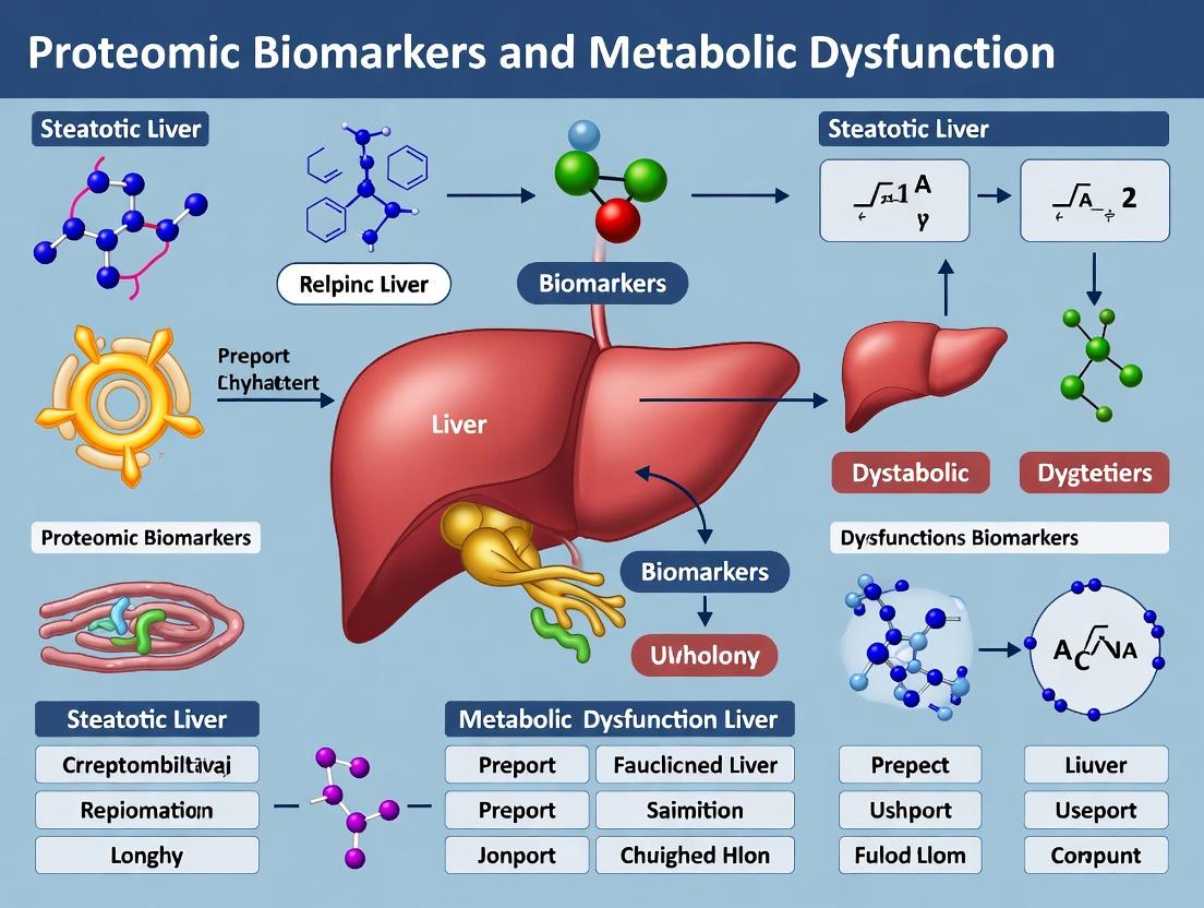
Unlocking Precision Medicine: Proteomic Biomarkers for Metabolic Dysfunction-Associated Steatotic Liver Disease (MASLD)
This article provides a comprehensive analysis of proteomic biomarkers for Metabolic Dysfunction-Associated Steatotic Liver Disease (MASLD), formerly known as NAFLD.
Unlocking Precision Medicine: Proteomic Biomarkers for Metabolic Dysfunction-Associated Steatotic Liver Disease (MASLD)
Abstract
This article provides a comprehensive analysis of proteomic biomarkers for Metabolic Dysfunction-Associated Steatotic Liver Disease (MASLD), formerly known as NAFLD. Designed for researchers, scientists, and drug development professionals, it explores the foundational biology linking hepatic proteome alterations to metabolic dysfunction, details cutting-edge methodologies for biomarker discovery and validation, addresses critical troubleshooting in assay development, and evaluates comparative performance against existing diagnostic tools. The synthesis aims to accelerate the translation of proteomic discoveries into reliable clinical diagnostics and targeted therapeutic strategies.
From Fat to Fibrosis: Decoding the Hepatic Proteome in MASLD Pathogenesis
The terminology for liver disease associated with metabolic dysfunction has evolved significantly. The shift from NAFLD (Non-Alcoholic Fatty Liver Disease) to MASLD (Metabolic Dysfunction-Associated Steatotic Liver Disease) reflects a more precise, pathophysiology-centered definition, inclusive of patients with concurrent alcohol intake or other liver diseases. This redefinition has direct implications for patient stratification, trial design, and biomarker discovery in proteomic research.
Table 1: Nomenclature Transition from NAFLD to MASLD and Associated Criteria
| Term | Acronym | Key Diagnostic Criteria | Exclusion Criteria |
|---|---|---|---|
| Non-Alcoholic Fatty Liver Disease | NAFLD | Hepatic steatosis >5% | Significant alcohol use, other liver diseases |
| Metabolic Dysfunction-Associated Steatotic Liver Disease | MASLD | Hepatic steatosis + ≥1 of 5 cardiometabolic risk factors | None (can coexist with other liver diseases) |
| Metabolic Dysfunction-Associated Steatohepatitis | MASH | MASLD criteria + hepatocyte injury (ballooning) + inflammation | --- |
| MetALD | MetALD | MASLD criteria + ‘significant’ alcohol intake (140-350 g/wk for women, 210-420 g/wk for men) | --- |
Clinical Staging and Histopathological Definitions
The progression of MASLD is staged based on histology from liver biopsy, which remains the diagnostic reference. The key stages are defined by the presence and degree of steatosis, lobular inflammation, hepatocyte ballooning, and fibrosis.
Table 2: Histopathological Features Defining the MASLD Spectrum (NAS/CRN & SAF Score)
| Feature | Score | Definition |
|---|---|---|
| Steatosis | 0 | <5% |
| 1 | 5-33% | |
| 2 | 34-66% | |
| 3 | >66% | |
| Lobular Inflammation | 0 | No foci |
| 1 | <2 foci per 200x field | |
| 2 | 2-4 foci per 200x field | |
| 3 | >4 foci per 200x field | |
| Hepatocyte Ballooning | 0 | None |
| 1 | Few balloon cells | |
| 2 | Many/prominent balloon cells | |
| Fibrosis Stage | 0 | None |
| 1 | Perisinusoidal or periportal | |
| 1A | Mild, perisinusoidal | |
| 1B | Moderate, perisinusoidal | |
| 1C | Periportal only | |
| 2 | Perisinusoidal & periportal | |
| 3 | Bridging fibrosis | |
| 4 | Cirrhosis |
Proteomic Biomarker Discovery: Core Experimental Protocol
This protocol outlines a targeted proteomic workflow for identifying and validating serum biomarkers to distinguish simple steatosis (MASL) from metabolic steatohepatitis (MASH).
Protocol 3.1: Serum Sample Preparation for LC-MS/MS
Objective: To isolate and digest proteins from human serum for liquid chromatography-tandem mass spectrometry (LC-MS/MS) analysis. Materials: Patient serum samples, ProteoMiner protein enrichment kit, 100mM ammonium bicarbonate buffer, dithiothreitol (DTT), iodoacetamide (IAA), sequencing-grade trypsin/Lys-C mix, C18 desalting columns, speed vacuum concentrator. Procedure:
- Depletion & Enrichment: Use ProteoMiner kit per manufacturer’s instructions to reduce high-abundance proteins (e.g., albumin, IgG) and enrich low-abundance species.
- Reduction & Alkylation: Resuspend enriched proteins in 100µL 100mM AmBic. Add DTT to 10mM, incubate 45 min at 55°C. Cool, add IAA to 20mM, incubate 30 min in dark at 25°C.
- Digestion: Add trypsin/Lys-C mix at 1:50 (enzyme:protein) ratio. Incubate overnight at 37°C with gentle agitation.
- Desalting: Acidify digest with 1% formic acid. Desalt using C18 column per manufacturer’s protocol. Elute peptides with 60% acetonitrile/0.1% formic acid.
- Concentration: Dry eluted peptides using a speed vacuum concentrator. Store at -80°C until LC-MS/MS analysis.
Protocol 3.2: LC-MS/MS Data Acquisition and SWATH Analysis
Objective: To perform data-independent acquisition (DIA) for quantitative proteomic profiling. Materials: Nanoflow LC system (e.g., Eksigent ekspert nanoLC 425), TripleTOF 6600+ MS, C18 trap and analytical columns, mobile phases (A: 0.1% FA in water, B: 0.1% FA in ACN). Procedure:
- Chromatography: Reconstitute peptides in 2% ACN/0.1% FA. Load 2µg onto a trap column (5µm, 5 x 0.3 mm) at 5µL/min for 10 min. Separate on analytical column (3µm, 150 mm x 75µm) with a 90-min gradient from 5-30% B at 300 nL/min.
- MS Acquisition (SWATH): Use a high-resolution TOF-MS scan (350-1250 m/z, 250 ms accumulation) followed by sequential 32 variable-width Q1 isolation windows (covering 400-1000 m/z) for MS/MS. Accumulation time 50 ms per window.
- Data Generation: Acquire data in triplicate for each sample. Use a sample-randomized order to minimize batch effects.
Protocol 3.3: Bioinformatics and Biomarker Validation Pipeline
Objective: To process SWATH data, identify differentially expressed proteins, and select candidates for orthogonal validation. Materials: SWATH processing software (e.g., DIA-NN, Spectronaut), R or Python statistical environment, ELISA/immunoassay kits for candidate proteins. Procedure:
- Spectral Library Building: Create a project-specific spectral library from pooled sample data-dependent acquisition (DDA) runs or use a comprehensive human library.
- Peptide Quantification: Process SWATH files using DIA-NN (v1.8) with default settings for high accuracy. Map peptides to UniProt human database.
- Statistical Analysis: Perform Shapiro-Wilk normality test. Use Mann-Whitney U test or Student’s t-test for case-control (MASL vs. MASH) comparison. Apply Benjamini-Hochberg correction for multiple testing (FDR <0.05). Proteins with ≥1.5-fold change and FDR<0.05 are considered significant.
- Pathway Analysis: Input significant proteins into IPA or STRING-db for pathway (e.g., inflammation, fibrosis) and network analysis.
- Orthogonal Validation: Select top 3-5 candidate biomarkers. Measure their levels in an independent validation cohort using ELISA or Olink immunoassays. Perform ROC curve analysis to assess diagnostic accuracy for MASH (vs. MASL).
Visualization of Key Pathways and Workflows
Diagram 1: Key Pathogenic Pathways in MASH Progression
Diagram 2: Proteomic Biomarker Discovery Workflow
The Scientist's Toolkit: Research Reagent Solutions
Table 3: Essential Research Reagents for MASLD Proteomic Studies
| Reagent/Material | Supplier Examples | Function in MASLD Research |
|---|---|---|
| ProteoMiner Protein Enrichment Kit | Bio-Rad | Equalizes protein concentrations in serum/plasma by reducing high-abundance proteins, critical for detecting low-abundance biomarkers. |
| Sequencing-Grade Modified Trypsin/Lys-C | Promega, Thermo Fisher | High-precision enzymatic digestion of proteins into peptides for mass spectrometry analysis. |
| C18 Desalting Spin Columns | Pierce, Nest Group | Removal of salts and detergents from digested peptide samples prior to LC-MS/MS. |
| Human XL Cytokine & Fibrosis Panel (Olink) | Olink Proteomics | Multiplex, high-sensitivity immunoassay for quantifying inflammatory and fibrogenic proteins in small sample volumes. |
| Human FGF21/CK-18/PIIINP ELISA Kits | BioVendor, R&D Systems | Orthogonal, quantitative validation of key candidate protein biomarkers identified by discovery proteomics. |
| DIA-NN Software | Vadim Demichev Lab | Open-source software for processing data-independent acquisition (DIA/SWATH) MS data, enabling high-throughput protein quantification. |
| Luminex MAGPIX System | Luminex Corp. | Multiplexing platform for validating panels of protein biomarkers in large patient cohorts. |
| Human Liver Proteome Spectral Library | ProteomeXchange, SCIEX | Curated reference library of human liver protein MS spectra to improve peptide identification accuracy in SWATH analysis. |
This document provides detailed protocols and application notes for investigating the interplay between insulin signaling, lipotoxic stress, and inflammation in metabolic dysfunction-associated steatotic liver disease (MASLD). The focus is on generating reproducible data to identify and validate proteomic biomarkers within these core, dysregulated pathways, supporting target discovery and therapeutic development.
Protocol: Integrated In Vitro Model of Lipotoxicity and Insulin Resistance in Hepatocytes
Aim: To induce and quantify insulin signaling dysfunction in the context of lipotoxic and inflammatory stress in a human hepatocyte cell line (e.g., HepG2, HulH-7).
Materials & Reagents:
- Cells: Human hepatocyte cell line.
- Lipotoxic Media: Complete growth media supplemented with 0.5 mM sodium palmitate (conjugated to 2% fatty acid-free BSA).
- Inflammatory Stimulus: Human recombinant TNF-α (10-20 ng/mL) and/or IL-1β (5-10 ng/mL).
- Insulin Stimulus: Human recombinant insulin (100 nM).
- Lysis Buffer: RIPA buffer supplemented with phosphatase and protease inhibitors.
- Key Assays: Phospho-AKT (Ser473) ELISA or Western blot, Pro-inflammatory cytokine secretion (ELISA for IL-6, IL-8), Intracellular lipid accumulation (Oil Red O staining or BODIPY 493/503 fluorescence).
Procedure:
- Cell Culture & Seeding: Maintain hepatocytes in standard culture. Seed cells at appropriate density in multi-well plates for assays.
- Disease Modeling (48-72h): Treat cells with:
- Control: Complete media + 2% BSA.
- Lipotoxic (LT): Lipotoxic media (Palmitate/BSA).
- Inflammatory (INF): Complete media + cytokines.
- Combined (LT+INF): Lipotoxic media + cytokines.
- Insulin Challenge (20 min): After disease modeling, stimulate all conditions with 100 nM insulin or vehicle control for 20 minutes.
- Termination & Sample Collection:
- For Signaling: Aspirate media, rinse with cold PBS, and lyse cells in ice-cold lysis buffer for phospho-protein analysis.
- For Secretomics: Collect conditioned media, centrifuge to remove debris, and store at -80°C for cytokine ELISA.
- For Lipid Analysis: Fix cells for Oil Red O staining or incubate with BODIPY dye.
- Downstream Analysis: Perform targeted protein analysis (Western/ELISA) and functional readouts.
Protocol: Phospho-Proteomic Enrichment and LC-MS/MS for Insulin Signaling Pathway Mapping
Aim: To profile global changes in tyrosine and serine/threonine phosphorylation in response to insulin under lipotoxic conditions.
Materials & Reagents:
- Phospho-Enrichment Kits: TiO2 or immobilized metal affinity chromatography (IMAC) magnetic beads.
- Lysis Buffer: Urea-based lysis buffer (8M Urea, 50 mM Tris-HCl pH 8.0) with benzonase, phosphatase, and protease inhibitors.
- Reduction/Alkylation: Dithiothreitol (DTT) and iodoacetamide (IAA).
- Digestion: Trypsin/Lys-C mix.
- Desalting: C18 solid-phase extraction tips or columns.
- LC-MS/MS System: Nanoflow HPLC coupled to high-resolution tandem mass spectrometer (e.g., Q-Exactive, timsTOF).
Procedure:
- Cell Lysis & Protein Prep: Lyse treated cells (from Protocol 2.0) in urea buffer. Determine protein concentration. Reduce with DTT, alkylate with IAA.
- Digestion: Dilute urea to <2M with Tris buffer. Digest with trypsin/Lys-C overnight at 37°C. Quench with acid.
- Phosphopeptide Enrichment: Desalt digested peptides. Follow manufacturer protocol for TiO2/IMAC enrichment. Elute phosphopeptides.
- LC-MS/MS Analysis: Reconstitute samples in LC loading buffer. Separate using a C18 nano-column gradient (90-120 min). Acquire data in data-dependent acquisition (DDA) mode with higher-energy collisional dissociation (HCD).
- Data Analysis: Process raw files using search engines (MaxQuant, Spectronaut) against human UniProt database. Use PTM-specific search parameters. Normalize label-free quantitation (LFQ) intensities.
Table 1: Representative Phospho-Proteomic Data (Simulated from Recent Studies)
| Protein (Gene) | Phosphosite | Insulin/Control (Fold Change) | Insulin/Lipotoxic (Fold Change) | Pathway |
|---|---|---|---|---|
| IRS1 | S307 (Inhibitory) | 1.0 | 3.5 | Insulin Signaling Negative Feedback |
| AKT1 | S473 (Activation) | 12.8 | 2.1 | Insulin Signaling Effector |
| mTOR | S2448 (Activation) | 5.2 | 1.8 | Nutrient Sensing/Growth |
| JNK1 | T183/Y185 (Activation) | 1.2 | 6.7 | Stress/Inflammation |
| STAT3 | S727 (Activation) | 1.5 | 4.3 | Inflammatory Signaling |
The Scientist's Toolkit: Key Research Reagent Solutions
Table 2: Essential Reagents for Metabolic Dysfunction Signaling Research
| Reagent Category | Example Product/Assay | Primary Function in Research |
|---|---|---|
| Metabolic Stress Inducers | Sodium Palmitate-BSA Conjugate; Recombinant TNF-α/IL-1β | Mimics lipotoxic and inflammatory environment of MASLD in vitro. |
| Pathway Activation Sensors | Phospho-Specific Antibodies (pAKT, pIRS, pJNK); PathHunter β-Arrestin Recruitment Assays | Detect activation/inhibition states of key signaling nodes. |
| Cytokine Profiling | V-PLEX Proinflammatory Panel 1 (Meso Scale Discovery); Luminex Multiplex Assays | Quantify secretome changes to gauge inflammatory output. |
| Lipid Accumulation Probes | BODIPY 493/503; LipidTOX Stains | Visualize and quantify intracellular lipid droplets. |
| Phospho-Proteomics | TiO2 Mag Sepharose (Cytiva); PTMScan Kits (CST) | Enrich for low-abundance phosphopeptides for MS analysis. |
| High-Content Imaging | CellMask Stains; Opera Phenix Plus System | Multiparametric analysis of cell health, morphology, and staining. |
Visualization of Pathways and Workflows
Title: Insulin Signaling Disruption by Lipotoxicity & Inflammation
Title: Integrated Experimental Workflow for Biomarker Discovery
Application Notes: Role in Metabolic Dysfunction-Associated Steatotic Liver Disease (MASLD)
The search for reliable, non-invasive biomarkers for MASLD progression—from simple steatosis to metabolic dysfunction-associated steatohepatitis (MASH) and fibrosis—remains a central challenge. This application note details the utility and interplay of key protein biomarkers within this proteomic landscape.
Table 1: Key Proteomic Biomarkers in MASLD Spectrum
| Biomarker | Primary Cellular Source | Physiological Role | Association in MASLD | Representative Concentrations (Serum) |
|---|---|---|---|---|
| Cytokeratin-18 (CK-18) M30/M65 | Hepatocytes (epithelial intermediate filaments) | Structural integrity. Caspase-cleaved (M30) indicates apoptosis; full-length (M65) indicates total cell death. | Strongly correlated with hepatocyte apoptosis and MASH activity. M30 fragment is a leading biomarker for NASH diagnosis. | Healthy: M30 ~ 150 U/L; MASH: M30 > 300 U/L. M65 levels often 2-3x higher in MASH vs. steatosis. |
| Fibroblast Growth Factor 21 (FGF21) | Liver (primary), adipocytes, pancreas | Endocrine hormone regulating glucose/lipid metabolism, insulin sensitivity, and adaptive starvation response. | Markedly elevated in MASLD as a compensatory hepatokine. Correlates with hepatic steatosis and insulin resistance but not specific for MASH. | Healthy: ~150-250 pg/mL; MASLD: often > 400 pg/mL; can exceed 1000 pg/mL. |
| Adiponectin | Adipocytes (white adipose tissue) | Insulin-sensitizing, anti-inflammatory, anti-steatotic adipokine. Enhances fatty acid oxidation, inhibits hepatic gluconeogenesis. | Levels are inversely correlated with hepatic steatosis, inflammation, and insulin resistance. Hypoadiponectinemia is a hallmark of metabolic dysfunction. | Healthy: 5-10 μg/mL (higher in females). MASLD/MASH: Often reduced by 30-50%. |
| Other Notable Players | ||||
| PNPLA3 (I148M variant) | Hepatocytes | Lipid droplet remodeling (patatin-like phospholipase). | Genetic risk factor; variant protein accumulation on lipid droplets promotes steatosis and fibrosis. | Genotypic (not circulating protein). |
| HSD17B13 | Hepatocytes | Lipid metabolism enzyme (retinol dehydrogenase). | Loss-of-function variants are protective against progression from steatosis to MASH/fibrosis. | Genotypic/protein expression. |
Table 2: Diagnostic Performance of Biomarker Panels
| Panel/Algorithm | Components | Clinical Utility | Reported AUC for MASH (NAS ≥4) |
|---|---|---|---|
| NIS4 | miR-34a-5p, α2-Macroglobulin, YKL-40, HbA1c | Non-invasive identification of at-risk NASH (NAS≥4 + F≥1). | 0.80 - 0.85 |
| FAST Score | CK-18 (M30), AST, Platelets | Rule-in/rule-out for progressive NASH (NAS≥4 + F≥2). | 0.80 (Rule-in: >0.67; Rule-out: <0.35) |
| ELF Test | HA, PIIINP, TIMP-1 | Assessment of liver fibrosis stage (≥F2, ≥F3). | ~0.82 for ≥F2; ~0.90 for ≥F3 |
Experimental Protocols
Protocol 1: Assessment of Hepatocyte Apoptosis via CK-18 M30 ELISA Objective: Quantify caspase-cleaved CK-18 (M30) in human serum/plasma as a marker of hepatocyte apoptosis.
- Sample Prep: Collect blood in serum separator tubes. Allow clotting (30 min, RT), centrifuge (2000 x g, 10 min). Aliquot and store serum at -80°C. Avoid repeated freeze-thaw.
- ELISA Procedure: Use a commercial M30 Apoptosense ELISA or equivalent.
- Bring all reagents and samples to RT.
- Add standards, controls, and samples (50 µL/well) to the antibody-coated microplate.
- Add detection antibody (50 µL/well). Incubate 4h on shaker (300 rpm) at RT.
- Wash plate 5x with 300 µL/well of provided wash buffer.
- Add Streptavidin-HRP conjugate (100 µL/well). Incubate 30 min on shaker at RT.
- Wash plate 5x.
- Add TMB substrate (100 µL/well). Incubate 15 min in the dark.
- Stop reaction with 1M H2SO4 (100 µL/well).
- Read absorbance at 450 nm (reference 620-650 nm) within 30 min.
- Analysis: Generate a 4-parameter logistic standard curve. Report concentrations in U/L.
Protocol 2: Measurement of Adiponectin Multimers by ELISA Objective: Specifically measure high-molecular-weight (HMW) adiponectin, the most bioactive form.
- Sample Prep: Serum/plasma (EDTA) collected as in Protocol 1. For HMW analysis, a pre-treatment step with a protease (e.g., Proteinase K) to digest non-HMW forms may be required per kit instructions.
- ELISA Procedure: Use a commercial HMW Adiponectin ELISA.
- Add standards and samples to wells (typically 10-20 µL). Incubate.
- Wash. Add detection antibody. Incubate.
- Wash. Add HRP-conjugated secondary antibody. Incubate.
- Wash. Add chromogenic substrate. Incubate in the dark.
- Stop reaction.
- Read absorbance at 450 nm.
- Analysis: Calculate HMW adiponectin concentration from standard curve. Total adiponectin can be measured using a separate kit for ratio calculation (HMW/Total).
Protocol 3: Western Blot for Hepatic FGF21 Expression in Rodent Models Objective: Semi-quantify hepatic FGF21 protein levels in liver tissue lysates from MASLD models (e.g., MCD diet, ob/ob mice).
- Tissue Lysate Prep: Homogenize ~30 mg liver tissue in RIPA buffer with protease/phosphatase inhibitors. Centrifuge at 12,000 x g, 20 min, 4°C. Collect supernatant, determine protein concentration (BCA assay).
- Gel Electrophoresis: Load 20-40 µg protein per lane on a 4-20% Tris-Glycine SDS-PAGE gel. Run at 120V for 90 min.
- Transfer: Transfer to PVDF membrane at 100V for 60 min (ice-cooled).
- Blocking & Probing: Block membrane in 5% non-fat milk in TBST for 1h. Incubate with primary antibodies (anti-FGF21, 1:1000; anti-β-Actin, 1:5000) in blocking buffer overnight at 4°C. Wash (TBST, 3 x 10 min). Incubate with HRP-conjugated secondary antibody (1:5000) for 1h at RT. Wash.
- Detection: Use enhanced chemiluminescence (ECL) substrate. Image on a chemiluminescence imager. Analyze band density relative to β-Actin.
Mandatory Visualizations
Biomarker Interplay in MASLD Pathogenesis
Workflow for Biomarker Analysis & Integration
The Scientist's Toolkit: Research Reagent Solutions
| Item | Function & Application in MASLD Proteomics |
|---|---|
| M30/M65 CK-18 ELISA Kits | Quantify caspase-cleaved (apoptosis) and total (cell death) CK-18. Critical for apoptosis-focused biomarker studies. |
| Multiplex Adipokine Panels | Simultaneously measure adiponectin (total/HMW), leptin, resistin, etc., to profile adipose tissue communication. |
| FGF21 ELISA (Animal/Human) | Species-specific kits to measure this key hepatokine in preclinical models and clinical samples. |
| Protease & Phosphatase Inhibitor Cocktails | Essential for stabilizing protein extracts from liver tissue or cells, preventing biomarker degradation. |
| RIPA Lysis Buffer | For efficient extraction of total protein from liver tissue for western blotting of targets like FGF21, PNPLA3. |
| Human Fibrosis/Inflammation Multiplex Assays | Measure panels of markers (e.g., HA, TIMP-1, PIIINP, IL-6, TNF-α) to correlate with fibrotic progression. |
| Simple Plex/Nanofluidic Immunoassay Platforms | Enable high-sensitivity, low-volume quantification of biomarkers from precious sample sets (e.g., rodent serum). |
| Anti-PNPLA3 (I148M) Antibodies | For immunohistochemistry or western blot to study the localization and expression of this genetic risk factor protein. |
Application Notes
This application note is framed within the broader thesis of discovering proteomic biomarkers for metabolic dysfunction-associated steatotic liver disease (MASLD). The selection of the optimal biospecimen—serum, plasma, or liver tissue—is critical for the accurate identification, validation, and clinical translation of candidate biomarkers. The primary goal is to identify a minimally invasive "liquid biopsy" capable of reliably detecting and staging MASLD, thereby reducing dependency on invasive liver biopsy.
Serum Proteomics
- Context: Serum is the liquid fraction remaining after blood coagulation. It lacks clotting factors (like fibrinogen) but is rich in proteins and peptides released from platelets and cells during clotting.
- Advantages for MASLD: Readily available from routine blood draws; high clinical utility; reflects a broad systemic physiological state, including inflammation and systemic metabolic dysfunction.
- Challenges: The clotting process introduces high-abundance, high-variability proteins (e.g., platelet-derived factors), increasing analytical noise and potentially masking low-abundance, liver-specific signals. Pre-analytical variability (clot time, temperature) is a major confounder.
Plasma Proteomics
- Context: Plasma is the liquid fraction of anticoagulated blood, containing all circulating proteins, including clotting factors.
- Advantages for MASLD: More accurately represents the in vivo circulating proteome with less contribution from platelets. Use of different anticoagulants (EDTA, citrate, heparin) allows for tailored protocols. Generally considered more reproducible for biomarker discovery.
- Challenges: The presence of high-abundance anticoagulant proteins can interfere with some mass spectrometry (MS) assays. Choice of anticoagulant must be consistent and declared, as it influences the proteomic profile.
Tissue Proteomics
- Context: Direct analysis of proteins extracted from liver tissue, typically obtained via biopsy.
- Advantages for MASLD: Provides the ground truth of disease pathology within the target organ. Enables spatial proteomics to distinguish zonated protein expression, inflammation foci, and fibrotic areas. Critical for mechanistic understanding and for validating the origin of circulating biomarkers.
- Challenges: Highly invasive, limiting serial sampling and patient willingness. Represents a single snapshot of a heterogeneous organ. Prone to sampling error.
Comparative Summary Table
| Parameter | Serum | Plasma (EDTA) | Tissue (Liver) |
|---|---|---|---|
| Invasiveness | Minimally invasive | Minimally invasive | Highly invasive (biopsy) |
| Key Composition | Coagulation cascade proteins, platelet factors, cytokines | Intact circulating proteome + anticoagulant proteins | Full cellular proteome, structural proteins |
| Primary Advantage | Clinical standard, high translational potential | Reproducible, reflects in vivo state more accurately | Direct disease pathology, mechanistic insights |
| Primary Disadvantage | High pre-analytical variability from clotting | Interference from anticoagulants in assays | Invasiveness, sampling bias, heterogeneity |
| Role in MASLD Thesis | Biomarker validation in clinical cohorts | Primary discovery matrix for liquid biopsy | Gold-standard correlation and pathway discovery |
Experimental Protocols
Protocol 1: Standardized Pre-Analytical Processing for Plasma/Serum in MASLD Studies
Objective: To minimize pre-analytical variability in liquid biospecimens for MS-based proteomics.
- Blood Draw: Collect whole blood via venipuncture using a consistent technique.
- For Plasma: Draw into pre-chilled K₂EDTA tubes. Invert gently 8-10 times. Process within 30 minutes at 4°C.
- For Serum: Draw into serum separator tubes. Allow to clot upright at room temperature for exactly 30 minutes.
- Centrifugation: Spin at 2,000 x g for 15 minutes at 4°C in a refrigerated centrifuge.
- Aliquotting: Carefully pipette the supernatant (plasma or serum) into pre-labeled cryovials, avoiding the buffy coat or clot material. Use low-protein-binding pipette tips.
- Storage: Flash-freeze aliquots in liquid nitrogen and store at -80°C. Avoid repeated freeze-thaw cycles.
Protocol 2: High-Abundance Protein Depletion and Clean-Up for LC-MS/MS
Objective: To enrich low-abundance candidate biomarkers by removing highly abundant proteins (e.g., albumin, IgG).
- Thawing: Thaw plasma/serum aliquots on ice.
- Depletion: Use a commercial immunoaffinity column (e.g., MARS Human 14, Agilent). Dilute sample 1:5 with provided buffer and load onto the column per manufacturer's instructions.
- Desalting/Buffer Exchange: Desalt the flow-through fraction using a 3kDa molecular weight cut-off (MWCO) filter or a C18 solid-phase extraction (SPE) cartridge.
- Protein Quantification: Measure protein concentration of the depleted sample using a colorimetric assay (e.g., BCA assay).
- Digestion: Reduce (5mM DTT, 30min, 56°C), alkylate (15mM iodoacetamide, 30min, dark), and digest with trypsin (1:50 enzyme-to-protein ratio, 37°C, overnight).
- Peptide Clean-up: Desalt digested peptides using C18 StageTips. Elute with 80% acetonitrile/0.1% formic acid. Dry in a vacuum concentrator.
Protocol 3: Data-Independent Acquisition (DIA) Mass Spectrometry Analysis
Objective: For reproducible, comprehensive quantification of the plasma/serum proteome.
- LC-MS Setup: Resuspend peptides in 0.1% formic acid. Separate on a 25cm C18 column using a 90-minute linear gradient (3-30% acetonitrile) at 300 nL/min.
- Mass Spectrometry: Use a Q-Exactive HF or Orbitrap Astral mass spectrometer.
- DIA Method: Full MS scan (350-1200 m/z, R=60,000). DIA windows: 24-32 variable windows covering the m/z range with a 1 m/z overlap. MS2 resolution: 30,000.
- Data Analysis: Process raw files using Spectronaut or DIA-NN. Use a comprehensive spectral library (e.g., generated from pooled samples via DDA or a publicly available human library). Filter results at 1% FDR.
Visualizations
Liquid Biopsy Development Workflow for MASLD
Key MASLD Pathways & Biomarker Release
The Scientist's Toolkit: Research Reagent Solutions
| Item | Function / Relevance |
|---|---|
| K₂EDTA Plasma Tubes | Preferred anticoagulant for plasma proteomics; minimizes ex vivo protein degradation and platelet activation. |
| Immunoaffinity Depletion Column (Hu14) | Removes 14 high-abundance plasma proteins (e.g., albumin, IgG), increasing depth of coverage for low-abundance biomarkers. |
| Trypsin, Sequencing Grade | Protease used for specific digestion of proteins into peptides for LC-MS/MS analysis. |
| TMTpro 16-plex / TMT 11-plex | Tandem Mass Tag reagents for multiplexed quantitative proteomics, enabling high-throughput comparison of MASLD cohorts. |
| Phosphatase & Protease Inhibitor Cocktails | Essential for tissue homogenization to preserve post-translational modifications and prevent protein degradation. |
| C18 StageTips / Spin Columns | For desalting and cleaning up peptide mixtures prior to MS, improving signal-to-noise. |
| DIA-NN Software | Open-source software for processing DIA-MS data, enabling high-throughput, reproducible quantification. |
| Liquid Chromatography System (nanoFlow) | Provides high-resolution separation of complex peptide mixtures prior to MS detection. |
| Orbitrap Mass Spectrometer | High-resolution, high-mass-accuracy MS instrument essential for confident protein identification and quantification. |
| Human Proteome Spectral Library | Curated reference of peptide spectra required for DIA data analysis, crucial for biomarker identification. |
Within the pursuit of proteomic biomarkers for metabolic dysfunction-associated steatotic liver disease (MASLD), single-omics approaches provide fragmented insights. Genomics identifies susceptibility loci and mutations, metabolomics captures the dynamic end-products of cellular processes, but proteomics delivers the essential middle layer: the functional effectors and direct biomarkers of disease activity. This Application Note details protocols and workflows for integrative multi-omics analysis, positioning proteomics as the central hub for validating genomic discoveries and explaining metabolomic phenotypes in MASLD research.
Application Notes: Multi-Omics Data Integration in MASLD
1. From GWAS Hit to Functional Protein Biomarker Genome-wide association studies (GWAS) have identified risk variants (e.g., in PNPLA3, TM6SF2). Proteomics bridges the gap between genetic association and mechanistic understanding by quantifying the resultant protein expression, post-translational modifications (PTMs), and protein-protein interactions.
2. Explaining Metabolomic Perturbations Metabolomic profiling of liver tissue or serum from MASLD patients reveals alterations in lipid species, bile acids, and glycolysis intermediates. Proteomic analysis of the enzymes, transporters, and regulators responsible for these metabolic pathways provides causal explanation. For example, elevated hepatic diacylglycerols (metabolomics) can be linked to the quantified depletion of SAMe synthetase (MAT1A) protein (proteomics).
Table 1: Correlation of Omics Data Types in MASLD Research
| Omics Layer | Data Type | MASLD Insight Example | Complementarity with Proteomics |
|---|---|---|---|
| Genomics | SNP (rs738409 in PNPLA3) | Genetic risk for steatosis and fibrosis. | Proteomics quantifies PNPLA3 protein abundance and its truncation/mutation status, linking genotype to molecular phenotype. |
| Transcriptomics | RNA-Seq Data | Differential gene expression of fibrotic pathways (TGF-β, COL1A1). | Proteomics measures actual collagen deposition and TGF-β pathway activation via phosphoproteomics, confirming translational regulation. |
| Metabolomics | LC-MS Lipidomics | Increase in hepatic ceramide and phosphatidylcholine species. | Proteomics identifies and quantifies key enzymes in sphingolipid synthesis (e.g., SPTLC2) and phospholipid transporters. |
| Proteomics | TMT-MS/Phospho-Proteomics | ↓ MAT1A, ↑ PSRC1, phospho-activation of JNK. | Serves as the functional readout linking genetic & transcriptomic changes to metabolomic dysregulation. |
Detailed Protocols
Protocol 1: Integrated Tissue Workflow for MASLD Biomarker Discovery
Objective: To extract genomic, proteomic, and metabolomic data from a single liver biopsy core.
Materials:
- MASLD Patient Biopsy Core: Flash-frozen in liquid N₂.
- Cryostat: Pre-cooled to -20°C.
- AllPrep DNA/RNA/Protein Mini Kit (Qiagen): For co-isolation.
- Methanol/Chloroform Solvents: LC-MS grade for metabolite/protein precipitation.
- RIPA Lysis Buffer (with phosphatase/protease inhibitors): For protein extraction.
- TMTpro 16plex Reagent Set (Thermo Fisher): For multiplexed quantitative proteomics.
- HILIC & C18 LC Columns: For metabolomics and proteomics, respectively.
- High-Resolution Tandem Mass Spectrometer (e.g., Orbitrap Astral).
Procedure:
- Sectioning: Using a cryostat, serially section one 30mg biopsy core (10 µm thickness).
- Macrodissection: Visually identify and separate steatotic vs. non-steatotic regions.
- Sequential Extraction: a. Metabolite Extraction: Transfer 10 sections to 500 µL cold 80% methanol. Homogenize. Centrifuge (15,000xg, 15min, 4°C). Collect supernatant for LC-MS metabolomics. b. Nucleic Acid/Protein Extraction: Pellet from step (a) is processed with the AllPrep kit per manufacturer's instructions, yielding DNA, RNA, and a protein fraction.
- Proteomic Sample Preparation: Digest protein fraction (50 µg) with trypsin/Lys-C. Label peptides with TMTpro reagents. Pool and fractionate by high-pH reverse-phase HPLC.
- LC-MS/MS Analysis:
- Metabolomics: Analyze methanol extract via HILIC-MS (negative/positive ion mode).
- Proteomics: Analyze TMT-labeled peptides via C18 nanoLC-MS/MS (120min gradient).
- Data Integration: Map proteomic data (differentially expressed proteins) onto KEGG pathways (e.g., fatty acid oxidation). Overlay metabolomic data (altered pathway substrates/products) and genomic risk alleles.
Protocol 2: Phosphoproteomic Workflow to Elucidate Signaling Drivers
Objective: To identify kinase-driven signaling networks linking MASLD genetic risk to metabolic dysfunction.
Materials:
- Tissue or Cell Lysate: From PNPLA3 I148M knock-in model.
- Phosphatase Inhibitors (PhosSTOP, Roche): Critical for phosphoproteomics.
- Fe-IMAC or TiO₂ Magnetic Beads: For phosphopeptide enrichment.
- EDTA and LC-MS grade Water: For chelation and washing.
- LC-MS equipped with EThcD Fragmentation: For improved phosphosite localization.
Procedure:
- Lysis: Lyse tissue in urea-based buffer (8M urea, 50mM Tris-HCl, pH 8.0) with PhosSTOP and protease inhibitors.
- Digestion & Desalting: Reduce, alkylate, and digest lysate with trypsin. Desalt peptides with C18 stage tips.
- Phosphopeptide Enrichment: Resuspend peptides in 80% acetonitrile/1% TFA. Incubate with Fe-IMAC beads for 30 min. Wash beads with 80% ACN/1% TFA, then 80% ACN/0.1% FA. Elute phosphopeptides with 1% NH₄OH.
- LC-MS/MS Analysis: Analyze eluate via C18 nanoLC coupled to MS with EThcD activation. Database search with phosphosite localization probability (e.g., using PTM-Score in Byonic).
- Upstream Kinase Prediction: Use tools like Kinase-Substrate Enrichment Analysis (KSEA) to link altered phosphosites to kinase activity (e.g., JNK, AKT).
Visualizations
Title: The Central Role of Proteomics in Multi-Omics Integration
Title: Sequential Multi-Omics Extraction from Single Biopsy
Title: Multi-Omics Elucidation of PNPLA3 Mechanism
The Scientist's Toolkit: Key Research Reagent Solutions
Table 2: Essential Materials for Integrated MASLD Omics Studies
| Item | Function & Role in Integration | Example Product/Catalog |
|---|---|---|
| AllPrep DNA/RNA/Protein Mini Kit | Enables simultaneous isolation of all three molecular classes from a single tissue sample, crucial for direct correlation. | Qiagen, 80004 |
| TMTpro 16plex Isobaric Label Reagents | Allows multiplexed quantitative comparison of up to 16 samples in one MS run, increasing throughput and reducing quantitative variability. | Thermo Fisher, A44520 |
| Phosphatase Inhibitor Cocktail (e.g., PhosSTOP) | Preserves the native phosphoproteome during lysis, essential for capturing kinase signaling events. | Roche, 4906845001 |
| Fe-IMAC or TiO₂ Magnetic Beads | Selective enrichment of phosphopeptides from complex digests, dramatically increasing coverage of phosphosites. | Thermo Fisher, 88826 / 88821 |
| HILIC Chromatography Columns | Optimal separation of polar metabolites (e.g., sugars, amino acids, nucleotides) for metabolomic LC-MS profiling. | Waters, BEH Amide Column |
| SAMe ELISA Kit | Targeted validation of MAT1A protein function loss, a key proteomic-metabolomic link in MASLD (SAMe depletion). | Abcam, ab242238 |
| JNK (phospho-T183/Y185) Antibody | Validates phosphoproteomic predictions of JNK pathway activation via Western blot or immunohistochemistry. | Cell Signaling, 4668 |
| PANOMICS Multi-omics Data Integration Software | Platform for statistical and pathway-based integration of genomic, proteomic, and metabolomic datasets. | Revvity Signals |
From Discovery to Assay: Cutting-Edge Proteomic Workflows for MASLD Biomarker Development
In the search for diagnostic and prognostic biomarkers for metabolic dysfunction-associated steatotic liver disease (MASLD) and its progressive form, metabolic dysfunction-associated steatohepatitis (MASH), comprehensive proteomic profiling is essential. The integration of discovery platforms like LC-MS/MS, SOMAscan, and Olink Proximity Extension Assay (PEA) enables the identification of novel protein signatures linked to hepatic steatosis, inflammation, and fibrosis. This application note details protocols and comparative analyses of these platforms within the context of a thesis focused on uncovering circulating and hepatic proteomic drivers of metabolic liver dysfunction.
Table 1: Comparative Overview of Key Proteomic Discovery Platforms
| Feature | LC-MS/MS (Discovery Proteomics) | SOMAscan (Aptamer-Based) | Olink PEA (Antibody-Based) |
|---|---|---|---|
| Principle | Liquid chromatography tandem mass spectrometry | Slow Off-rate Modified Aptamers (SOMAmers) | Proximity Extension Assay (paired antibodies) |
| Assay Type | Untargeted/Targeted | Multiplexed affinity binding | Multiplexed affinity binding |
| Typical Sample Volume | 10-100 µL (plasma/serum) | 65-150 µL (plasma/serum) | 1-3 µL (plasma/serum) |
| Throughput | Low to medium | High | High |
| Dynamic Range | ~4-5 orders of magnitude | >10 orders of magnitude | >10 orders of magnitude |
| Multiplexing Capacity | 1000s (untargeted), 100s (targeted) | ~7,000 protein assays (v4) | Up to 3,072 (Explore) |
| Key Metric for MASLD | Identifies novel, unanticipated proteins; quantifies proteoforms. | Broad screening for pathway analysis. | High specificity/sensitivity for low-abundance cytokines & hormones. |
| Primary Output | Peptide spectra, protein identification/quantification. | Relative Fluorescence Unit (RFU). | Normalized Protein eXpression (NPX) on log2 scale. |
Table 2: Representative Data from a Simulated MASLD Pilot Study (Plasma)
| Platform | Proteins Measured | Differentially Expressed Proteins (MASH vs Control) | Key Pathways Enriched (Example) |
|---|---|---|---|
| LC-MS/MS | ~800 quantified | 124 (p<0.01) | Complement activation, Fatty acid beta-oxidation |
| SOMAscan 7k | ~7,000 | 842 (FDR<0.05) | Inflammation (IL-6, TNF signaling), Fibrosis (TGF-β, PDGF) |
| Olink Inflammation Panel | 92 | 28 (FDR<0.05) | Cytokine signaling (IL-6, IL-10, MCP-1), Chemotaxis |
Experimental Protocols
Protocol 1: LC-MS/MS for Plasma Proteomics in MASLD
Title: Untargeted Plasma Proteome Profiling for Biomarker Discovery.
Key Research Reagent Solutions:
- Depletion Column (e.g., MARS Hu-14): Removes high-abundance proteins to enhance detection of low-abundance biomarkers.
- Reduction/Alkylation Reagents (DTT, IAA): Break and cap disulfide bonds for complete denaturation and digestion.
- Trypsin/Lys-C Mix: Protease for specific cleavage into peptides for MS analysis.
- C18 Desalting/Solid-Phase Extraction Tips: Purify and concentrate digested peptides.
- LC-MS Grade Solvents (Water, Acetonitrile): High-purity solvents for reproducible chromatography.
- Internal Standard (Heavy-labeled peptides): For absolute quantification in targeted assays (e.g., SRM/PRM).
Methodology:
- Sample Preparation: Deplete 20 µL of plasma using a MARS-14 column. Reduce with 10mM DTT (30 min, 56°C), alkylate with 25mM IAA (30 min, dark, RT). Quench with excess DTT.
- Digestion: Dilute sample in 50mM TEAB buffer. Digest with Trypsin/Lys-C (1:50 enzyme:protein) overnight at 37°C.
- Peptide Clean-up: Acidify with 1% TFA. Desalt using C18 tips, elute in 70% ACN/0.1% FA. Dry in vacuum concentrator.
- LC-MS/MS Analysis: Reconstitute in 2% ACN/0.1% FA. Inject 2 µg onto a nano-flow UHPLC system coupled to a high-resolution tandem mass spectrometer (e.g., Q-Exactive HF-X).
- Chromatography: 120-min gradient (3-35% ACN) on a C18 column.
- MS Settings: Data-Dependent Acquisition (DDA) mode. Full MS scan (350-1500 m/z, R=120,000), followed by top 20 MS/MS scans (HCD fragmentation, R=15,000).
- Data Processing: Process raw files using software (e.g., MaxQuant, Proteome Discoverer) against the human UniProt database. Use label-free quantification (LFQ) for differential analysis.
Protocol 2: SOMAscan Assay for Serum Proteomic Profiling
Title: High-Throughput Serum Proteomic Analysis via SOMAmer Affinity.
Key Research Reagent Solutions:
- SOMAscan Assay Kit (e.g., 7k): Contains all SOMAmers, buffers, and controls for multiplexed analysis.
- Biotinylated SOMAmers: Protein-binding reagents with a unique fluorescent DNA tag.
- Streptavidin-Coated Beads: Capture biotinylated SOMAmer-protein complexes.
- Polyanionic Competitor Reagents: Reduce non-specific binding.
- Control SOMAmers (Hybridization, Normalization): For intra- and inter-assay QC.
Methodology:
- Sample Dilution & Denaturation: Dilute 65 µL of serum 3-fold in appropriate buffer. Heat denature at 55°C for 10 minutes to expose epitopes.
- SOMAmer Incubation: Incubate denatured sample with the SOMAmer reagent mix for binding equilibrium (specific time/temp per kit protocol).
- Complex Capture & Wash: Bind SOMAmer-protein complexes to streptavidin beads. Wash extensively to remove non-specifically bound proteins.
- Elution & Quantification: Elute SOMAmers from beads. Quantify each SOMAmer via hybridization to its complementary sequence on a custom DNA microarray or by qPCR (older kits). Signal is reported as Relative Fluorescence Units (RFU).
- Data Normalization: Apply adaptive normalization using internal controls to generate final RFU values for statistical analysis.
Protocol 3: Olink PEA for Targeted Inflammation Panel
Title: Ultrasensitive Measurement of Inflammatory Proteins via PEA.
Key Research Reagent Solutions:
- Olink Target 96/384 Panel (e.g., Inflammation): Kit containing matched antibody pairs (PEA probes) for each target.
- PEA Probes (DNA-conjugated antibodies): Paired antibodies bind to the same target, enabling DNA extension.
- Extension & Amplification Master Mix: Contains polymerase for extension and PCR reagents for pre-amplification.
- Microfluidic qPCR Chip (Biomark HD) or NGS Reagents: For final readout.
- Internal Controls (Incubation, Extension, Amplification): Monitor each assay step.
Methodology:
- Sample & Probe Incubation: Dilute 1 µL of plasma to 3 µL. Incubate with a panel of 92 paired PEA probes in a 96-well plate for overnight binding at 4°C.
- Extension: Add extension master mix. When two probes are in proximity on the target protein, their DNA tails hybridize and are extended by a DNA polymerase, creating a unique, protein-specific DNA barcode.
- Pre-Amplification: Perform a limited-cycle PCR to amplify all DNA barcodes simultaneously.
- Quantification (qPCR or NGS): For 96-plex panels, quantify the DNA barcode via microfluidic qPCR (Fluidigm Biomark HD). For Explore panels, use NGS.
- Data Processing: Data is normalized using internal and inter-plate controls. Results are reported in Normalized Protein eXpression (NPX) units on a log2 scale, where a 1 NPX increase represents a doubling in protein concentration.
Pathway and Workflow Diagrams
Title: Proteomic Discovery Workflow for MASLD Research
Title: Proteomic Biomarkers in MASLD Pathogenesis
Application Notes: The Clinical Translation Imperative in Steatotic Liver Disease
The discovery and validation of protein biomarkers for metabolic dysfunction-associated steatotic liver disease (MASLD) and its progressive form, MASH (metabolic dysfunction-associated steatohepatitis), represent a critical frontier in hepatology. Initial discovery-phase studies using untargeted proteomics often yield expansive biomarker panels with high diagnostic potential in cohort studies. However, the transition from these complex multi-analyte panels to single, or small multiplex, routinizable assays is a major bottleneck in clinical translation. This document outlines the strategic application of targeted mass spectrometry (MS) and enzyme-linked immunosorbent assay (ELISA) methodologies to bridge this gap, enabling the development of robust, cost-effective, and scalable diagnostic assays suitable for clinical laboratories and large-scale trials.
Key Rationale for Transition:
- Throughput & Cost: Targeted MS (e.g., LC-MRM/MS) and ELISA offer higher sample throughput and lower per-sample cost than discovery proteomics, essential for validation studies involving thousands of samples.
- Standardization: These platforms have well-defined protocols, calibrators, and controls, facilitating inter-laboratory reproducibility.
- Regulatory Path: ELISA, in particular, has a clear regulatory pathway for IVD (In Vitro Diagnostic) approval, while targeted MS assays can be developed as laboratory-developed tests (LDTs).
- Quantitative Precision: Both platforms provide absolute or relative quantitative data with high precision and accuracy over a dynamic range relevant to serum/plasma biomarkers.
Strategic Workflow: The progression involves using targeted MS to verify and prioritize the most promising candidates from discovery panels, followed by the development of high-throughput ELISA or similar immunoassays for ultimate clinical deployment.
Table 1: Key Protein Biomarker Candidates for MASLD/MASH Progression.
| Biomarker | Biological Function | Associated Pathology | Reported Fold-Change (MASH vs Control) | Optimal Platform for Translation |
|---|---|---|---|---|
| Cytokeratin-18 (CK-18) M30 fragment | Epithelial cell apoptosis marker | Hepatocyte apoptosis, inflammation | 2.5 - 5.8 | ELISA (Commercial kits available) |
| Pro-C3 (N-terminal propeptide of type III collagen) | Collagen formation & turnover | Fibrosis stage | 1.8 - 4.2 | ELISA (Commercial kits available) |
| FGF21 (Fibroblast Growth Factor 21) | Metabolic regulator | Insulin resistance, steatosis | 1.5 - 3.0 | ELISA / Targeted MS |
| PNPLA3 (I148M variant protein) | Lipid droplet remodeling | Genetic risk, steatosis progression | Variant-specific | Targeted MS (for variant quantification) |
| LEAP2 (Liver-Expressed Antimicrobial Peptide 2) | Ghrelin system modulator | Metabolic dysfunction, inflammation | 0.3 - 0.6 (down) | Targeted MS / ELISA (in dev.) |
Experimental Protocols
Protocol 3.1: Targeted LC-MRM/MS Assay for Verification of Candidate Biomarkers
Objective: To develop a multiplex, quantitative assay for the verification of 5-10 prioritized protein candidates in human plasma.
Materials (Research Reagent Solutions Toolkit):
- Biological Matrix: EDTA or citrate human plasma (depleted of top 14 high-abundance proteins).
- Internal Standards: Stable Isotope-Labeled Standard (SIL) peptides for each target protein (AQUA peptides).
- Digestion Reagents: Sequencing-grade trypsin/Lys-C, urea, DTT, iodoacetamide, ammonium bicarbonate.
- Solid-Phase Extraction: C18 desalting cartridges or plates.
- LC-MS System: Nano-flow or micro-flow HPLC coupled to a triple quadrupole mass spectrometer.
- Software: Skyline for method development and data analysis.
Method:
- Sample Preparation: Dilute 20 µL of depleted plasma with 50 mM ABC. Reduce with 10 mM DTT (30 min, 60°C), alkylate with 20 mM iodoacetamide (30 min, RT in dark).
- Protein Digestion: Dilute urea concentration to <1M. Add trypsin/Lys-C at 1:25 (w/w) enzyme-to-protein ratio. Incubate overnight at 37°C.
- Peptide Cleanup: Acidify digest with formic acid (FA) to pH <3. Desalt using C18 SPE. Elute peptides in 60% acetonitrile (ACN), 0.1% FA. Dry in a vacuum concentrator.
- Spike-in of SIL Standards: Reconstitute peptide digests in 0.1% FA containing a known amount (e.g., 50 fmol/µL) of each SIL peptide.
- LC-MRM/MS Analysis:
- Chromatography: Use a reversed-phase C18 column (e.g., 150 mm x 0.3 mm). Gradient: 2-35% ACN in 0.1% FA over 30 min.
- Mass Spectrometry: Operate in positive ion mode. For each target peptide, define 3-5 optimal precursor→product ion transitions. Set dwell times to achieve ~10 points per peak.
- Data Analysis: Import raw data into Skyline. Integrate peak areas for native and SIL peptide transitions. Calculate the ratio of native/SIL peak area for quantification using a calibration curve from serial dilutions of the SIL peptides.
Protocol 3.2: Development of a Sandwich ELISA for a Novel Biomarker
Objective: To develop a quantitative sandwich ELISA for a novel candidate, e.g., LEAP2, validated initially by targeted MS.
Materials (Research Reagent Solutions Toolkit):
- Capture & Detection Antibodies: Pair of high-affinity, monoclonal antibodies against non-overlapping epitopes of the target protein.
- Coating Buffer: 0.1 M Carbonate-Bicarbonate buffer, pH 9.6.
- Blocking Buffer: 1% BSA or 5% non-fat dry milk in PBS-T (PBS with 0.05% Tween-20).
- Detection System: Biotinylated detection antibody, Streptavidin-Horseradish Peroxidase (SA-HRP), TMB (3,3',5,5'-Tetramethylbenzidine) substrate.
- Stop Solution: 1 M H₂SO₄.
- Microplate Reader: Capable of measuring absorbance at 450 nm (with 570 nm reference).
Method:
- Coating: Dilute capture antibody in coating buffer. Add 100 µL/well to a 96-well microplate. Incubate overnight at 4°C.
- Blocking: Wash plate 3x with PBS-T. Add 200 µL/well of blocking buffer. Incubate for 1-2 hours at RT.
- Sample & Standard Incubation: Prepare serial dilutions of recombinant target protein (standard) in assay buffer (e.g., PBS-T with 1% BSA). Dilute plasma samples 1:10-1:50. Add 100 µL of standard or sample per well. Incubate for 2 hours at RT.
- Detection Antibody Incubation: Wash plate 5x. Add 100 µL/well of biotinylated detection antibody (diluted in assay buffer). Incubate for 1 hour at RT.
- Enzyme Conjugate Incubation: Wash plate 5x. Add 100 µL/well of SA-HRP (appropriate dilution). Incubate for 30 min at RT, protected from light.
- Substrate Development & Stop: Wash plate 7x. Add 100 µL/well of TMB substrate. Incubate for 5-15 min until blue color develops. Stop reaction by adding 50 µL/well of 1 M H₂SO₄ (turns yellow).
- Measurement & Analysis: Read absorbance at 450 nm within 30 minutes. Generate a 4-parameter logistic (4PL) standard curve and interpolate sample concentrations.
Visualizations
Diagram 1: From Discovery to Routine Assay Pathway
Diagram 2: Targeted LC-MRM/MS Workflow
Diagram 3: Key Signaling Pathways in MASLD
The Scientist's Toolkit: Essential Research Reagents
Table 2: Key Research Reagent Solutions for Biomarker Translation.
| Reagent/Tool | Function | Example in Protocol |
|---|---|---|
| Top 14 Immunodepletion Column | Removes high-abundance proteins (e.g., albumin, IgG) to enhance detection of low-abundance biomarkers. | Plasma pre-processing for LC-MRM/MS. |
| Stable Isotope-Labeled (SIL) Peptides | Internal standards for absolute quantification; identical chemical properties but distinct mass. | Spike-in control in Targeted MS Protocol 3.1. |
| Skyline Software | Open-source tool for developing, optimizing, and analyzing targeted MS methods and data. | Data analysis for LC-MRM/MS runs. |
| Matched Antibody Pair (Mab-Mab) | Two monoclonal antibodies binding distinct epitopes on the target protein, essential for sandwich assay specificity. | Capture & detection antibodies in ELISA Protocol 3.2. |
| Recombinant Protein Standard | Highly purified, quantified protein for generating the standard curve in an immunoassay. | LEAP2 standard for ELISA calibration. |
| MS-Quality Trypsin/Lys-C | Protease for highly specific, reproducible protein digestion into measurable peptides. | Protein digestion step in Protocol 3.1. |
| Streptavidin-HRP Conjugate | Amplification system linking biotinylated detection antibody to enzymatic signal generation. | Signal generation in ELISA Protocol 3.2. |
Data-Independent Acquisition mass spectrometry (DIA-MS) represents a paradigm shift in proteomic profiling, offering a systematic and unbiased alternative to traditional Data-Dependent Acquisition (DDA). For the discovery and validation of proteomic biomarkers in metabolic dysfunction-associated steatotic liver disease (MASLD), reproducibility across laboratories and instrument platforms is paramount. DIA-MS addresses critical limitations by fragmenting all ions within pre-defined, sequential m/z windows, creating comprehensive, digitally archivable spectral libraries. This technical note details the application of DIA-MS within the context of MASLD biomarker research, providing protocols and resources to enhance reproducibility and robustness in longitudinal and multi-site studies.
Table 1: Comparison of DDA-MS and DIA-MS Performance Metrics in Proteomic Studies
| Metric | DDA-MS (Typical Performance) | DIA-MS (Typical Performance) | Implication for MASLD Biomarker Research |
|---|---|---|---|
| Protein Identification Reproducibility (Coefficient of Variation) | 20-40% | 10-20% | Enables reliable tracking of subtle proteome shifts across patient cohorts. |
| Median CV for Quantitative Precision | >15% | <10% | Critical for quantifying low-abundance regulatory proteins in metabolic pathways. |
| Missing Values (Across Multi-run Experiments) | High (Stochastic) | Low (Systematic) | Reduces data imputation bias in longitudinal studies of disease progression. |
| Depth of Proteome Coverage (Single Shot) | ~2,500 proteins | ~4,000+ proteins | Enhances detection of hepatokines, inflammatory mediators, and mitochondrial proteins. |
| Inter-laboratory Concordance | Moderate | High | Facilitates cross-validation of candidate biomarkers in independent cohorts. |
Table 2: Key Reagents and Materials for DIA-MS Workflow in Liver Tissue
| Item | Function | Example Product/Catalog Number |
|---|---|---|
| Tissue Lysis Buffer (e.g., RIPA with protease/phosphatase inhibitors) | Efficient extraction and solubilization of proteins from fibrotic liver tissue. | T-PER Tissue Protein Extraction Reagent |
| Protein Quantitation Assay | Accurate normalization of protein load across samples. | Pierce BCA Protein Assay Kit |
| Reducing/Alkylating Agents | Denaturation and cysteine blocking for consistent digestion. | Dithiothreitol (DTT), Iodoacetamide (IAA) |
| Protease (Sequencing Grade) | Specific, reproducible protein digestion to peptides. | Trypsin (Porcine, Modified) |
| Solid-Phase Extraction Tips/Columns | Desalting and cleanup of peptide digests prior to MS. | C18 StageTips or Spin Columns |
| Retention Time Calibration Standards | Alignment of LC runs for accurate quantification. | iRT Kit (Biognosys) |
| DIA-MS Spectral Library | Public or custom-built reference for liver proteome. | Pan-Human Library, or custom MASLD library. |
| Chromatographic Column | High-resolution peptide separation. | C18, 75µm x 25cm, 1.6µm beads |
| LC-MS Grade Solvents | Minimize background noise and ion suppression. | 0.1% Formic Acid in Water/ACN |
Experimental Protocols
Protocol 1: DIA-MS Workflow for Murine or Human Liver Tissue
Objective: To generate reproducible, quantitative proteomic profiles from liver tissue biopsies for biomarker discovery.
Materials: Frozen liver tissue sections (~5-10 mg), reagents as listed in Table 2, liquid chromatography-tandem mass spectrometry (LC-MS/MS) system capable of DIA acquisition (e.g., Thermo Fisher Q Exactive HF-X, Sciex 7600, or Bruker timsTOF Pro).
Procedure:
- Tissue Homogenization & Protein Extraction:
- Homogenize tissue in 500 µL of ice-cold lysis buffer using a bead mill or Dounce homogenizer.
- Centrifuge at 16,000 x g for 15 min at 4°C. Transfer supernatant to a new tube.
- Quantify protein concentration using the BCA assay. Normalize all samples to a common concentration (e.g., 1 µg/µL).
In-Solution Tryptic Digestion:
- Reduce 50 µg of protein with 5 mM DTT at 56°C for 30 min.
- Alkylate with 15 mM IAA at room temperature in the dark for 30 min.
- Digest with trypsin at a 1:50 (enzyme:protein) ratio overnight at 37°C.
- Acidify digest with 1% trifluoroacetic acid (TFA) to stop digestion.
Peptide Cleanup:
- Desalt peptides using C18 StageTips. Elute peptides with 40-80% acetonitrile in 0.1% formic acid.
- Lyophilize peptides in a vacuum concentrator and reconstitute in 2% acetonitrile/0.1% formic acid for MS analysis.
LC-MS/MS Analysis:
- Chromatography: Separate peptides on a reversed-phase C18 column over a 90-minute gradient (e.g., 2-30% ACN).
- Mass Spectrometry (DIA Acquisition):
- Full MS scan: 350-1200 m/z, resolution 120,000.
- DIA scans: Isolate and fragment peptides in variable m/z windows (e.g., 24-32 windows covering 400-1000 m/z). Use 1-2 m/z overlap.
- Set HCD collision energy to 25-30% with a resolution of 30,000.
Data Analysis:
- Use a project-specific or public spectral library (e.g., from liver tissue DDA runs).
- Process raw files with DIA analysis software (Spectronaut, DIA-NN, or Skyline).
- Perform statistical analysis to identify differentially expressed proteins between control and MASLD samples.
Protocol 2: Building a Liver-Specific Spectral Library for DIA
Objective: To create a comprehensive spectral library that maximizes proteome coverage for MASLD studies.
Procedure:
- Library Sample Preparation: Generate a pooled "library" sample representing the biological diversity of your study (e.g., mix equal amounts of peptide digests from control, steatotic, and NASH samples).
- DDA-MS Library Acquisition: Analyze the pooled sample using a DDA method with high-resolution MS1 and MS2 scans. Use variable isolation windows and include gas-phase fractionation to increase depth.
- Library Generation: Search DDA files against a protein sequence database (e.g., UniProt Human or Mouse) using search engines (MaxQuant, MSFragger). Filter results at 1% FDR.
- Library Export: Export the consensus spectral library in the appropriate format (e.g.,
.tsvfor DIA-NN,.kitfor Spectronaut) containing peptide sequences, charges, fragment ions, and retention times.
Visualizations
Diagram 1: DIA-MS Experimental & Analysis Workflow
Diagram 2: Key MASLD Pathways & DIA-MS Biomarker Detection
Diagram 3: Conceptual Comparison of DDA vs DIA Acquisition
Application Notes
Within the broader thesis on discovering proteomic biomarkers for metabolic dysfunction-associated steatotic liver disease (MASLD), single-cell and spatial proteomics are revolutionizing our understanding of intra-tissue heterogeneity. These technologies move beyond bulk tissue analysis to map distinct cellular phenotypes and their spatial neighborhoods, which is critical for identifying cell-type-specific biomarker signatures and pathogenic mechanisms.
Key Insights:
- Cellular Atlas of Steatosis: Single-cell proteomics, primarily using mass cytometry (CyTOF) and high-dimensional flow cytometry, has delineated immune and non-parenchymal cell subpopulations in steatotic livers. For example, a recent study identified a 12-fold increase in a pro-inflammatory CD44hi macrophage subset in murine NASH models compared to healthy controls, directly correlating with fibrosis stage (r=0.89).
- Spatial Biology of Disease Zones: Imaging Mass Cytometry (IMC) and multiplexed immunofluorescence (mIF) reveal the spatial organization of inflammation and injury. Data shows that hepatocyte apoptosis (cleaved caspase-3+) is not random but is concentrated within specific inflammatory niches, defined as being within a 50µm radius of a crown-like structure of CD68+ macrophages. Over 70% of apoptotic events occur in these niches.
- Biomarker Discovery: Spatial analysis has uncovered that the expression of the biomarker candidate PLIN2 in lipid-laden hepatocytes is modulated by signals from adjacent activated hepatic stellate cells (α-SMA+), with a negative correlation (r = -0.65) observed in zones of early fibrosis.
Quantitative Summary of Single-Cell & Spatial Proteomics Findings in MASLD:
Table 1: Key Cellular Alterations Quantified by Single-Cell Proteomics in MASLD Progression
| Cell Population | Marker Panel (Example) | Change in Steatosis vs. Healthy | Change in NASH vs. Steatosis | Associated Process |
|---|---|---|---|---|
| Inflammatory Macrophages | CD68+, CD44hi, CD11c+ | +180% | +320% | Inflammation, Fibrogenesis |
| Restorative Macrophages | CD68+, CD163+, MERTK+ | +50% | -40% | Tissue Repair |
| Activated HSCs | α-SMA+, PDGFRβ+, Collagen-I+ | +110% | +450% | Fibrosis |
| CD8+ T Cells | CD8+, Granzyme B+, PD-1+ | +75% | +220% | Cytotoxicity, Immune Exhaustion |
| Damaged Hepatocytes | PLIN2hi, CYP2E1+, Cleaved Caspase-3+ | +200% | +500% | Lipotoxicity, Apoptosis |
Table 2: Spatial Relationships Quantified by Multiplexed Imaging in NASH Tissue
| Spatial Feature | Measurement Method | Quantitative Finding | Biological Implication |
|---|---|---|---|
| Macrophage Crown-Like Structure (CLS) Density | IMC / mIF; structures per mm² | 4.2 ± 1.1 in NASH vs. 0.1 in Healthy | Core unit of inflammatory niche. |
| Hepatocyte Apoptosis Proximity to CLS | Distance analysis from cleaved caspase-3+ to CD68+ CLS | 72% within 50µm radius | CLS are major drivers of parenchymal injury. |
| HSC Activation Gradient | α-SMA intensity vs. distance from portal vein | R² = 0.78 for negative correlation in NASH | Fibrosis initiates in periportal zones. |
| Immune Cell Infiltrate | Number of CD45+ cells within 100µm of central vein | 350% increase in NASH vs. Steatosis | Venocentric inflammation is a late event. |
Detailed Experimental Protocols
Protocol 1: High-Dimensional Phenotyping of Liver Non-Parenchymal Cells by Mass Cytometry (CyTOF)
Objective: To obtain a single-cell proteomic map of immune and stromal cell populations from a digested steatotic liver.
Materials: See "The Scientist's Toolkit" below.
Procedure:
- Liver Dissociation: Perfuse mouse or human liver tissue with collagenase IV (2 mg/mL) and DNase I (0.1 mg/mL) in a recirculating system at 37°C for 15 min. Mechanically dissociate, filter through a 70µm strainer, and centrifuge (300 x g, 5 min, 4°C).
- Immune Cell Enrichment: Resuspend pellet in 30% Percoll solution. Centrifuge (500 x g, 20 min, no brake) to separate hepatocytes (pellet) from non-parenchymal cells (interface). Collect interface.
- Cell Staining for CyTOF:
- Viability Staining: Resuspend cells in 1 mL of 1:1000 Cell-ID Intercalator-Ir in PBS. Incubate 15 min at RT.
- Surface Staining: Wash with Cell Staining Media (CSM). Block with Fc receptor block (10 min, RT). Incubate with preconjugated metal-tagged antibody cocktail (see Toolkit) for 30 min at RT. Wash twice with CSM.
- Fixation: Fix cells with 1.6% PFA for 10 min at RT. Wash with CSM.
- Intracellular Staining (Optional): Permeabilize with ice-cold 100% methanol for 15 min on ice. Wash with CSM, then stain with intracellular antibody cocktail in CSM for 30 min at RT. Wash.
- DNA Staining: Resuspend in 1:1000 Cell-ID Intercalator-Ir in PBS. Incubate overnight at 4°C.
- Acquisition: Wash cells twice in CSM and twice in deionized water. Resuspend in deionized water with 10% EQ calibration beads. Acquire on a Helios or CyTOF 2 system at ~300-500 events/sec.
- Data Analysis: Normalize data using bead standards. Use dimensionality reduction (t-SNE, UMAP) and clustering (PhenoGraph) in software like Cytobank or OMIQ.
Protocol 2: Multiplexed Imaging of Steatotic Liver Tissue Using Imaging Mass Cytometry (IMC)
Objective: To spatially map 30+ protein markers on a formalin-fixed, paraffin-embedded (FFPE) liver section to define disease niches.
Materials: See "The Scientist's Toolkit" below.
Procedure:
- Tumor Microarray (TMA) or Section Preparation: Cut 4µm FFPE sections onto adhesive slides. Bake at 60°C for 1 hour.
- Deparaffinization & Antigen Retrieval: Deparaffinize in xylene and rehydrate through graded ethanol. Perform heat-induced epitope retrieval in Tris-EDTA buffer (pH 9.0) for 20 min in a pressure cooker.
- Antibody Staining:
- Block with 3% BSA/10% normal goat serum for 1 hour.
- Incubate with a cocktail of metal-tagged primary antibodies (diluted in blocking buffer) overnight at 4°C in a humid chamber.
- Wash thoroughly with TBS-Tween (0.1%).
- Note: For FFPE, antibodies are conjugated to lanthanide metals via polymer tags (e.g., Maxpar).
- DNA Staining: Stain with 1:200 Cell-ID Intercalator-Ir (125 nM) in PBS for 5 min. Rinse with deionized water and air dry.
- IMC Acquisition:
- Load slide onto the Hyperion Imaging System.
- Define the region of interest (ROI) using the software.
- The laser (UV, 193nm) ablates spots (1µm diameter) sequentially. The ablated material is carried by argon gas into the CyTOF mass spectrometer.
- Acquire data across all predefined metal channels.
- Data Processing & Analysis:
- Convert raw files to MCD format using MCD Viewer.
- Use software (e.g., MCD Viewer, HistoCAT, or steinbock) for channel alignment, segmentation of cells/nuclei based on DNA and membrane markers, and extraction of single-cell expression data.
- Perform spatial analysis (neighborhood analysis, distance metrics).
Diagrams
Single-Cell Proteomics Workflow
Fibrosis Signaling in Steatotic Liver
Spatial Proteomics Analysis Pipeline
The Scientist's Toolkit: Research Reagent Solutions
Table 3: Essential Materials for Single-Cell & Spatial Proteomics in Liver Research
| Item | Function & Application | Example Product(s) |
|---|---|---|
| Collagenase IV & DNase I | Enzymatic digestion of liver tissue to create a single-cell suspension. | Worthington CLS-4; Sigma DN25 |
| Percoll Solution | Density gradient medium for enrichment of non-parenchymal cells (NPCs). | Cytiva 17-0891-01 |
| Cell Viability Stains | Distinguish live/dead cells for data quality control. | Cell-ID Intercalator-Ir (Fluidigm); Zombie NIR (BioLegend) |
| Mass Cytometry Antibody Conjugation Kits | To tag primary antibodies with rare-earth metal isotopes for CyTOF/IMC. | Maxpar X8 Antibody Labeling Kit (Standard BioTools) |
| Preconjugated Metal-Tagged Antibody Panels | For phenotyping liver immune/stromal cells without conjugation. | Maxpar Direct Immune Profiling Panel (Standard BioTools) |
| Metal Isotopes | Lanthanide metals used as tags for antibodies. | Purchased as chloride salts (e.g., 141Pr, 156Gd, 165Ho, 175Lu) |
| EQ Calibration Beads | Contains a known mixture of metals for instrument calibration and signal normalization. | EQ Four Element Calibration Beads (Standard BioTools) |
| Hyperion Tissue Staining Kit | Optimized reagents for antibody staining of FFPE tissues for IMC. | Standard BioTools |
| Cell Segmentation Software | To identify individual cells in multiplexed images based on nuclear/membrane markers. | ilastik; CellProfiler; MCD Viewer |
| Spatial Analysis Platforms | For quantitative analysis of cell neighborhoods and spatial relationships. | HistoCAT; steinbock; PhenoptrReports |
Within the context of proteomic biomarker research for metabolic dysfunction-associated steatotic liver disease (MASLD), the translation of discovery-phase biomarkers to clinical application is paramount. This Application Notes and Protocols document details methodologies for utilizing proteomic signatures to stratify patient populations and monitor pharmacodynamic responses in clinical trials for MASLD therapeutics. Effective stratification enriches trial cohorts with patients more likely to exhibit treatment response, while dynamic response monitoring provides early evidence of target engagement and biological efficacy.
Key Proteomic Biomarkers for MASLD Patient Stratification
Recent studies have identified circulating protein biomarkers that reflect distinct pathogenic processes in MASLD progression, from steatosis to metabolic dysfunction-associated steatohepatitis (MASH) and fibrosis. These markers enable stratification beyond standard clinical parameters.
Table 1: Key Proteomic Biomarkers for MASLD Patient Stratification
| Biomarker | Biological Function | Associated MASLD Phenotype | Reported Concentration Range (Plasma) | Primary Utility |
|---|---|---|---|---|
| Cytokeratin-18 (CK-18) M30 fragment | Epithelial cell apoptosis marker | MASH, Significant Fibrosis (F≥2) | 200-600 U/L in advanced disease | Distinguishing MASH from simple steatosis; Prognostic for fibrosis progression. |
| Pro-C3 (N-terminal type III collagen propeptide) | Formation of type III collagen | Active Fibrogenesis | 15-40 ng/mL in significant fibrosis | Identifying patients with active, progressive fibrosis. |
| FGF21 | Metabolic hormone regulating glucose/lipid metabolism | Early Metabolic Dysfunction, Insulin Resistance | 100-500 pg/mL (elevated in MASLD) | Stratifying patients with pronounced hepatic metabolic stress. |
| LEAP-1 (Hepcidin) | Iron homeostasis regulator | MASH with inflammatory activity | 20-80 ng/mL (often depressed) | Identifying dysregulated iron metabolism linked to oxidative stress. |
| sCD163 (Soluble CD163) | Macrophage activation marker | Hepatic Inflammation (MASH) | 1.8-4.5 mg/L in MASH | Quantifying Kupffer cell/macrophage activation. |
Detailed Experimental Protocols
Protocol 3.1: Serum Biomarker Quantification for Stratification (Luminex Multiplex Assay)
Objective: To simultaneously quantify a panel of stratification biomarkers (e.g., FGF21, sCD163, Pro-C3) from baseline patient serum samples.
Materials:
- Research Reagent Solutions (See Toolkit Table 1).
- Patient serum samples (fasted, stored at -80°C).
- Luminex MAGPIX or FLEXMAP 3D system.
- Microplate shaker, magnetic microplate separator, plate washer.
Procedure:
- Assay Setup: Thaw serum samples on ice. Prepare all standards and controls according to the custom multiplex kit manufacturer's instructions.
- Bead Incubation: Add 50 µL of mixed magnetic bead cocktail to each well of a 96-well plate. Wash beads twice with wash buffer using a magnetic separator.
- Sample/Standard Addition: Add 50 µL of standard, control, or diluted (1:2 in assay buffer) serum sample to appropriate wells. Seal plate and incubate for 2 hours at room temperature on a shaker.
- Detection Antibody Incubation: After washing three times, add 50 µL of biotinylated detection antibody cocktail. Incubate for 1 hour with shaking.
- Streptavidin-PE Incubation: Wash three times, then add 50 µL of Streptavidin-Phycoerythrin (SA-PE). Incubate for 30 minutes protected from light.
- Reading: Wash three times, resuspend beads in 100 µL reading buffer. Analyze on the Luminex instrument. Use instrument software to generate a 5-parameter logistic (5PL) standard curve and calculate sample concentrations.
Protocol 3.2: Pharmacodynamic Monitoring via Quantitative Proteomics (LC-MS/MS)
Objective: To identify and quantify changes in the serum/plasma proteome following therapeutic intervention to assess pharmacodynamic response.
Materials:
- Research Reagent Solutions (See Toolkit Table 2).
- Paired patient plasma samples (Baseline, Week 12).
- High-pH reversed-phase fractionation columns.
- Nanoflow LC system coupled to high-resolution tandem mass spectrometer (e.g., Q-Exactive HF, timsTOF).
Procedure:
- High-Abundance Protein Depletion: Deplete 20 µL of each plasma sample using a MARS-14 or SuperMix immunoaffinity column to remove top abundant proteins.
- Protein Digestion: Reduce depleted plasma with 10mM DTT, alkylate with 50mM iodoacetamide, and digest with sequencing-grade trypsin (1:50 w/w) overnight at 37°C.
- Peptide Clean-up and Fractionation: Desalt peptides using C18 solid-phase extraction. Fractionate pooled baseline samples using high-pH reversed-phase chromatography into 8-12 fractions to increase depth.
- LC-MS/MS Analysis: Reconstitute peptides in 0.1% formic acid. Load onto a C18 nanoLC column. Elute peptides with a 90-minute gradient. Acquire data in data-dependent acquisition (DDA) mode for discovery, or parallel reaction monitoring (PRM) for targeted quantification of candidate PD markers.
- Data Analysis: Process raw files using software (e.g., MaxQuant, Skyline). For DDA, match spectra to a human protein database. For PRM, quantify peak areas for specific target peptides. Normalize data and perform statistical analysis (e.g., paired t-test) to identify significant proteomic changes post-treatment.
Visualizations
Patient Stratification Workflow for MASLD Trials
Pharmacodynamic Response Monitoring Logic
The Scientist's Toolkit: Research Reagent Solutions
Table 1: Key Reagents for Immunoassay-Based Stratification
| Item | Function & Explanation | Example Product/Catalog |
|---|---|---|
| Custom Luminex Multiplex Panel | Magnetic bead-based immunoassay for simultaneous quantification of 5-10 protein biomarkers from low sample volume. Essential for high-throughput stratification. | R&D Systems Human Metabolic Panel 2, Milliplex MAP Human Fibrosis Panel. |
| Pro-C3 (Competitive) ELISA Kit | Specific quantification of the N-terminal propeptide of type III collagen, a direct marker of active fibrogenesis. Critical for fibrosis patient selection. | Nordic Bioscience Pro-C3 ELISA (Cat# 1700). |
| M30 Apoptosense ELISA | Specifically measures the caspase-cleaved CK-18 fragment (M30), a validated marker of hepatocyte apoptosis in MASH. | VLVbio M30 Apoptosense ELISA. |
| Multispecies (e.g., Human/Mouse) FGF21 ELISA | For translational studies, allows quantification of FGF21 in both preclinical models and human clinical samples to bridge efficacy. | Biovendor Human/Mouse FGF21 ELISA. |
Table 2: Key Materials for Proteomic PD Monitoring
| Item | Function & Explanation | Example Product/Catalog |
|---|---|---|
| Human 14 Multiple Affinity Removal System (MARS-14) Column | Immunoaffinity column for depletion of 14 high-abundance plasma proteins (e.g., Albumin, IgG), dramatically improving depth of LC-MS/MS proteomic analysis. | Agilent Hu-14, 4.6 x 100 mm (Cat# 5188-6560). |
| Sequencing-Grade Modified Trypsin | High-purity, proteomics-grade enzyme for reproducible and complete protein digestion into peptides for mass spectrometry analysis. | Promega Trypsin Gold (Cat# V5280). |
| Tandem Mass Tag (TMT) 16plex Reagents | Isobaric labeling reagents allowing multiplexed quantitative comparison of up to 16 different samples (e.g., baseline, multiple time points) in a single LC-MS/MS run. | Thermo Scientific TMTpro 16plex Kit. |
| Pierce Quantitative Colorimetric Peptide Assay | Rapid, accurate determination of peptide concentration post-digestion and cleanup, crucial for equal loading in LC-MS/MS or multiplex labeling. | Thermo Scientific (Cat# 23275). |
| C18 Solid Phase Extraction (SPE) Plates | 96-well format plates for high-throughput desalting and cleanup of peptide samples prior to LC-MS/MS, removing detergents and salts. | Waters Oasis HLB µElution Plate. |
Navigating the Noise: Overcoming Pre-Analytical and Technical Hurdles in MASLD Proteomics
In the pursuit of proteomic biomarkers for metabolic dysfunction-associated steatotic liver disease (MASLD), the pre-analytical phase is a critical determinant of data integrity and reproducibility. Variations introduced during sample collection, processing, and storage can profoundly alter the proteome, leading to false biomarker discovery and invalid conclusions. This document provides detailed application notes and standardized protocols to minimize pre-analytical variability in MASLD research.
Table 1: Impact of Common Pre-Analytical Variables on Proteomic Biomarker Stability in Blood/Serum for MASLD Research
| Pre-Analytical Variable | Recommended Protocol | Observed Proteomic Alteration (>20% change) | Key Biomarkers Affected (Examples) |
|---|---|---|---|
| Blood-to-Serum Clot Time | 30 min at RT (22-25°C) | Extended time (>60 min): ↑ platelet-derived proteins (e.g., PF4, β-thromboglobulin) | Fibrinogen chains, Complement factors |
| Centrifugation Force/Time | 2,000 x g for 10 min at 4°C | Insufficient force: ↑ cellular proteins from residual platelets | ALT, AST, GLDH (from hemolysis) |
| Sample Freezing Delay | ≤60 min at 4°C | Delay >2h at RT: ↑ degradation peptides, ↓ apolipoproteins | ApoA-I, ApoB, Adiponectin |
| Freeze-Thaw Cycles | ≤2 cycles (avoid) | >3 cycles: ↑ protein aggregation & fragmentation | Fetuin-A, Cytokeratin-18 fragments |
| Long-Term Storage | -80°C in single-use aliquots | -20°C storage: progressive degradation over months | Leptin, TGF-β1, HSPs |
| Needle Gauge (Blood Draw) | 21G or larger | >21G (smaller): shear stress-induced hemolysis | Hemoglobin subunits (spurious signal) |
Detailed Experimental Protocols
Protocol 3.1: Standardized Plasma Collection for MASLD Proteomics (K2EDTA)
- Objective: To obtain platelet-poor plasma minimizing ex vivo platelet activation and protease activity.
- Materials: 21G needle, K2EDTA vacuum tubes, chilled centrifuge (4°C), low-protein-binding microtubes.
- Procedure:
- Perform venipuncture with minimal stasis (<1 min).
- Fill K2EDTA tube to the marked volume; invert gently 8-10 times.
- Place tube in crushed ice slurry (0-4°C) immediately.
- Within 30 minutes, centrifuge at 2,000 x g for 15 minutes at 4°C.
- Carefully aspirate the upper plasma layer (avoiding the buffy coat) using a pipette.
- Perform a second centrifugation of the aspirated plasma at 2,500 x g for 10 minutes at 4°C to remove residual platelets.
- Aliquot into cryovials (50-100 µL) and flash-freeze in liquid nitrogen.
- Transfer to -80°C for long-term storage. Record freeze time.
Protocol 3.2: Liver Tissue Biopsy Processing for Proteomic Analysis
- Objective: To preserve the in vivo proteomic and phosphoproteomic state of liver tissue.
- Materials: Disposable biopsy needles, aluminum foil, liquid N₂, pre-cooled cryovials, RNase-free conditions.
- Procedure:
- Immediately upon extraction, blot biopsy on sterile filter paper to remove blood.
- Snap-Freezing Method: Place tissue on a small, labeled piece of aluminum foil. Submerge foil directly into a liquid nitrogen bath for 10-15 seconds.
- Transfer the frozen tissue to a pre-cooled (-80°C) cryovial.
- Store at -80°C. For optimal phosphoproteomic preservation, consider in situ freezing with clamps pre-cooled in liquid N₂.
- For Homogenization: Under liquid N₂, pulverize tissue using a pre-cooled mortar and pestle. Transfer powder to lysis buffer containing phosphatase and protease inhibitors.
Protocol 3.3: Routine Quality Control (QC) for Pre-Analytical Integrity
- Objective: To assess sample quality prior to costly proteomic analysis.
- Method: Perform immunoassay or capillary electrophoresis on a pilot aliquot.
- Acceptance Criteria:
- Hemolysis Index: Measure free hemoglobin (absorbance 414 nm). Reject if >0.2 AU.
- Lipemia Index: Measure light scatter (absorbance 660 nm). Note for downstream clean-up.
- Sample Stability Marker: Measure known labile protein (e.g., intact Adiponectin by ELISA). Signal loss >15% from reference pool indicates degradation.
Visualized Workflows & Pathways
Diagram 1: Pre-Analytical Workflow for MASLD Biomarker Studies
Diagram 2: Impact of Variability on Key MASLD Pathways
The Scientist's Toolkit: Research Reagent Solutions
Table 2: Essential Materials for Minimizing Pre-Analytical Variability
| Item | Function & Rationale |
|---|---|
| Stabilizer Tubes (e.g., containing protease/phosphatase inhibitors) | Immediately stabilizes the proteome and phosphoproteome at draw, preventing ex vivo degradation and signaling changes. Critical for phospho-protein biomarkers. |
| Pre-Cooled (4°C) Centrifuge Rotors | Maintains sample at 4°C during processing, slowing enzymatic activity. Essential for reproducible plasma preparation. |
| Low-Protein-Binding Microtubes/Cryovials | Minimizes adsorptive loss of low-abundance proteins and peptides, improving detection sensitivity. |
| Liquid Nitrogen Dewar for Snap-Freezing | Enforces rapid thermal quenching of tissue biopsies, preserving the in vivo molecular state for accurate omics analysis. |
| Automated Aliquotter | Ensures rapid, consistent aliquoting to avoid repeated freeze-thaw cycles and reduces inter-operator variability. |
| Bar-Coded, LIMS-Compatible Tubes | Enables unambiguous sample tracking from collection to analysis, crucial for audit trails in large cohort studies. |
| Hemolysis/Lipemia Index Detector | Provides rapid, objective QC to reject or flag compromised samples before resource-intensive proteomics. |
| Validated, Pre-Defined SOPs | The most critical "tool." Documented, trained protocols ensure consistency across personnel and time, reducing technical noise. |
Combatting High-Abundance Protein Interference (e.g., Albumin, IgG)
1. Introduction The identification of robust proteomic biomarkers for metabolic dysfunction-associated steatotic liver disease (MASLD) and its progressive form, MASH, is crucial for diagnosis, stratification, and therapeutic monitoring. A primary analytical challenge in plasma/serum proteomics is the presence of highly abundant proteins (HAPs) like albumin and immunoglobulins (IgG), which constitute ~80% of the total protein mass. This dominance severely masks the detection of lower-abundance, potentially disease-relevant proteins and peptides, leading to false negatives and reduced dynamic range. Effective depletion of these interferents is therefore a critical first step in biomarker discovery pipelines.
2. Key Depletion Strategies: Quantitative Performance Comparison The efficacy of HAP depletion is measured by the depth of the subsequent proteomic analysis. The following table summarizes data from recent studies comparing common platforms in the context of liver disease research.
Table 1: Performance Comparison of Major HAP Depletion Methods in Plasma/Serum Proteomics
| Method / Kit | Target Proteins | % Total Protein Removed | Identified Proteins (Post-Depletion) | Key Advantages | Key Limitations |
|---|---|---|---|---|---|
| Immunoaffinity (e.g., MARS-14, ProteoPrep) | Top 2-20 HAPs (Alb, IgG, IgA, etc.) | 85-95% | 300-500+ | High specificity, reproducible, fast. | Co-depletion of bound LAPs, antibody leaching, high cost. |
| ProteoMiner (CPLL) | Broad-spectrum, equalizing | ~97% (enrichment of LAPs) | 400-600+ | Enriches low-abundance species, reduces dynamic range. | Lower total protein recovery, sample complexity. |
| Agarose-Con A/HSA (LC) | Glycoproteins/Albumin | 50-70% | 200-350 | Low cost, targets specific classes. | Moderate depletion efficiency, less comprehensive. |
| Organic Solvent Precipitation (e.g., ACN) | Albumin, Lipoproteins | 60-80% | 150-250 | Extremely low cost, high throughput. | Poor reproducibility, significant co-precipitation of LAPs. |
| High-Throughput LC (HSA/IgG) | Albumin, IgG | 70-80% | 250-400 | Automatable, good for specific targets. | Requires HPLC/FPLC system, moderate efficiency. |
3. Detailed Application Protocols
Protocol 3.1: Immunoaffinity Depletion using Spin Columns for MASLD Plasma Profiling Objective: Remove the top 12-14 high-abundance proteins from human plasma prior to LC-MS/MS. Materials: Human MASLD/Control plasma; ProteoPrep Immunoaffinity Albumin & IgG Depletion Kit (or equivalent); PBS (pH 7.4); 0.22 µm spin filter; low-protein-binding microcentrifuge tubes. Procedure:
- Thaw plasma samples on ice and centrifuge at 16,000 x g for 10 min at 4°C to remove particulates.
- Dilute 20 µL of plasma with 80 µL of provided binding buffer.
- Equilibrate a spin column with 400 µL of binding buffer; centrifuge at 1,000 x g for 1 min. Discard flow-through.
- Apply the diluted plasma to the center of the column bed. Incubate at room temperature for 5 min.
- Centrifuge at 1,000 x g for 2 min. Collect the flow-through as the depleted fraction.
- Wash the column with 100 µL of binding buffer, centrifuge, and combine with the initial flow-through.
- Concentrate and buffer-exchange the depleted fraction using a 10kDa MWCO centrifugal filter. Determine protein concentration via BCA assay.
- Process 50 µg of depleted protein for tryptic digestion and LC-MS/MS analysis.
Protocol 3.2: Low-Abundance Protein Enrichment using Combinatorial Peptide Libraries (CPLL) Objective: Compress the dynamic range of serum proteins to enhance detection of low-abundance biomarkers. Materials: ProteoMiner (CPLL) kit; serum sample; 20 mM NaPhosphate, 0.15M NaCl, pH 7.4 (Binding Buffer); Elution Regimen solutions (Acidic, Organic, Denaturing); spin columns. Procedure:
- Wash CPLL beads three times with 1 mL of Binding Buffer.
- Incubate 100 µL of clarified serum with the bead slurry in a final volume of 1 mL Binding Buffer for 2 hours at RT with gentle agitation.
- Transfer to a spin column. Wash beads with 5 mL Binding Buffer, followed by 5 mL water.
- Sequential Elution: Elute bound proteins in a stepwise manner to reduce complexity: a. Mild Acidic Elution: 200 µL of 0.1 M Glycine-HCl, pH 2.5. Collect fraction (F1). b. Organic Elution: 200 µL of 50% Acetonitrile, 0.1% TFA. Collect fraction (F2). c. Denaturing Elution: 200 µL of 9M Urea, 2% CHAPS, pH 8.0. Collect fraction (F3).
- Neutralize F1 and F2 immediately with 1M Tris-HCl, pH 8.0. Pool or analyze fractions separately.
- Desalt and concentrate each fraction before downstream proteomic processing.
4. The Scientist's Toolkit: Essential Research Reagents
Table 2: Key Reagent Solutions for HAP Depletion in Liver Proteomics
| Reagent / Material | Function & Application |
|---|---|
| Immunoaffinity Spin Columns (e.g., MARS, ProteoPrep) | Antibody-coated resin for specific, rapid removal of top HAPs. Ideal for initial deep-depletion. |
| Combinatorial Hexapeptide Library (ProteoMiner) | Large, diverse bead-based library for equalizing protein concentrations by capturing all species to capacity. |
| Human Albumin & IgG Agarose | For custom, low-cost depletion of the two most abundant proteins via batch or column incubation. |
| High-Abundance Protein Multi-Analyte ELISA | To quantitatively verify depletion efficiency for Albumin, IgG, IgA, Transferrin, etc. |
| Stable Isotope-Labeled Standard (SIS) Peptides | For MS-based absolute quantification to control for variable peptide recovery post-depletion. |
| Protease Inhibitor Cocktail (Serum-Specific) | Prevents protein degradation during the depletion process, preserving native biomarker profiles. |
| Low-Protein-Binding Filters & Tubes | Minimizes adsorptive losses of already scarce low-abundance proteins during sample handling. |
5. Pathway & Workflow Visualizations
Title: Proteomic Workflow for MASLD Biomarker Discovery
Title: The Signal Masking Effect of HAPs in Proteomics
Data Normalization Strategies for Inflammatory and Metabolic Confounders
Within the broader thesis on discovering and validating proteomic biomarkers for metabolic dysfunction-associated steatotic liver disease (MASLD), robust data normalization is paramount. Mass spectrometry and immunoassay-based proteomic workflows are highly susceptible to technical and biological variation. Inflammatory states (e.g., elevated IL-6, TNF-α) and systemic metabolic dysregulation (e.g., insulin resistance, dyslipidemia) act as potent confounders, masking true disease-specific signals. These Application Notes detail protocols and strategies to identify, measure, and normalize for these confounders to enhance the specificity of candidate biomarkers for hepatic steatosis, inflammation, and fibrosis.
Key Confounders and Their Quantitative Impact
The following table summarizes primary confounders, their measurable proxies, and reported impact on plasma/serum proteomic variance.
Table 1: Primary Inflammatory and Metabolic Confounders in MASLD Proteomics
| Confounder Category | Specific Analytes/Proxies | Typical Assay | Reported % of Plasma Proteome Variance (Range) | Direction of Effect on Common Biomarkers |
|---|---|---|---|---|
| Systemic Inflammation | CRP, IL-6, TNF-α, SAA1 | Multiplex immunoassay, ELISA | 15-25% | ↑ Acute-phase reactants (e.g., SAA, CRP) ↓ Albumin, Adiponectin |
| Insulin Resistance | Fasting Insulin, HOMA-IR, Adiponectin | Chemiluminescence, ELISA | 10-20% | ↑ PAI-1, Fetuin-A, RBP4 ↓ SHBG, IGFBP-1 |
| Dyslipidemia | Triglycerides, LDL-C, HDL-C | Clinical chemistry analyzer | 5-15% | ↑ ApoC-III, ApoE ↓ ApoA-I |
| Adipose Tissue Mass | BMI, Leptin, Adiponectin | DEXA, ELISA | 8-18% | ↑ Leptin, Resistin, PAI-1 ↓ Adiponectin |
| Cardiometabolic Risk | (Composite) | — | 12-28% (combined) | Covaries with inflammation & IR proteins |
Experimental Protocols for Confounder Assessment
Protocol 3.1: Multiplex Quantification of Inflammatory Mediators
Objective: Simultaneously measure 10 key inflammatory cytokines/chemokines in serum/plasma samples from a MASLD cohort.
- Reagents: Human High-Sensitivity T Cell Panel (13-plex) kit, assay buffer, wash buffer, calibrator standards, detection antibodies.
- Sample Prep: Thaw EDTA-plasma samples on ice. Centrifuge at 10,000 x g for 10 min at 4°C to remove precipitates. Dilute samples 1:2 in provided assay buffer.
- Assay Run:
- Load 25 µL of standard, control, or diluted sample per well of a pre-coated magnetic plate.
- Add 25 µL of bead mixture. Seal and incubate for 2 hours at room temperature (RT) on a plate shaker (850 rpm).
- Wash plate 2x with 100 µL wash buffer using a magnetic plate washer.
- Add 25 µL detection antibody. Incubate for 1 hour at RT on shaker.
- Wash 2x. Add 25 µL Streptavidin-PE. Incubate for 30 minutes at RT, protected from light.
- Wash 2x. Resuspend beads in 100 µL drive fluid. Read on a multiplex reader (e.g., Luminex).
- Data Analysis: Generate a 5-parameter logistic (5PL) standard curve for each analyte. Apply curve fit to calculate sample concentrations. Flag values outside the assay's dynamic range for re-run at adjusted dilution.
Protocol 3.2: Normalization of LC-MS/MS Proteomic Data Using Internal and External Standards
Objective: Apply a multi-step normalization to raw MS peak areas to remove technical and specified biological variance.
- Sample Preparation: Deplete top 14 high-abundance proteins from 20 µL plasma using an affinity column. Reduce, alkylate, and digest with trypsin (1:50 w/w) overnight at 37°C. Desalt peptides using C18 stage tips.
- Spike-in Standards: Add a known amount (e.g., 10 fmol/µL) of a stable isotope-labeled (SIL) peptide standard mix (e.g., Pierce Retention Time Calibration Mix) to each sample prior to LC-MS/MS.
- LC-MS/MS Acquisition: Inject 2 µg peptide on a nanoLC system coupled to a Q-Exactive HF or timsTOF Pro. Use a 120-min gradient. Acquire data in data-independent acquisition (DIA) mode.
- Normalization Workflow:
- Step 1 - Internal Reference Normalization: Align runs using spiked-in SIL peptide retention times.
- Step 2 - Median Normalization: Scale all protein abundances so the median intensity is equal across all runs.
- Step 3 - Confounder Adjustment: Using pre-measured confounder values (Table 1), perform linear regression (e.g.,
lm(Protein ~ CRP + HOMA-IR + BMI, data)in R). Retain the model residuals as the "confounder-normalized" protein abundance. - Step 4 - Batch Correction: If samples were processed in multiple batches, apply ComBat or remove batch as a random effect in a mixed linear model.
Visualized Workflows and Pathways
Diagram Title: Proteomic Data Normalization Workflow for MASLD
Diagram Title: Confounding Effects on MASLD Proteomic Biomarkers
The Scientist's Toolkit: Research Reagent Solutions
Table 2: Essential Reagents and Kits for Confounder Assessment & Normalization
| Item Name | Vendor (Example) | Function in Protocol | Critical Notes |
|---|---|---|---|
| Human ProcartaPlex High-Sensitivity Panel | Thermo Fisher | Multiplex quantification of IL-6, TNF-α, IL-1β, etc. | Validated for serum/plasma; choose panels aligning with confounders. |
| Mercodia Human Insulin ELISA | Mercodia | Specific measurement of insulin for HOMA-IR calculation. | Less cross-reactivity with proinsulin than some chemiluminescence assays. |
| Pierpeptide Retention Time Calibration Mix | Thermo Fisher | Spiked-in internal standards for LC-MS retention time alignment. | Essential for longitudinal study normalization. |
| Multiple Affinity Removal Column (Hu-14) | Agilent | Depletes top 14 high-abundance plasma proteins (e.g., Albumin, IgG). | Increases depth of proteome coverage; requires HPLC system. |
| Sequencing Grade Modified Trypsin | Promega | Protein digestion for bottom-up proteomics. | Consistent activity is critical for reproducible peptide yield. |
| CombiPlex Metabolic Syndrome Array | R&D Systems | Combined measurement of leptin, adiponectin, PAI-1, etc. | Efficient for capturing key metabolic confounder proteins. |
| Mass Spectrometry Immunoassay (MSIA) DIPS for CRP | Thermo Fisher | Automated, high-throughput immunoaffinity enrichment for MS readout. | Provides precise quantification of key confounders via MS. |
| Normal Human Plasma/Serum Pool | BioIVT | Preparation of in-house quality control (QC) samples. | Run QC repeatedly across batches to monitor technical variance. |
Biological variability driven by diet, circadian rhythms, and comorbidities presents a significant challenge in the discovery and validation of proteomic biomarkers for metabolic dysfunction-associated steatotic liver disease (MASLD). This variability can confound data interpretation, masking true disease signals and reducing the reproducibility of findings. These Application Notes provide a structured framework and detailed experimental protocols to systematically account for and leverage this variability within the context of proteomic biomarker research for MASLD.
The Impact of Biological Variability on Proteomic Studies
Table 1: Documented Impact of Key Variability Factors on Proteomic and Metabolic Readouts in Liver Research
| Variability Factor | Key Affected Pathways/Proteins | Reported Effect Size (Range) | Primary Confounding Effect |
|---|---|---|---|
| High-Fat Diet (HFD) vs. Chow | PPARα, SREBP-1c, FASN, ApoB100 | Protein abundance changes: 1.5 to 8-fold (Rodent models) | Masks early disease-specific signatures; alters inflammation markers. |
| Time-Restricted Feeding (TRF) | BMAL1, REV-ERBα, CYP7A1, PEPCK | Amplitude of oscillation: 1.8 to 3.2-fold (Hepatic proteome) | Misalignment of sampling can lead to false negative/positive biomarker calls. |
| Comorbidity (e.g., T2DM) | FGF21, Fetuin-A, PEDF, SELENOP | Additive/synergistic change: 2 to 5-fold vs. MASLD alone (Human serum) | Obscures MASLD-specific biomarkers; introduces comorbid-associated proteins. |
| Circadian Phase (ZT0 vs ZT12) | Enzymes of β-oxidation, gluconeogenesis, glycogenesis | Cyclic variation: 1.5 to 4-fold (Murine liver) | Time-of-day of sample collection drastically alters proteomic landscape. |
| Microbiome Composition | PON1, LPS-binding proteins, BA synthetic enzymes | Dependent on dysbiosis index | Introduces systemic inflammatory and metabolic noise. |
Research Reagent Solutions Toolkit
Table 2: Essential Reagents and Kits for Controlling and Studying Biological Variability
| Item Name | Vendor Examples | Function in Context |
|---|---|---|
| Pair-Fed Control Diet Kits | Research Diets Inc., Envigo | Precisely matches caloric intake of HFD group to isolate diet composition effects. |
| Circadian Entrainment Chambers | Campden Instruments, TSE Systems | Controls light/dark cycles and allows for timed behavioral monitoring in rodent models. |
| Automated Precision Samplers | Culex, Promethion (Sable Systems) | Enables longitudinal, low-volume blood sampling without stress-induced artifacts. |
| Multiplex Immunoassay Panels (Metabolic) | Milliplex (Merck), Luminex | Simultaneously quantifies cytokines, adipokines, and metabolic hormones from single sample. |
| Stable Isotope-Labeled Amino Acids (SILAC) Diet | Cambridge Isotope Labs, Silantes | Enables in-vivo metabolic labeling for precise temporal proteomic turnover studies. |
| Fecal Microbiota Transplant (FMT) Kits | BioIVT, Charles River | Standardizes or manipulates gut microbiome across animal cohorts. |
| Liver-Specific CRISPRa/i Systems | Santa Cruz Biotech, Vector Builder | Enables targeted perturbation of circadian genes (e.g., Clock, Bmal1) in vivo. |
Core Experimental Protocols
Protocol 1: Controlling for Diet-Induced Variability in Rodent MASLD Models
Objective: To isolate the proteomic signature of MASLD progression from the confounding effects of diet composition and caloric intake.
- Cohort Design: Establish four groups (n≥10): (A) Chow ad libitum, (B) Chow pair-fed to HFD group, (C) Methionine-Choline Deficient (MCD) diet ad libitum, (D) High-Fat High-Sucrose (HFHS) diet ad libitum.
- Pair-Feeding Procedure: Daily, measure the average food consumption of Group D (HFHS). The following day, provide that exact mass of chow diet to Group B. Adjust weekly.
- Sampling: At endpoint (8-16 weeks), sacrifice animals in a randomized order within a 2-hour morning window (e.g., ZT2-ZT4). Collect serum, plasma, and liver lobes snap-frozen and fixed.
- Proteomic Analysis: Perform TMT-based LC-MS/MS on liver lysates. Normalize data using the pair-fed group (B) as a reference to subtract non-specific diet effects from the HFHS proteome.
Protocol 2: Circadian-Phase Resolved Human Serum Collection
Objective: To standardize biomarker sampling across the circadian cycle in human subjects.
- Subject Recruitment & Phenotyping: Recruit MASLD patients and healthy controls with matched age/BMI. Exclude shift workers. Document comorbidities (T2DM, CVD) and medications.
- Circadian Rhythm Assessment: Participants wear actigraphs for 7 days. Salivary melatonin or cortisol profiles are collected on the final day to determine dim-light melatonin onset (DLMO).
- Stratified Phlebotomy: Schedule blood draws relative to each individual's DLMO (e.g., -2h, +6h, +12h). Use an indwelling catheter for multiple draws within 24h to minimize stress.
- Sample Processing: Process serum within 30 minutes. Aliquot and store at -80°C. Annotate metadata precisely: "Hours from DLMO," sleep log, and last meal time.
- Data Analysis: Use cosinor regression or JTK_CYCLE algorithm to identify and filter out proteins with significant circadian oscillation before cross-group biomarker analysis.
Protocol 3: Comorbidity Stratification and Deconvolution Analysis
Objective: To identify MASLD-specific biomarkers distinct from those associated with common comorbidities.
- Clinical Cohort Stratification: From a biobank, stratify subjects into: MASLD only, T2DM only, MASLD+T2DM, and healthy controls. Match groups for age, sex, and BMI where possible.
- Proteomic Profiling: Utilize a high-throughput platform (e.g., Olink Explore or SomaScan) to quantify >1000 proteins in plasma.
- Statistical Deconvolution:
a. Perform ANOVA across all four groups.
b. Apply linear modeling:
Protein Abundance ~ MASLD_status + T2DM_status + (MASLD_status * T2DM_status). c. Identify proteins where the main effect of MASLD is significant (p<0.01) and the interaction term is non-significant (p>0.05). These are candidate "pure" MASLD biomarkers. - Validation: Validate candidates using targeted MS (PRM/SRM) in an independent, prospectively collected cohort.
Visualization of Key Concepts and Workflows
Title: Triad of Biological Variability in MASLD Research
Title: Workflow for Diet-Controlled Proteomic Analysis
Title: Deconvolving MASLD-Specific Biomarkers from Comorbidities
1. Introduction and Context Within proteomic biomarker research for metabolic dysfunction-associated steatotic liver disease (MASLD), the simultaneous quantification of multiple protein targets is crucial. Multiplex immunoassays enable the profiling of inflammatory cytokines, adipokines, hepatokines, and metabolic regulators from limited patient serum or tissue lysate samples. This document provides application notes and detailed protocols for rigorously validating multiplex panels to ensure data reliability for downstream biomarker discovery and validation phases of a MASLD research thesis.
2. Core Performance Parameters: Definitions and Benchmarks For MASLD proteomics, panels must accurately measure proteins across concentrations spanning homeostatic and disease-exacerbated levels. Key parameters are defined with target benchmarks derived from recent literature (2023-2024).
Table 1: Target Performance Benchmarks for MASLD Biomarker Panels
| Parameter | Definition | Target Benchmark (Typical) | MASLD-Specific Consideration |
|---|---|---|---|
| Specificity | Ability to measure the target analyte without interference from similar molecules. | Signal in non-target well < 3% of target signal. | Critical for differentiating isoforms (e.g., IL-1α vs. IL-1β, cytokeratin-18 fragments). |
| Dynamic Range | The span of concentrations over which an assay provides quantitative results. | 3-4 logs (e.g., 1 pg/mL – 10 ng/mL). | Must cover low-level adipokines (e.g., adiponectin ~ng/mL) and high-level acute-phase proteins (e.g., CRP ~μg/mL). |
| Cross-Reactivity | Non-specific binding or detection of unintended analytes due to antibody pair similarity. | < 0.1% for most homologs. | Check within protein families (e.g., FGF19/21/23; TGF-β superfamily). |
| Lower Limit of Quantification (LLOQ) | Lowest concentration quantified with acceptable precision (CV <20%) and accuracy (80-120% recovery). | At or below lowest expected physiological level. | Determines ability to detect early, subtle proteomic shifts. |
| Intra-/Inter-Assay Precision | Repeatability (within-run) and reproducibility (between-run, between-operator, between-days). | CV < 10% (Intra), < 15% (Inter). | Essential for longitudinal studies of disease progression. |
3. Detailed Validation Protocols
Protocol 3.1: Specificity and Cross-Reactivity Check Objective: To confirm assay specificity and quantify cross-reactivity with homologous proteins or expected interfering substances. Materials: Multiplex assay kit (e.g., magnetic bead-based Luminex or MSD U-PLEX), recombinant target antigen, recombinant homologous/structurally similar antigens, sample diluent, assay buffer. Procedure:
- Prepare Specificity Solutions: For each capture bead region, prepare solutions containing:
- a) The intended recombinant target antigen at a mid-range concentration (e.g., 500 pg/mL).
- b) Each potential cross-reactant (e.g., homologous protein, high-abundance serum protein) at a high concentration (e.g., 10-100 ng/mL).
- c) A combination of (a) and (b).
- Run Assay: Process specificity solutions alongside standard curve and blank samples according to the manufacturer's protocol.
- Analysis: Calculate apparent concentration in wells containing only the cross-reactant. Determine cross-reactivity percentage:
(Apparent Concentration of Cross-Reactant / Actual Concentration of Cross-Reactant) * 100. Signal recovery in combination wells should be 85-115%.
Protocol 3.2: Establishing Dynamic Range and LLOQ Objective: To empirically determine the quantifiable range and the lowest reliable concentration. Materials: High-accuracy recombinant protein master standard, sample matrix (e.g., pooled control serum or appropriate buffer). Procedure:
- Prepare Standard Curve: Serially dilute the master standard in the desired sample matrix across a broad range (e.g., 0.1 - 10,000 pg/mL). Use at least 8 non-zero points.
- Run Assay in Replicate: Analyze the standard curve in a minimum of 3 independent runs.
- Analyze Data: Fit the mean signal to a 5-parameter logistic (5PL) curve. The dynamic range is defined by the concentrations corresponding to the upper and lower asymptotes. Calculate the LLOQ as the lowest concentration with a CV <20% and mean recovery within 80-120%.
Protocol 3.3: Parallelism (Matrix Effect) Assessment Objective: To validate that endogenous analytes in complex MASLD samples (serum, plasma, liver homogenate) behave similarly to recombinant standards in buffer. Procedure:
- Obtain High-Analyte Sample: Use a patient sample with elevated analyte levels or spike a pool.
- Perform Serial Dilution: Create a series of 2-4 fold dilutions of the sample using the standard diluent.
- Run Assay: Measure analyte concentration in each dilution.
- Analysis: Plot measured concentration vs. dilution factor. The curve should be linear and pass near the origin. Percent recovery between dilutions should be 80-120%. Significant divergence indicates matrix interference.
4. The Scientist's Toolkit: Research Reagent Solutions
Table 2: Essential Materials for Multiplex Panel Optimization in MASLD Research
| Item | Function & Importance |
|---|---|
| Magnetic Bead-Based Multiplex Kits | Enable simultaneous quantification of 30-50+ analytes from a single 25-50 μL sample, conserving precious MASLD cohort sera. |
| High-Purity Recombinant Protein Standards | Crucial for generating accurate standard curves. MASLD-specific panels require standards for metabolic targets (e.g., FGF21, GDF15, Fetuin-A). |
| Matrix-Matched Diluent / Calibrator Diluent | Provides a background similar to study samples (e.g., serum, plasma) to minimize matrix effects in standard curves. |
| Multiplex Assay Validation Buffer | Used for sample dilution and bead washing; often contains blockers to reduce non-specific binding. |
| Quality Control (QC) Pools | Commercially available or internally prepared serum pools with low, mid, and high analyte levels. Monitored in every run for inter-assay precision. |
| Plate Shaker with Temperature Control | Ensures consistent antigen-antibody binding kinetics during incubation steps. |
| Automated Magnetic Washer | Critical for reproducible bead washing to reduce background and maximize signal-to-noise ratio. |
| Data Analysis Software (e.g., xPONENT, Discovery Workbench) | Specialized software for curve fitting, interpolation of concentrations, and calculation of validation parameters. |
5. Visualization of Workflows and Relationships
Diagram 1: Multiplex Panel Optimization Workflow for MASLD Biomarkers
Diagram 2: Key MASLD Biomarker Classes for Multiplex Panels
Benchmarking Performance: How Do Proteomic Biomarkers Stack Up Against Current Standards?
This application note is framed within a broader thesis on the role of advanced proteomic biomarkers in metabolic dysfunction-associated steatotic liver disease (MASLD) research. The transition from simple steatosis to metabolic dysfunction-associated steatohepatitis (MASH) and fibrosis is a critical determinant of patient outcomes. While traditional clinical scores (FIB-4, NFS) and serum protein tests (ELF) are established, high-throughput proteomic panels promise a more granular, mechanism-based assessment of disease activity and stage. This document provides a direct comparison and detailed protocols for their application in a research and drug development setting.
Table 1: Comparative Overview of Non-Invasive Liver Fibrosis Assessment Tools
| Feature | FIB-4 Index | NFS (NAFLD Fibrosis Score) | ELF Test | Proteomic Panels (e.g., NIS4+, HD Proteomics) |
|---|---|---|---|---|
| Primary Components | Age, AST, ALT, Platelets | Age, BMI, Hyperglycemia, AST/ALT ratio, Albumin, Platelets | HA, PIIINP, TIMP-1 | Multi-protein signatures (e.g., A2MG, HA, PIIINP, YKL-40, CK-18 fragments, numerous novel candidates) |
| Output | Single numerical score | Single numerical score | Single numerical score (and individual biomarkers) | Multi-parametric score & individual protein abundances |
| Key Clinical Cut-offs | <1.30 (low), >2.67 (high) | <-1.455 (low), >0.676 (high) | <7.7 (low), >9.8 (high) | Proprietary algorithms (e.g., high vs. low probability of NASH+NAS≥4+F≥1) |
| Primary Strengths | Simple, inexpensive, widely accessible. Good for ruling out advanced disease. | Incorporates metabolic components (BMI, diabetes). | Directly measures ECM turnover. FDA-cleared for fibrosis staging. | High biological resolution, reveals pathways, potential for sub-phenotyping, dynamic for treatment response. |
| Primary Limitations | Age-dependent, poor specificity in elderly, insensitive to early fibrosis/steatohepatitis. | Requires BMI/diabetes status, less accurate in intermediate scores. | Cost, limited mechanistic insight beyond fibrosis. | Cost, complex analysis, lack of standardization, predominantly research-use only. |
| AUROC for Advanced Fibrosis (≥F2) | ~0.70-0.80 | ~0.70-0.85 | ~0.80-0.90 | Reported up to 0.90-0.95 in discovery cohorts |
Table 2: Example Proteomic Biomarker Candidates in MASLD/MASH Research
| Biomarker | Associated Biological Process | Potential Utility |
|---|---|---|
| Cytokeratin-18 (CK-18) M30 | Hepatocyte Apoptosis | Discriminating steatosis from MASH |
| YKL-40 (CHI3L1) | Inflammation, Fibrogenesis, Tissue Remodeling | Disease activity and fibrosis stage |
| FGF-21 | Metabolic Stress Response | Early metabolic dysfunction indicator |
| LEAP-1 (Hepcidin) | Iron Metabolism, Inflammation | Linked to insulin resistance and severity |
| SMASH Proteomic Panel | Multiple Pathways (Apoptosis, Inflammation, etc.) | Differentiating MASH from non-MASH |
Experimental Protocols
Protocol 3.1: Sample Processing for Serum/Plasma Proteomic Analysis Objective: To prepare human serum/plasma samples for multiplex proteomic assays (e.g., Olink, SomaScan) or LC-MS/MS.
- Collection: Draw blood into serum separator or EDTA/K2EDTA plasma tubes. Process within 2 hours.
- Processing: Centrifuge at 1500-2000 x g for 10-15 minutes at 4°C. Aliquot supernatant into low-protein-binding microtubes.
- Storage: Flash-freeze aliquots in liquid nitrogen and store at -80°C. Avoid repeated freeze-thaw cycles (>2 cycles).
- Depletion (Optional for LC-MS/MS): For deep profiling, use a Human Top 14 Abundant Protein Depletion Spin Column per manufacturer's protocol to enhance detection of low-abundance proteins.
- Dilution: Thaw samples on ice. Dilute as required by the specific multiplex platform (e.g., 1:10 for Olink).
Protocol 3.2: Validation of Biomarker Panels Using ELISA Objective: To quantify individual protein biomarkers (e.g., HA, PIIINP, TIMP-1, YKL-40) for ELF score calculation or proteomic panel verification.
- Plate Preparation: Coat 96-well plate with capture antibody in carbonate-bicarbonate buffer (pH 9.6) overnight at 4°C.
- Blocking: Wash 3x with PBS + 0.05% Tween-20 (PBST). Block with 5% BSA in PBST for 1-2 hours at room temperature (RT).
- Sample & Standard Incubation: Add diluted serum/plasma samples and a serial dilution of recombinant protein standard in duplicate. Incubate 2 hours at RT or overnight at 4°C.
- Detection: Wash, add biotinylated detection antibody for 1-2 hours. Wash, add streptavidin-HRP for 30 minutes.
- Signal Development: Wash, add TMB substrate. Incubate 10-20 minutes in the dark. Stop reaction with 2M H2SO4.
- Analysis: Read absorbance at 450nm. Generate a 4-parameter logistic standard curve to interpolate sample concentrations.
Protocol 3.3: Computational Calculation of FIB-4 and NFS Objective: To calculate traditional clinical scores from routine clinical data.
- Data Extraction: Obtain patient age (years), AST (U/L), ALT (U/L), Platelet count (10^9/L). For NFS, additionally require BMI (kg/m²), presence of impaired fasting glycemia/diabetes (yes/no), and Albumin (g/dL).
- Calculation:
- FIB-4:
(Age × AST) / (Platelets × √ALT) - NFS:
-1.675 + (0.037 × Age) + (0.094 × BMI) + (1.13 × IFG/Diabetes [yes=1, no=0]) + (0.99 × AST/ALT ratio) - (0.013 × Platelets) - (0.66 × Albumin)
- FIB-4:
- Interpretation: Use cut-offs in Table 1 to stratify patients into low, indeterminate, and high-risk categories.
Pathway & Workflow Visualizations
Title: Proteomic Biomarkers Map to MASLD Disease Pathways
Title: Comparative Experimental Workflow for Liver Biomarkers
The Scientist's Toolkit: Research Reagent Solutions
| Item | Function/Application in MASLD Biomarker Research |
|---|---|
| EDTA Plasma Collection Tubes | Standardized anticoagulant for plasma proteomic studies, minimizing pre-analytical variation. |
| Human Top 14 Protein Depletion Column | Removes high-abundance proteins (e.g., albumin, IgG) to improve depth of LC-MS/MS-based discovery. |
| Proximity Extension Assay (PEA) Kit (e.g., Olink) | Enables high-sensitivity, high-specificity multiplex quantification of hundreds of proteins from minimal sample volume. |
| Aptamer-Based Multiplex Assay (e.g., SomaScan) | Measures ~7000 protein analytes for deep-discovery proteomic screening and signature identification. |
| Duplex/Quadplex ELISA Kits (HA, PIIINP, TIMP-1, YKL-40) | For precise, validated quantification of key ECM and inflammatory biomarkers for ELF and panel validation. |
| Recombinant Protein Standards | Essential for generating accurate standard curves in immunoassays for absolute quantification. |
| LC-MS/MS Grade Solvents (Acetonitrile, Formic Acid) | Critical for optimal peptide separation and ionization in mass spectrometry-based proteomics. |
| Stable Isotope Labeled (SIL) Peptide Standards | For absolute quantification (PRM/SRM) of candidate protein biomarkers in verification studies. |
Within the broader thesis on proteomic biomarkers for metabolic dysfunction-associated steatotic liver disease (MASLD) research, the accurate non-invasive diagnosis of non-alcoholic steatohepatitis (NASH) and significant fibrosis (stage ≥F2) remains a critical unmet need. This application note details the performance metrics—Area Under the Curve (AUC), sensitivity, and specificity—of contemporary diagnostic modalities, with a focus on serum proteomic biomarkers and imaging techniques, and provides standardized protocols for their validation.
Quantitative Performance Data of Diagnostic Modalities
Table 1: Diagnostic Accuracy of Selected Blood-Based Biomarker Panels
| Biomarker Panel / Algorithm | Target Condition | AUC (95% CI) | Sensitivity (%) | Specificity (%) | Key Constituents |
|---|---|---|---|---|---|
| NIS4 | NASH (NAFLD Activity Score ≥4, fibrosis ≥F1) | 0.80 (0.76–0.84) | 77 | 87 | miR-34a-5p, α2-Macroglobulin, YKL-40, HbA1c |
| ELF Test | Significant Fibrosis (≥F2) | 0.87 (0.84–0.90) | 80 | 90 | TIMP-1, PIIINP, HA |
| FAST Score | NASH with ≥F2 Fibrosis | 0.86 (0.83–0.89) | 89 | 88 | AST, HA, CAP (FibroScan) |
| PRO-C3 (ADAPT) | Significant Fibrosis (≥F2) | 0.85 (0.82–0.88) | 92 | 74 | PRO-C3, Age, Diabetes, Platelets |
Table 2: Diagnostic Accuracy of Imaging-Based Techniques
| Imaging Technique | Target Condition | AUC (95% CI) | Sensitivity (%) | Specificity (%) | Principle |
|---|---|---|---|---|---|
| VCTE (FibroScan) with CAP | Significant Fibrosis (≥F2) | 0.84 (0.81–0.87) | 85 | 80 | Liver Stiffness Measurement (LSM) + Controlled Attenuation Parameter |
| MRI-PDFF | Hepatic Steatosis (≥S1) | 0.99 (0.98–1.00) | 94 | 97 | Proton Density Fat Fraction |
| MRE | Significant Fibrosis (≥F2) | 0.92 (0.90–0.94) | 88 | 90 | Magnetic Resonance Elastography |
Detailed Experimental Protocols
Protocol 1: Validation of a Proteomic Biomarker Panel for NASH (e.g., NIS4)
Objective: To validate the performance of a multi-analyte blood test for identifying NASH in an at-risk cohort.
Materials:
- Patient serum/plasma samples (fasting, biobanked at -80°C).
- Validated ELISA kits for protein biomarkers (e.g., α2-Macroglobulin, YKL-40).
- qPCR assay for miRNA-34a-5p.
- Clinical chemistry analyzer for HbA1c.
- Pre-specified algorithm calculator.
Procedure:
- Cohort Definition: Enroll patients with suspected MASLD. Liver biopsy (the reference standard) is performed per clinical protocol and scored centrally by blinded hepatopathologists using the NASH CRN system.
- Sample Processing: Draw fasting blood. Centrifuge to isolate serum/plasma. Aliquot and freeze at -80°C within 2 hours. Avoid freeze-thaw cycles.
- Biomarker Assay:
- Perform ELISA assays for protein biomarkers in duplicate according to manufacturer's instructions.
- Isolate total RNA from serum. Perform reverse transcription and quantitative PCR (qPCR) for miR-34a-5p, normalized to a spiked-in synthetic miRNA.
- Measure HbA1c using standard clinical chemistry methods.
- Score Calculation: Input the calibrated biomarker values into the proprietary algorithm to compute a single index score.
- Statistical Analysis: Compare the index score against the histological diagnosis. Perform ROC analysis to determine AUC, optimal cut-point, sensitivity, and specificity. Calculate 95% confidence intervals.
Protocol 2: Magnetic Resonance Elastography (MRE) for Significant Fibrosis
Objective: To assess liver stiffness using MRE as a non-invasive marker of significant fibrosis (≥F2).
Materials:
- 3T or 1.5T MRI scanner with MRE hardware (active driver system).
- Standard 2D or 3D GRE-based MRE sequence.
- Post-processing software for stiffness map (elastogram) generation.
Procedure:
- Patient Preparation: Patients fast for a minimum of 4 hours prior to the exam to reduce blood flow variability.
- Driver Placement: Place the passive acoustic driver on the body wall over the right lobe of the liver.
- Sequence Acquisition: Position the patient in the scanner. Acquire standard anatomical localizers. Perform the MRE sequence with the active driver generating continuous 60 Hz vibrations. Acquire wave images in 4 phase offsets.
- Elastogram Processing: Transfer wave images to the processing workstation. Use automated inversion algorithm to generate a quantitative stiffness map (in kilopascals, kPa). Exclude areas with significant wave interference or artifacts.
- Region of Interest (ROI) Analysis: A blinded radiologist places a large ROI within the liver parenchyma on the stiffness map, avoiding edges, major vessels, and artifacts. Record the mean liver stiffness value.
- Validation: Compare the mean liver stiffness value with the stage of fibrosis from a concurrent liver biopsy (reference standard). Perform ROC analysis.
Visualizations
Biomarker Validation Workflow for NASH (71 chars)
Key Pathways Driving Liver Fibrosis (53 chars)
The Scientist's Toolkit: Research Reagent Solutions
Table 3: Essential Research Reagents for Proteomic Biomarker Studies in NASH
| Item | Function & Application | Example/Provider |
|---|---|---|
| Pro-C3 ELISA | Quantifies type III collagen formation, a direct marker of fibrogenic activity. Essential for ADAPT algorithm. | Nordic Bioscience (PRO-C3) |
| Human TIMP-1, HA, PIIINP ELISAs | Measures components of the Enhanced Liver Fibrosis (ELF) panel. Key for ECM turnover assessment. | Siemens Healthineers, R&D Systems |
| miRNA Isolation & qPCR Kits | For extraction and quantification of circulating miRNAs (e.g., miR-34a-5p) from serum/plasma. | Qiagen, Thermo Fisher Scientific |
| Multiplex Proteomic Assay Panels | High-throughput quantification of dozens to hundreds of serum proteins for biomarker discovery (e.g., Olink, SomaScan). | Olink, Somalogic |
| Phantom for MRE Calibration | Standardized gelatin-based phantom for quality control and calibration of MRE stiffness measurements. | Resoundant Inc., CIRS Inc. |
| Liver Biopsy RNA Stabilizer | Preserves RNA integrity in liver tissue samples for subsequent transcriptomic analysis (e.g., RNAlater). | Thermo Fisher Scientific |
| Steatosis/Fibrosis Staining Kits | For histological validation (H&E, Masson's Trichrome, Picrosirius Red). | Abcam, Sigma-Aldrich |
Within the thesis on proteomic biomarkers in metabolic dysfunction-associated steatotic liver disease (MASLD), prognostic validation is a critical translational step. Moving beyond diagnostic or staging biomarkers, the focus shifts to identifying and verifying protein signatures that predict the future course of disease, specifically the risk of progression to metabolic dysfunction-associated steatohepatitis (MASH), fibrosis stages F3-F4, and the occurrence of liver-related events (LREs) such as hepatic decompensation or hepatocellular carcinoma (HCC).
Key Prognostic Biomarker Candidates from Recent Research
Current research, validated in longitudinal cohorts, highlights several proteomic panels with prognostic utility.
Table 1: Validated Proteomic Panels for MASLD Prognosis
| Biomarker Panel/Name | Components (Proteins) | Predicted Outcome | Validation Cohort (Example) | Performance (AUC/HR) |
|---|---|---|---|---|
| ADAPT Risk Score | ADIPOQ, AFP, FGF21, GDF15, PLIN1 | 3-Year Risk of Fibrosis Progression (≥1 stage) | LITMUS Consortium | AUC: 0.83 |
| PRO-C3-Based Scores | PRO-C3 (N-terminal type III collagen propeptide) | Liver-Related Events & Mortality | Nash Clinical Research Network | HR: 2.5 per SD increase |
| ELF Test | HA, PIIINP, TIMP-1 | Progression to Advanced Fibrosis | Numerous real-world cohorts | AUC: 0.78-0.82 |
| Cytokeratin-18 (M30/M65) | Caspase-cleaved (M30) & total (M65) CK-18 | Disease Activity & Fibrosis Progression | PIVENS Trial Sub-study | AUC for MASH: 0.70-0.75 |
Detailed Experimental Protocol: Prognostic Validation in a Longitudinal Cohort
This protocol outlines the steps for validating a candidate proteomic biomarker panel for predicting liver-related events.
Title: Longitudinal Prognostic Validation of a Proteomic Panel for MASLD
Objective: To determine the association between baseline levels of a multi-protein panel and the subsequent incidence of liver-related events (composite of hepatic decompensation, HCC, liver transplantation, or liver-related mortality) over a 5-year follow-up.
Materials & Reagents:
- Cohort: Archived baseline serum/plasma samples from a well-characterized MASLD cohort (e.g., patients with biopsy-proven MASLD, baseline F0-F2 fibrosis).
- Outcome Data: Precisely adjudicated clinical follow-up data (minimum 5 years).
- Assay Platform: Proximity Extension Assay (Olink), SOMAscan, or validated ELISA kits for target proteins.
- Statistical Software: R (v4.3+) with packages
survival,survminer,timeROC,ggplot2.
Procedure:
- Sample Selection & Blinding: Select all patients from the cohort biobank who met inclusion criteria and have completed follow-up. Assign unique, anonymized lab IDs. Ensure technicians are blinded to all clinical outcomes.
- Protein Quantification:
- Thaw samples on ice, centrifuge at 10,000xg for 10 minutes at 4°C.
- Follow the manufacturer's protocol for the chosen high-throughput proteomic platform (e.g., Olink Target 96 or 384 panel) to quantify protein levels in relative fluorescence units (RFU) or normalized protein expression (NPX). Include internal controls and inter-plate calibrators.
- For ELISA-based validation, perform assays in duplicate. Calculate mean concentration.
- Data Preprocessing: Normalize protein expression data using platform-specific methods (e.g., intra- and inter-plate normalization). Log2-transform data if necessary to achieve normal distribution.
- Statistical Analysis:
a. Primary Analysis (Time-to-Event): Use Cox proportional-hazards regression. The dependent variable is time from baseline sample to first LRE or censoring. The primary independent variable is the continuous protein panel score (or individual proteins). Adjust for key clinical covariates (age, sex, baseline fibrosis stage, diabetes status). Report Hazard Ratios (HR) with 95% Confidence Intervals (CI).
b. Discrimination Analysis: Calculate the time-dependent Area Under the Receiver Operating Characteristic Curve (AUC) at 3 and 5 years using the
timeROCpackage to assess the model's ability to discriminate between patients who will vs. will not experience an LRE. c. Calibration: Assess calibration by comparing predicted vs. observed risk of LRE at 5 years (e.g., using a calibration plot). d. Reclassification: Evaluate if the biomarker panel improves risk stratification over standard clinical models (e.g., FIB-4) using Net Reclassification Improvement (NRI) and Integrated Discrimination Improvement (IDI).
Deliverables: A validated prognostic score (algorithm) linking baseline protein levels to absolute risk of LREs at 3/5 years.
Visualization of Pathways and Workflow
Prognostic Biomarker-Driven Risk Stratification
Two-Phase Prognostic Validation Workflow
The Scientist's Toolkit: Key Research Reagent Solutions
Table 2: Essential Reagents for Proteomic Prognostic Studies
| Reagent / Material | Provider Examples | Function in Prognostic Validation |
|---|---|---|
| Proximity Extension Assay (PEA) Panels | Olink Proteomics | High-throughput, multiplex quantification of hundreds of proteins from minimal sample volume; ideal for discovery and panel development. |
| Aptamer-Based Multiplex Assays (SOMAscan) | SomaLogic | Measures ~7000 proteins; useful for large-scale discovery of novel prognostic protein signatures. |
| Validated ELISA Kits (PRO-C3, CK-18, Adiponectin) | Nordic Bioscience, Peptidylinks, R&D Systems | Targeted, absolute quantification for independent verification and clinical assay translation. |
| Multiplex Immunoassay Platforms (Luminex, MSD) | Luminex Corp., Meso Scale Discovery | Flexible, mid-plex validation of custom protein panels with good dynamic range. |
| Stable Isotope-Labeled Peptide Standards | Sigma-Aldrich, JPT Peptide Tech. | Essential for mass spectrometry-based absolute quantification (SRM/PRM) of candidate biomarkers. |
| Biomarker-Specific Quality Control Sera | NIST, commercial providers | Ensures inter-laboratory reproducibility and longitudinal data consistency for clinical-grade assays. |
| Cohort Management & Biobanking Software | Freezerworks, OpenSpecimen | Tracks longitudinal clinical metadata, sample aliquots, and freeze-thaw cycles, critical for linking baseline proteomics to future outcomes. |
Application Notes: Context within Proteomic Biomarker Research for Metabolic Dysfunction-Associated Steatotic Liver Disease (MASLD)
The integration of non-invasive proteomic biomarkers into MASLD clinical and research pathways presents a paradigm shift. The core thesis posits that a multi-omics, proteomics-driven approach can deconstruct the heterogeneous pathophysiology of MASLD, leading to precise biomarkers. This analysis evaluates the economic and clinical utility of these biomarkers relative to the histological gold standard, liver biopsy.
Table 1: Comparative Analysis of Liver Biopsy vs. Serum Proteomic Panels
| Parameter | Percutaneous Liver Biopsy | Non-Invasive Proteomic Biomarker Panels (e.g., NIS4, PRO-C3) |
|---|---|---|
| Diagnostic Accuracy (NASH Detection) | Sensitivity: ~80-90% Specificity: ~90-95% (Reference standard) | Sensitivity: 66-80% Specificity: 77-88% (Varies by panel/cut-off) |
| Procedure Cost (USD, Estimated) | $2,500 - $5,000 (Includes procedure, pathology, potential hospitalization) | $200 - $800 (Lab processing fee for ELISA/LC-MS) |
| Major Complication Rate | 0.5% - 3.0% (Pain, bleeding, rare mortality) | < 0.1% (Venipuncture related) |
| Result Turnaround Time | 5 - 14 days | 1 - 7 days |
| Fibrosis Stage Correlation (vs. Biopsy) | Reference | AUROC: 0.77-0.87 for F≥2; 0.80-0.92 for F≥3 |
| Suitability for Serial Monitoring | Poor (High risk, cost, sampling variability) | Excellent (Low risk, cost, high patient compliance) |
| Information Gained | Histology (steatosis, ballooning, inflammation), Fibrosis stage, Possible concomitant diagnoses | Disease activity scores, Fibrosis propensity, Pathway-specific signals (e.g., apoptosis, inflammation) |
Table 2: Cost-Benefit Decision Matrix for Biomarker Application
| Clinical/Research Scenario | Recommended Strategy | Rationale and Supporting Data |
|---|---|---|
| Primary Care / Tier 1 Screening | Replace Biopsy. Use biomarker panels (e.g., FIB-4/ELF combination). | Cost-effective for identifying low-risk patients (NPV >90%). Avoids >80% of unnecessary referrals. |
| Secondary Care / Staging in At-Risk Cohort | Complement Biopsy. Biomarker triage; biopsy for indeterminate/high-risk biomarker profiles. | Reduces biopsy volume by 30-50%. PRO-C3+ACT test for NASH shows PPV ~0.90 for active disease. |
| Clinical Trial Enrollment (NASH with F2-F3) | Complement Biopsy. Biomarker pre-screening, confirmatory biopsy. | Speeds recruitment, reduces screen-fail costs. Biomarkers ensure activity (e.g., ALT, CK-18, PRO-C3) alongside fibrosis. |
| Longitudinal Therapy Monitoring | Replace Biopsy. Use serial biomarker measurements (e.g., every 6 months). | PRO-C3 reduction correlates with histological improvement (r=0.55, p<0.001). Enables adaptive trial designs. |
| Deep Phenotyping for Research | Complement Biopsy. Core biopsy split for histology & multi-omics (proteomics/transcriptomics). | Integrates spatial histology with molecular pathways. Identifies novel biomarker-to-tissue linkages. |
Experimental Protocols
Protocol 1: Validation of Serum Proteomic Biomarkers Against Histological Endpoints
Objective: To correlate serum levels of candidate protein biomarkers (e.g., CK-18 fragments, PRO-C3, FGF21) with histological scores from paired liver biopsies.
Materials: See "The Scientist's Toolkit" below.
Procedure:
- Cohort Selection: Enroll patients with suspected MASLD scheduled for clinically indicated liver biopsy. Obtain informed consent. Collect serum samples (fasting) within 30 days of biopsy.
- Sample Processing: Centrifuge blood at 1500 x g for 15 min at 4°C. Aliquot serum and store at -80°C. Avoid freeze-thaw cycles.
- Histological Assessment: Biopsy slides are assessed by a central hepatopathologist, blinded to biomarker data, using the NASH CRN scoring system (steatosis 0-3, lobular inflammation 0-3, ballooning 0-2) and fibrosis stage (0-4).
- Biomarker Quantification:
- ELISA Method: Perform commercially available ELISAs (e.g., for PRO-C3, CK-18 M30/M65) in duplicate according to manufacturer instructions. Use a 4-parameter logistic curve for interpolation from standard concentrations.
- LC-MS/MS Proteomics (Discovery/Validation): Deplete high-abundance serum proteins. Digest with trypsin. Use tandem mass tag (TMT) labeling for multiplexed analysis. Perform LC-MS/MS on a high-resolution instrument (e.g., Orbitrap). Analyze data with software (e.g., Proteome Discoverer, MaxQuant). Normalize peptide abundances.
- Statistical Analysis: Use Spearman's rank correlation for biomarker vs. histological grade. Calculate AUROC for biomarker's ability to discriminate disease states (e.g., NASH vs. non-NASH, significant fibrosis F≥2). Determine optimal cut-off via Youden's index.
Protocol 2: Integrated Multi-Omic Analysis from a Single Liver Biopsy Core
Objective: To extract maximal molecular (proteomic, transcriptomic) and histological data from a single biopsy core to build integrated signatures.
Materials: RNAlater, RIPA buffer with protease/phosphatase inhibitors, formalin, paraffin, laser capture microdissection (LCM) system.
Procedure:
- Biopsy Handling: Immediately upon extraction, place a ~15-20 mm segment of the fresh biopsy core into RNAlater for transcriptomics/proteomics. Place the remaining core (~10-15mm) into formalin for standard histology.
- Histology-Driven Microdissection (Optional, for Spatial Proteomics):
- Cryopreserve a small portion of the RNAlater-stabilized tissue. Cut cryosections (5-10 µm).
- Stain with H&E or specific antibodies. Use LCM to isolate specific regions (e.g., ballooned hepatocytes vs. normal parenchyma).
- Protein Extraction from Stabilized Tissue/Beads: Homogenize the RNAlater-stabilized tissue or LCM-captured cells in RIPA buffer. Centrifuge at 14,000 x g for 15 min at 4°C. Quantify protein supernatant via BCA assay.
- Proteomic Processing: Reduce, alkylate, and digest extracted proteins. Perform data-independent acquisition (DIA) LC-MS/MS for deep, reproducible quantification. Alternatively, use proximity extension assay (PEA) technology for high-throughput, multiplexed protein detection from minimal input.
- Data Integration: Align proteomic data with paired transcriptomic data (RNA-seq from same RNAlater sample) and histological scores. Use multi-optic factor analysis or pathway overrepresentation tools to identify co-regulated modules linked to specific histopathological features.
Pathway and Workflow Diagrams
Title: Clinical Decision Pathway Integrating Biomarkers & Biopsy
Title: Proteomic Biomarker Sources in MASLD Pathogenesis
The Scientist's Toolkit: Essential Research Reagent Solutions
| Item/Category | Function & Application | Example Product/Source |
|---|---|---|
| PRO-C3 ELISA Kit | Quantifies type III collagen formation, a direct marker of active fibrogenesis. Key for staging and monitoring. | Nordic Bioscience 'PRO-C3' (RUO) |
| M30/M65 Apoptosis ELISA Kits | Differentiate caspase-cleaved (M30) vs. total (M65) CK-18, indicating hepatocyte apoptosis & necrosis. | Peviva / Diasys M30/M65 ELISAs |
| Olink Explore Proximity Extension Assay (PEA) Panels | High-throughput, multiplexed (up to 3072 proteins) quantification of serum proteins with minimal sample volume. | Olink Target 96 or Explore panels |
| Tandem Mass Tag (TMT) Reagents | Isobaric labels for multiplexed quantitative proteomics via LC-MS/MS (e.g., 6- or 11-plex). | Thermo Fisher Scientific TMTpro |
| RNeasy/MagMAX Kit for RNA Isolation | High-quality RNA extraction from liver tissue or biofluids for integrated transcriptomic analysis. | Qiagen RNeasy; Thermo Fisher MagMAX |
| MSD or Luminex Multiplex Cytokine Panels | Multiplex quantification of inflammatory cytokines/chemokines (e.g., IL-6, TNF-a, MCP-1) in serum. | Meso Scale Discovery V-PLEX |
| Laser Capture Microdissection (LCM) System | Enables precise isolation of specific cell populations from tissue sections for spatially resolved omics. | Leica LMD7 or ArcturusXT |
| Phosphoproteome Enrichment Kits (TiO2/IMAC) | Enrichment of phosphorylated peptides for LC-MS/MS analysis to study kinase-driven signaling pathways. | Thermo Fisher Scientific TiO2 Mag Sepharose |
The discovery and translation of proteomic biomarkers for metabolic dysfunction-associated steatotic liver disease (MASLD) and its progressive form, MASH, require rigorous validation to achieve regulatory endorsement as diagnostic tools or drug development companion diagnostics. This Application Note details the parallel yet distinct frameworks of the U.S. Food and Drug Administration (FDA) and the European Medicines Agency (EMA) for analytical and clinical validation, contextualized within a proteomics research thesis.
Core Regulatory Validation Requirements: FDA vs. EMA
Analytical Validation
Analytical validation establishes that a test accurately and reliably measures the analyte(s) of interest.
Table 1: Comparative Analytical Validation Parameters
| Parameter | FDA (IVD/CDx) | EMA (CTR Annex IV) | Proteomic Biomarker Consideration |
|---|---|---|---|
| Accuracy | Comparison to a reference method or standard. | Closeness of agreement between measured and true value. | Use of stable isotope-labeled peptide standards (SIS) as internal references for MS-based assays. |
| Precision (Repeatability & Reproducibility) | CLSI EP05, EP15 guidelines. Multiple sites, days, operators, lots. | Repeatability, intermediate precision, reproducibility. | Critical for LC-MS/MS platform variability and antibody lot variability in immunoassays. |
| Specificity/Interference | Assessment from endogenous/exogenous substances. | Ability to assess unequivocally the analyte in the presence of components expected in the sample. | Evaluate cross-reactivity with related protein isoforms or fragments in serum/plasma. |
| Limit of Detection (LOD) / Quantitation (LOQ) | Defined protocol for establishing lowest detectable/quantifiable level. | Suitability for the intended use, defined lower limit of quantification (LLOQ). | Must be established for each target peptide/protein in complex matrices. LLOQ must be below clinically relevant thresholds. |
| Linearity/Range | Demonstration across the reportable range. | Defined interval where accuracy, precision, and linearity are acceptable. | Calibration curves using SIS peptides across physiological and pathological concentration ranges. |
| Reference Interval | Establishment of normal vs. abnormal values. | Not always mandatory for CDx; required for standalone diagnostics. | Cohort-specific determination from well-phenotyped MASLD and healthy control populations. |
| Stability | Sample (pre-analytical), reagent, and processed sample stability. | Stability under specified storage and handling conditions. | Evaluate stability of target proteins in blood/serum under various freeze-thaw cycles and storage temperatures. |
Clinical Validation
Clinical validation confirms that the test accurately identifies or predicts the clinical condition or patient phenotype of interest.
Table 2: Comparative Clinical Validation & Study Design Requirements
| Aspect | FDA Pathway | EMA Pathway | MASLD Biomarker Application |
|---|---|---|---|
| Intended Use | Clearly defined as Aid in Diagnosis, Prognosis, Monitoring, etc. | Clearly defined and aligned with the Clinical Development Plan for a therapy. | e.g., "Aid in identifying patients with MASH and significant fibrosis (F≥2)" |
| Clinical Performance | Sensitivity, Specificity, PPV, NPV established vs. a reference standard. | Diagnostic Sensitivity, Specificity, Predictive Values. | Reference standard is liver biopsy with NASH-CRN/SAF scoring. Non-invasive tests (ELF, VCTE) may serve as comparator. |
| Study Design | Prospective studies are strongly preferred. Retrospective specimen use may be acceptable with stringent controls. | Requires data from the target population in the intended clinical setting. | Prospective collection of serum/plasma paired with contemporaneous liver biopsy from at-risk cohorts. |
| Statistical Evidence | Pre-specified endpoints, statistical plan, power calculation. Control for confounding variables. | Pre-specified analysis plan. Demonstration of clinical utility. | Pre-specified primary endpoint (e.g., AUROC for diagnosing MASH). |
| Clinical Utility | For CDx: demonstration that test identifies patients who benefit from a specific therapeutic product (PMA). | Demonstration that use of the test improves patient management or outcomes relative to not using it. | Proof that biomarker test enables better patient stratification for clinical trials or alters management decisions. |
| Key Guidance | FDA Guidance: "Bioanalytical Method Validation," "Clinical and Analytical Validation of IVDs," CDx-specific guidances. | IVDR (In Vitro Diagnostic Regulation), EMA "Guideline on GCP compliance for biomarker studies." | Study must comply with GCP and IVDR requirements for performance evaluation. |
Experimental Protocols for Proteomic Biomarker Validation
Protocol 1: Targeted LC-MS/MS Assay for Candidate Protein Verification
Objective: To develop and analytically validate a multiplex, quantitative assay for candidate MASLD biomarker proteins in human serum. Workflow:
- Candidate Selection: From discovery-phase proteomics, select 5-10 candidate proteins associated with MASLD severity.
- Tryptic Digestion: Dilute 10 µL of serum 1:5 with 50 mM ammonium bicarbonate. Add 0.5 µg of sequencing-grade trypsin. Incubate at 37°C for 16 hours. Quench with formic acid (1% final concentration).
- SIS Peptide Spiking: Add a known molar amount of synthesized, heavy isotope-labeled (13C, 15N) SIS peptides for each target protein sequence to the digested sample as internal standards.
- LC-MS/MS Analysis:
- Chromatography: Use a reversed-phase C18 nano-column (75 µm x 25 cm) with a 30-minute linear gradient from 2% to 35% acetonitrile in 0.1% formic acid.
- Mass Spectrometry: Operate a triple quadrupole mass spectrometer in Multiple Reaction Monitoring (MRM) mode. Pre-define 3-5 optimal precursor-product ion transitions per peptide.
- Quantification: Calculate the peak area ratio (light endogenous peptide / heavy SIS peptide) for each transition. Use a 7-point calibration curve (prepared in digested, depleted serum) for absolute quantification.
Protocol 2: Retrospective Clinical Validation Using a Biobank Cohort
Objective: To assess the clinical performance of a verified protein panel against histological endpoints. Workflow:
- Cohort Selection: Identify a well-phenotyped biobank cohort of 300 patients with biopsy-proven MASLD. Samples must have informed consent for biomarker research.
- Blinded Analysis: Aliquot and randomize serum samples. Perform the targeted LC-MS/MS assay (Protocol 1) blinded to all clinical and histological data.
- Reference Standard: Use the central histology read (SAF score: Steatosis, Activity, Fibrosis) as the reference standard. Define the primary binary endpoint (e.g., MASH + F≥2 vs. simple steatosis/healthy).
- Statistical Analysis:
- Use logistic regression to combine protein measurements into a single score.
- Calculate the AUROC, sensitivity, specificity, PPV, and NPV at the optimal cut-point (Youden's index).
- Perform cross-validation (e.g., 1000x bootstrap) to correct for over-optimism.
- Report: Document all steps per STARD (Standards for Reporting Diagnostic Accuracy Studies) guidelines.
Diagrams (Generated with Graphviz)
Diagram Title: Biomarker Validation Pathway to Regulatory Submission
Diagram Title: Targeted LC-MS/MS Proteomic Assay Workflow
The Scientist's Toolkit: Key Research Reagent Solutions
Table 3: Essential Materials for Proteomic Biomarker Validation
| Item / Reagent | Function / Role in Validation | Example Product/Type |
|---|---|---|
| Stable Isotope-Labeled Standards (SIS) | Absolute quantification internal standards for MS; critical for accuracy and precision. | Synthetic peptides with [13C/15N] labeled Arg/Lys. |
| Quality Control (QC) Pools | Monitor assay precision and reproducibility across runs; prepared from study matrix. | Charcoal-stripped or disease-state serum pools. |
| Calibrators | Establish the quantitative standard curve for each analyte. | SIS peptides spiked into digested, immunoaffinity-depleted serum. |
| Sequencing-Grade Trypsin | Ensures complete, reproducible protein digestion for consistent peptide generation. | Modified, proteomics-grade trypsin (e.g., Trypsin Gold). |
| LC-MS Grade Solvents | Minimize background noise and ion suppression for robust signal detection. | Acetonitrile, Water, Formic Acid (Optima LC/MS grade). |
| Solid-Phase Extraction Plates | Sample clean-up and desalting post-digestion to improve LC-MS performance. | C18 or mixed-mode 96-well plates. |
| Reference Standard Material | For method comparison studies or commutability assessment (if available). | WHO International Standards or NIST SRMs. |
| Validated Immunoassays | Orthogonal method for clinical verification of a subset of protein targets. | ELISA or Luminex-based assays. |
Conclusion
Proteomic biomarkers represent a transformative frontier in MASLD management, offering unparalleled insights into disease pathogenesis, staging, and heterogeneity. The integration of robust discovery platforms with rigorous validation pipelines is bridging the gap between research and clinical application. While significant progress has been made in identifying candidates like CK-18 fragments and novel multiplex panels, challenges in standardization, biological variability, and demonstrating superior cost-effectiveness remain. Future directions must focus on large-scale, longitudinal cohorts to validate prognostic utility, the development of point-of-care technologies, and the integration of artificial intelligence for multi-omic data synthesis. For the research and drug development community, the priority is to move beyond single biomarkers towards context-of-use defined panels that accurately distinguish simple steatosis from progressive NASH, monitor treatment response, and ultimately guide personalized therapeutic interventions, thereby fulfilling the promise of precision hepatology.