Unlocking the Polar Metabolome: A Comprehensive Guide to GC-MS Derivatization for Metabolomics Researchers
This article provides researchers, scientists, and drug development professionals with a comprehensive guide to gas chromatography-mass spectrometry (GC-MS) with derivatization for polar metabolite analysis in metabolomics.
Unlocking the Polar Metabolome: A Comprehensive Guide to GC-MS Derivatization for Metabolomics Researchers
Abstract
This article provides researchers, scientists, and drug development professionals with a comprehensive guide to gas chromatography-mass spectrometry (GC-MS) with derivatization for polar metabolite analysis in metabolomics. Covering foundational concepts to advanced applications, the content explores why derivatization is essential for profiling polar compounds, details modern methodological workflows and reagent choices, offers solutions for common troubleshooting and optimization challenges, and validates the technique through comparative analysis with other platforms like LC-MS. The goal is to equip practitioners with the knowledge to robustly expand their analytical coverage to include crucial, yet challenging, polar metabolites in biomedical research.
Why Derivatize? The Essential Role of Chemical Modification in Polar Metabolite GC-MS Analysis
Within the broader thesis on GC-MS with derivatization for metabolomics, the analysis of polar metabolites represents a fundamental challenge. Conventional Gas Chromatography-Mass Spectrometry (GC-MS), while robust for volatile and non-polar compounds, fails to accurately profile key polar intermediates in central carbon metabolism (e.g., sugars, organic acids, amino acids, phosphorylated compounds). Their high polarity leads to poor volatility, strong adsorption to active sites in the inlet and column, and thermal degradation, resulting in tailing peaks, low sensitivity, and incomplete data.
Quantitative Challenges of Native Polar Metabolites
The following table summarizes the core physicochemical issues that impede conventional GC-MS analysis.
Table 1: Key Challenges of Native Polar Metabolites in Conventional GC-MS
| Challenge | Physicochemical Cause | Consequence for GC-MS Analysis |
|---|---|---|
| Low Volatility | Extensive hydrogen bonding and high dipole moments. | Inadequate vaporization in the GC inlet; no elution from column. |
| Thermal Lability | Presence of functional groups (-OH, -COOH, -PO₄) prone to decomposition. | Degradation into multiple artifacts before/during chromatography. |
| Strong Adsorption | Polar interactions with active sites (e.g., free silanols in column). | Severe peak tailing, loss of signal, and poor quantification. |
| Poor Chromatographic Resolution | Mixed mechanisms of interaction with stationary phase. | Co-elution, leading to misidentification and inaccurate integration. |
Solution Framework: Derivatization
The established solution is chemical derivatization, which masks polar functional groups to produce volatile, thermally stable analogues. Two primary strategies are employed: Silylation and Methylation/Acylation.
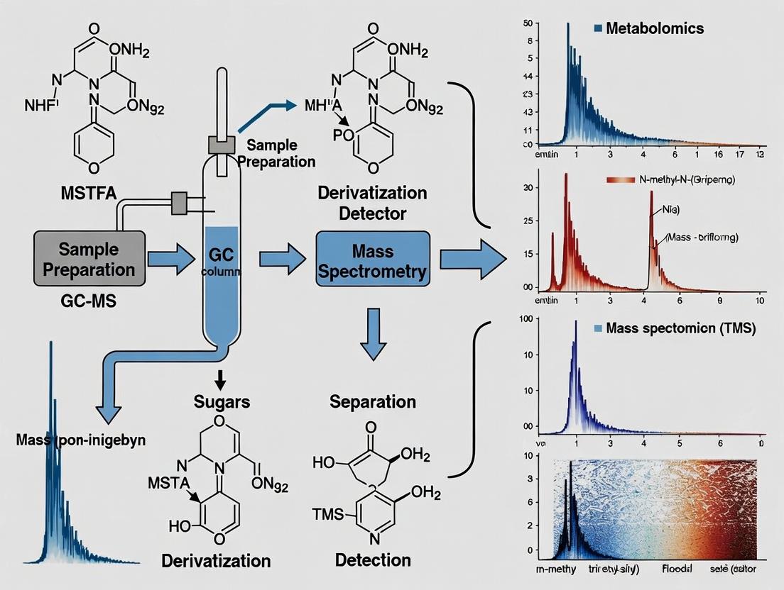
Experimental Protocol: Two-Step Derivatization for Polar Metabolomics
This detailed protocol is optimized for comprehensive polar metabolite coverage from a biological extract (e.g., from cell culture or plasma).
Protocol 1: Methoximation and Silylation
- Objective: To derivative carbonyl (aldehyde/ketone) and polar functional groups (e.g., -OH, -COOH, -NH₂) for GC-MS analysis.
- Materials: Dried metabolite extract, methoxyamine hydrochloride (MeOX) in pyridine, N-Methyl-N-(trimethylsilyl)trifluoroacetamide (MSTFA) with 1% TMCS.
- Procedure:
- Methoximation: Reconstitute the dried sample in 50 µL of 20 mg/mL MeOX in pyridine. Vortex thoroughly. Incubate at 30°C for 90 minutes with gentle shaking. This step converts reducing sugars and keto-acids into stable methoximes, preventing ring formation and yielding single chromatographic peaks.
- Silylation: Add 50 µL of MSTFA (+1% TMCS) to the reaction mixture. Vortex thoroughly. Incubate at 37°C for 30 minutes.
- Analysis: Centrifuge briefly and transfer the supernatant to a GC-MS vial. Analyze via GC-MS (typically using a 5% phenyl/95% dimethyl polysiloxane column, 30m x 0.25mm i.d., 0.25µm film thickness). Use a temperature ramp (e.g., 60°C to 325°C at 10°C/min).
- Notes: Pyridine must be anhydrous. Include process blanks and pooled quality control samples.
Protocol 2: Acid-Catalyzed Methylation (for Organic/Fatty Acids)
- Objective: Specifically derivative carboxylic acid groups to their methyl esters.
- Materials: Dried metabolite extract, 3N HCl in methanol, hexane.
- Procedure:
- Esterification: Add 100 µL of 3N HCl-methanol to the dried sample. Vortex thoroughly.
- Incubation: Heat at 80°C for 1 hour.
- Extraction: Cool to room temperature. Add 100 µL of hexane and 100 µL of water. Vortex vigorously for 30 seconds.
- Analysis: Centrifuge to separate phases. Collect the upper organic (hexane) layer containing the methyl esters for GC-MS analysis.
Visualization of Workflow and Impact
Title: Polar Metabolite GC-MS Problem & Derivatization Solution
Title: Two-Step Derivatization GC-MS Workflow
The Scientist's Toolkit: Key Research Reagent Solutions
Table 2: Essential Reagents for GC-MS Derivatization of Polar Metabolites
| Reagent / Material | Primary Function | Critical Consideration |
|---|---|---|
| Methoxyamine Hydrochloride (MeOX) | Converts carbonyl groups (aldehydes/ketones) to methoximes, preventing multiple anomeric forms for sugars. | Must be prepared in anhydrous pyridine; purity is critical for reproducible oximation. |
| N-Methyl-N-(trimethylsilyl)-trifluoroacetamide (MSTFA) | A powerful silyl donor; replaces active hydrogens in -OH, -COOH, -NH, -SH groups with trimethylsilyl (TMS) groups. | Often used with catalysts like TMCS (trimethylchlorosilane) to speed up reaction for sterically hindered groups. |
| Anhydrous Pyridine | Serves as the solvent for methoximation and silylation; basic and anhydrous to prevent reagent hydrolysis. | Must be sealed from moisture; under inert atmosphere for best results. |
| N,O-Bis(trimethylsilyl)trifluoroacetamide (BSTFA) | Alternative to MSTFA; another common silylation reagent. | Slightly different reactivity profile; choice may be metabolite-dependent. |
| Alkane Standard Mixture | A series of straight-chain hydrocarbons of known retention indices. | Used to calculate Retention Index (RI) for each metabolite, enabling library matching and compound identification. |
| Quality Control (QC) Pooled Sample | A pooled aliquot of all experimental samples. | Injected repeatedly throughout the run to monitor instrument performance and derivatization reproducibility. |
In gas chromatography-mass spectrometry (GC-MS) based metabolomics, the analysis of polar metabolites (e.g., organic acids, amino acids, sugars) presents a significant challenge. These compounds often exhibit low volatility, thermal instability, and poor chromatographic behavior, leading to adsorption, decomposition, and weak detectability. Derivatization, the chemical modification of analytes prior to analysis, is a cornerstone technique to overcome these limitations. This article, framed within a thesis on GC-MS with derivatization for polar metabolite analysis, details the core principles and provides application notes and protocols for the research community.
Principle 1: Enhancing Volatility
Polar functional groups (-OH, -COOH, -NH2) form strong intermolecular hydrogen bonds, resulting in high boiling points and low volatility. Derivatization replaces active hydrogens with non-polar, hydrophobic groups.
- Mechanism: Silylation (e.g., with MSTFA) replaces -H in -OH, -COOH, and -NH2 with a -Si(CH3)3 group. Alkylation (e.g., with chloroformates) replaces -H with an alkyl chain (e.g., -CH3, -C2H5).
- Outcome: The derivative has a higher vapor pressure, lower boiling point, and is efficiently transported in the GC carrier gas.
Principle 2: Improving Thermal Stability
Thermally labile metabolites (e.g., sugars, phosphorylated compounds) can degrade in the hot GC injector or column. Derivatization shields these vulnerable sites.
- Mechanism: The derivatizing group stabilizes the molecule against thermal decomposition by sativating reactive sites. For instance, silylation of the anomeric center of sugars prevents thermal degradation pathways.
- Outcome: Intact analyte molecules reach the detector, ensuring accurate quantification and identification.
Principle 3: Increasing Detectability and Selectivity
Derivatization enhances both the sensitivity and specificity of MS detection.
- Mechanism: It increases the molecular weight and can introduce fragments that yield characteristic, high-mass ions, moving signals away from chemical noise. It can also improve ionization efficiency in the MS source. Specific derivatives (e.g., pentafluorobenzyl) increase electron capture negative ionization (ECNI) response.
- Outcome: Lower limits of detection (LOD), improved signal-to-noise ratios, and the ability to perform trace analysis.
Table 1: Impact of Derivatization on Analytical Performance of Selected Metabolites
| Metabolite (Class) | Native Form Boiling Point (°C, est.) | Derivatized Form (Reagent) | Relative Peak Area Increase | LOD Improvement (Fold) | Key Reference Ion (m/z) |
|---|---|---|---|---|---|
| Lactic Acid (Acid) | ~122 (decomposes) | TMS ester (MSTFA) | 450x | 80x | 261 [M-15]⁺ |
| Glucose (Sugar) | Decomposes | Methoxime-TMS (MOX+MSTFA) | >1000x | 200x | 319 (base fragment) |
| Alanine (Amino Acid) | Sublimes/Decomp | tert-Butyldimethylsilyl (TBDMS) | 320x | 50x | 260 [M-57]⁺ |
| Succinic Acid (Diacid) | ~235 (decomposes) | Methyl ester (BF3/MeOH) | 200x | 40x | 115 (base fragment) |
Table 2: Comparison of Common Derivatization Reagents
| Reagent Class | Example Reagents | Target Groups | Key Advantages | Key Drawbacks |
|---|---|---|---|---|
| Silylation | MSTFA, BSTFA (+1% TMCS), TMSI | -OH, -COOH, -NH, -SH | Comprehensive, volatile derivatives, high yield. | Moisture-sensitive, derivatives can be hydrolytically unstable. |
| Alkylation | Chloroformates (ethyl, methyl), Diazomethane | -COOH, -OH | Fast reaction, can be performed in aqueous media (chloroformates). | Diazomethane is toxic/explosive. May not cover all polar groups. |
| Acylation | TFAA, PFPA, MBTFA | -NH2, -OH | Very stable derivatives, excellent for ECNI-MS. | Harsh conditions, may not be suitable for all metabolites. |
| Methoximation | Methoxyamine HCl | Aldehyde, Ketone (>C=O) | Protects sugars from ring tautomerization, creates two isomers per sugar for better separation. | Additional step required prior to silylation. |
Experimental Protocols
Protocol 1: Two-Step Methoximation and Silylation for Comprehensive Metabolite Profiling This protocol is standard for polar metabolite extraction from biological fluids (e.g., serum, urine).
I. Materials & Sample Preparation
- Lyophilized metabolite extract or 50-100 µL of biological sample.
- Internal Standard Solution: e.g., Succinic-d4 acid, Alanine-d4 (10 µg/mL in pyridine).
- Methoxyamine hydrochloride (MOX) solution: 20 mg/mL in anhydrous pyridine. Prepare fresh or store at -20°C under desiccant.
- Silylation Reagent: N-Methyl-N-(trimethylsilyl)trifluoroacetamide (MSTFA) with 1% trimethylchlorosilane (TMCS) as catalyst.
- Anhydrous pyridine.
- GC-MS vials and crimp caps with TFE septa.
II. Procedure
- Sample Preparation: Transfer dried extract to a clean 2 mL derivatization vial. Add 50 µL of internal standard solution and dry completely under a gentle stream of nitrogen or in a vacuum concentrator.
- Methoximation: Add 50 µL of MOX solution to the dried residue. Vortex vigorously for 30 seconds. Incubate at 37°C for 90 minutes with shaking (750 rpm).
- Silylation: After incubation, add 100 µL of MSTFA (+1% TMCS) to the reaction mixture. Vortex vigorously for 30 seconds. Incubate at 37°C for 30 minutes.
- Completion & Transfer: Let the vial cool to room temperature. Centrifuge briefly. Transfer 100-150 µL of the clear supernatant to a GC-MS vial for immediate analysis. Analyze within 24-48 hours for optimal results.
III. GC-MS Parameters (Example)
- GC: 30 m DB-5MS (or equivalent) capillary column (0.25 mm i.d., 0.25 µm film).
- Inlet: Split/splitless, 250°C, splitless mode (1 min).
- Carrier Gas: Helium, constant flow (1.0 mL/min).
- Oven Program: 60°C (hold 1 min), ramp 10°C/min to 325°C, hold 5 min.
- MS: Electron Impact (EI) at 70 eV. Source: 230°C. Quadrupole: 150°C. Solvent Delay: 5-6 min. Scan Range: m/z 50-600.
Visualizations
Title: Derivatization Principle Workflow for GC-MS
Title: Two-Step Derivatization Protocol Workflow
The Scientist's Toolkit: Essential Research Reagent Solutions
| Reagent/Material | Function & Rationale |
|---|---|
| N-Methyl-N-(trimethylsilyl)trifluoroacetamide (MSTFA) | The most common silyl donor for trimethylsilylation. Highly reactive, volatile byproducts. Often used with a catalyst (TMCS). |
| Methoxyamine Hydrochloride (MOX) | Converts carbonyls (aldehydes/ketones) to methoximes, preventing ring tautomerization in sugars and stabilizing α-keto acids. |
| tert-Butyldimethylchlorosilane (TBDMS) | Reagent for forming tert-butyldimethylsilyl derivatives. Produces more stable, higher molecular weight derivatives than TMS, often with characteristic fragmentation for amino acids and alcohols. |
| Anhydrous Pyridine | The solvent of choice for silylation. It acts as a base, scavenging the acid (HCl, TFA) produced during the reaction, driving the equilibrium to completion. Must be kept dry. |
| Deuterated Internal Standards (e.g., Succinic-d4 acid) | Added at the beginning of sample workup to correct for losses during derivatization, injection variability, and matrix effects in MS ionization. Essential for accurate quantification. |
| Chlorotrimethylsilane (TMCS) | Used as a catalyst (1%) in silylation reagents like MSTFA. Enhances the rate of derivatization, particularly for sterically hindered and tertiary functional groups. |
| Alkyl Chloroformates (e.g., Ethyl Chloroformate) | Enables fast, one-step esterification and acylation directly in aqueous samples (e.g., urine) for specific metabolite classes like organic acids, suitable for high-throughput analysis. |
This application note details the targeted analysis of key polar metabolite classes—organic acids, sugars, amino acids, and amines—using Gas Chromatography-Mass Spectrometry (GC-MS) with derivatization. Within the broader thesis of metabolomics research, this protocol is critical for converting non-volatile, thermally labile polar metabolites into volatile, stable derivatives suitable for GC-MS analysis, enabling comprehensive profiling in biomedical and pharmaceutical research.
Key Polar Metabolite Classes and Quantitative Ranges
The following table summarizes typical concentration ranges for key polar metabolite classes in human biological samples, as established in current literature.
Table 1: Representative Concentration Ranges of Key Polar Metabolite Classes in Human Plasma/Serum
| Metabolite Class | Example Metabolites | Typical Concentration Range | Key Biological Role |
|---|---|---|---|
| Organic Acids | Lactate, Succinate, Citrate | 50–5000 µM | Energy metabolism (TCA cycle), gut microbiota co-metabolism |
| Sugars & Sugar Phosphates | Glucose, Fructose-6-phosphate | 3–6 mM (Glucose), 10–100 µM (Phosphates) | Central energy currency, glycolysis, pentose phosphate pathway |
| Amino Acids | Glutamine, Alanine, Leucine | 50–1000 µM | Protein synthesis, nitrogen transport, neurotransmitters |
| Amines & Polyamines | Choline, Spermine, Spermidine | 1–50 µM | Cell proliferation, membrane integrity, methylation |
Detailed Experimental Protocol: GC-MS with Derivatization for Polar Metabolites
Materials and Reagents
- Sample: 50-100 µL of plasma, serum, or tissue homogenate supernatant.
- Internal Standard Solution: 10 µM d27-Myristic acid in pyridine (for quantification).
- Methoxyamination Reagent: 20 mg/mL Methoxyamine hydrochloride in anhydrous pyridine.
- Silylation Reagent: N-Methyl-N-(trimethylsilyl)trifluoroacetamide (MSTFA) with 1% trimethylchlorosilane (TMCS).
- Solvents: HPLC-grade methanol, chloroform, water.
- Equipment: GC-MS system with a 30m DB-5MS or equivalent column, chemical ionization capability, micro-centrifuge, speed vacuum concentrator, thermomixer.
Protocol Steps
1. Sample Extraction (Modified Bligh & Dyer): a. Add 200 µL of ice-cold methanol:chloroform (2:1 v/v) to 50 µL of sample in a 1.5 mL microcentrifuge tube. b. Spike with 10 µL of internal standard solution. c. Vortex vigorously for 10 minutes at 4°C. d. Centrifuge at 14,000 x g for 15 minutes at 4°C. e. Transfer 150 µL of the supernatant (polar phase) to a fresh glass derivatization vial. f. Dry completely using a speed vacuum concentrator (approx. 2 hours).
2. Methoxyamination: a. Add 50 µL of methoxyamination reagent to the dried extract. b. Cap tightly and vortex. c. Incubate at 30°C for 90 minutes in a thermomixer with agitation (750 rpm). This step protects carbonyl groups (in sugars, keto-acids) by converting them to methoximes.
3. Silylation: a. Add 80 µL of MSTFA (+1% TMCS) to the reaction vial. b. Vortex thoroughly. c. Incubate at 37°C for 30 minutes. This step replaces active hydrogens (from -OH, -COOH, -NH groups) with trimethylsilyl (TMS) groups, conferring volatility.
4. GC-MS Analysis: a. Inject 1 µL of the derivatized sample in split or splitless mode (as required by concentration). b. GC Parameters: Inlet temperature: 250°C; Helium flow: 1.0 mL/min; Oven program: 60°C hold for 1 min, ramp at 10°C/min to 325°C, hold for 10 min. c. MS Parameters: Transfer line: 280°C; Ion source: 230°C; Electron impact ionization: 70 eV; Scan range: 50-600 m/z. d. Use alkane series (C8-C30) for retention index calibration.
5. Data Processing: a. Use deconvolution software (e.g., AMDIS, ChromaTOF) to identify peaks by matching retention indices and mass spectra to reference libraries (e.g., NIST, FiehnLib, GMD). b. Quantify relative to the internal standard peak area.
Visualizing the Derivatization Workflow
GC-MS Derivatization Workflow for Polar Metabolites
Interconnecting Metabolic Pathways of Target Classes
Core Metabolic Pathway Interconnections
The Scientist's Toolkit: Essential Research Reagents
Table 2: Key Reagents for GC-MS Polar Metabolomics
| Reagent/Solution | Function in Protocol | Critical Note |
|---|---|---|
| Methoxyamine hydrochloride | Methoxyamination reagent. Converts aldehydes and ketones (in sugars, keto-acids) to stable methoxime derivatives, preventing ring formation and enabling single-peak detection. | Must be prepared fresh in anhydrous pyridine and protected from moisture. |
| MSTFA with 1% TMCS | Silylation reagent. Replaces active hydrogens in -OH, -COOH, -SH, -NH groups with trimethylsilyl (TMS) groups, increasing volatility and thermal stability. | TMCS acts as a catalyst. Reagent is moisture-sensitive; use anhydrous conditions. |
| Deuterated Internal Standards (e.g., d27-Myristic acid) | Added at extraction start. Corrects for variability in derivatization efficiency, sample loss, and instrument sensitivity drift. | Choose standards not endogenous to the sample. Use across all samples for robust quantification. |
| Alkane Series Standard Mix (C8-C30) | Injected separately. Used to calculate retention indices (RI) for each metabolite, enabling library matching independent of minor retention time shifts. | Essential for confident metabolite identification using RI-based libraries. |
| Anhydrous Pyridine | Solvent for methoxyamination. Provides an alkaline, anhydrous medium crucial for the reaction. Also helps solubilize sample residues. | Extreme hygroscopicity requires strict anhydrous handling and storage. |
Historical Context and Evolution of Derivatization Reagents in Metabolomics
Within the broader thesis of employing GC-MS with derivatization for comprehensive polar metabolite analysis in metabolomics research, understanding the historical trajectory of derivatization reagents is paramount. This evolution is driven by the core challenge in GC-MS: the analysis of highly polar, non-volatile, and thermally labile metabolites inherent to biological systems. Derivatization chemically modifies these analytes to enhance volatility, thermal stability, and chromatographic behavior. This application note details this historical context, provides contemporary protocols, and visualizes the key workflows.
Historical Progression and Quantitative Performance Data
The development of derivatization reagents has progressed through distinct generations, each improving upon the limitations of the last. The quantitative performance of common reagent classes is summarized below.
Table 1: Historical Evolution and Key Characteristics of Derivatization Reagent Classes
| Era/Generation | Primary Reagent Classes | Key Target Functional Groups | Major Advantages | Inherent Limitations |
|---|---|---|---|---|
| First (1960s-70s) | Silylation (e.g., TMS, MSTFA) | -OH, -COOH, -NH, -SH | Broad applicability, high volatility derivatives. | Moisture sensitivity, formation of multiple derivatives, instability. |
| Second (1980s-90s) | Acylation (e.g., Acetylation), Alkylation (e.g., Methylation) | -OH, -NH, -COOH | Improved stability for some analytes, selective reactions. | Less broad than silylation, harsher conditions for some. |
| Third (2000s-Present) | Chloroformates (e.g., ECF, MCF), Specialty Silyl (e.g., TBDMS) | -OH, -COOH, -NH (CFs); -OH, -COOH (TBDMS) | Aqueous/rapid reaction (CFs), Enhanced stability (TBDMS), chiral separation capabilities. | CFs: Not for all acids; TBDMS: Larger derivative mass. |
Table 2: Performance Comparison of Modern Derivatization Methods in Metabolomics
| Method | Derivatization Time | Typical Derivatives per Metabolite | MS Fragmentation | Stability of Derivatives | Compatibility with Automation |
|---|---|---|---|---|---|
| Methoxyamination + MSTFA | 60-90 min + 30 min | Often multiple (syn/anti oximes, TMS) | Abundant TMS-related ions | Moderate (hygroscopic) | Medium (requires drying steps) |
| Ethyl Chloroformate (ECF) | < 5 min | Single, well-defined | Distinct ethyl esters/amines | High | High (can be in aqueous medium) |
| MSTFA + 1% TMCS | 30-60 min | Often multiple | Abundant TMS-related ions | Low-Moderate | Medium |
Detailed Experimental Protocols
Protocol 1: Standard Two-Step Methoxyamination and Silylation for Polar Metabolomics
This is the classical and most widely used protocol for comprehensive polar metabolite profiling.
I. Materials & Reagent Solutions: The Scientist's Toolkit
- Methoxyamine hydrochloride: (20 mg/mL in pyridine). Converts carbonyls (aldehydes/ketones) to methoximes, reducing tautomerization and forming two derivatives (syn/anti).
- N-Methyl-N-(trimethylsilyl)trifluoroacetamide (MSTFA): Primary silyl donor. Replaces active hydrogens in -OH, -COOH, -NH, -SH groups with a trimethylsilyl (TMS) group.
- Retention Index Markers (e.g., n-Alkanes): A series of straight-chain hydrocarbons (C8-C30) analyzed separately to calculate retention indices for universal metabolite identification.
- Anhydrous Pyridine: Dry reaction solvent. Critical to prevent hydrolysis of silylation reagents.
- Internal Standards Mix: Deuterated or 13C-labeled analogs of key metabolites (e.g., d27-myristic acid, 13C5-proline) added at extraction to correct for technical variability.
II. Procedure:
- Sample Preparation: Lyophilize 50-100 µL of extracted polar metabolite fraction in a glass GC-MS vial.
- Methoxyamination: Add 50 µL of methoxyamine solution (20 mg/mL in pyridine) to the dried residue. Vortex vigorously. Incubate at 30°C for 90 minutes with shaking.
- Silylation: Add 100 µL of MSTFA (with or without 1% TMCS as a catalyst) to the reaction mixture. Vortex vigorously.
- Derivative Formation: Incubate at 37°C for 30 minutes.
- GC-MS Analysis: Cool the vial to room temperature, centrifuge briefly, and transfer an appropriate volume (e.g., 1 µL) for split/splitless injection. Use a temperature-programmed run (e.g., 60°C to 330°C) on a non-polar (5% phenyl) column.
Protocol 2: Rapid Aqueous-Phase Derivatization with Ethyl Chloroformate (ECF)
This protocol is suited for high-throughput analysis of amino and organic acids directly from aqueous extracts.
I. Materials & Reagent Solutions:
- Ethyl Chloroformate (ECF): Derivatizing agent for amines and carboxyls in aqueous medium.
- Pyridine: Catalyst for the reaction.
- Ethanol: Reacts with ECF in situ to form ethoxycarbonyl intermediates.
- Sodium Hydroxide (1M): Adjusts pH to optimize derivatization yield.
II. Procedure:
- Aqueous Sample: Place 200 µL of filtered biological extract (e.g., urine, cell culture media) in a glass vial.
- pH Adjustment: Add 100 µL of NaOH (1M) to adjust the pH to ~9-10.
- Derivatization: Sequentially add 200 µL of ethanol and 50 µL of pyridine. Finally, add 40 µL of ethyl chloroformate in two aliquots of 20 µL, with vigorous vortexing for 30 seconds after each addition.
- Extraction: Add 400 µL of chloroform to extract the derivatives. Vortex for 10 seconds.
- Phase Separation: Centrifuge at 3000 rpm for 5 minutes. The lower organic phase contains the target ethyl ester/ethoxycarbonyl derivatives.
- GC-MS Analysis: Recover the lower organic layer and inject 1-2 µL into the GC-MS.
Visualization of Workflows and Logical Relationships
Derivatization Strategy Selection for GC-MS Metabolomics
Classical Two-Step Derivatization Protocol Workflow
From Sample to Spectrum: A Step-by-Step GC-MS Derivatization Protocol for Metabolomics
Within a thesis investigating GC-MS with derivatization for polar metabolite analysis in metabolomics, the workflow from sample preparation to data acquisition is foundational. This protocol details the critical steps required to ensure reproducible, high-quality data for subsequent multivariate statistical analysis in biomarker discovery, toxicology, and drug development research.
Critical Workflow Protocol
Sample Quenching and Metabolite Extraction
Objective: To instantly halt metabolic activity and efficiently extract a broad spectrum of polar intracellular metabolites. Detailed Protocol:
- Cell Quenching: For adherent cells, rapidly aspirate culture medium and add pre-chilled (-20°C) methanol (40% v/v in water) directly to the culture plate. Place the plate on a cold metal block (-20°C) for 5 minutes.
- Metabolite Extraction: Scrape cells in the quenching solution and transfer the suspension to a pre-cooled microcentrifuge tube.
- Add chilled chloroform (to a final ratio of 1:3:1 water:methanol:chloroform). Vortex vigorously for 1 minute.
- Centrifuge at 14,000 x g for 15 minutes at 4°C to induce phase separation.
- Carefully transfer the upper aqueous phase (containing polar metabolites) to a new vial. The lower organic phase can be retained for lipid analysis.
- Dry the aqueous extract using a vacuum concentrator (e.g., SpeedVac) at room temperature. Store dried extracts at -80°C until derivatization.
Chemical Derivatization for GC-MS
Objective: To increase the volatility and thermal stability of polar metabolites (e.g., sugars, organic acids, amino acids). Detailed Protocol (Two-Step Methoximation and Silylation):
- Methoximation: Redissolve the dried extract in 50 µL of methoxyamine hydrochloride (20 mg/mL in pyridine). Vortex thoroughly.
- Incubate at 30°C for 90 minutes with shaking (750 rpm) to protect carbonyl groups by forming methoximes.
- Silylation: Add 100 µL of N-Methyl-N-(trimethylsilyl)trifluoroacetamide (MSTFA) with 1% trimethylchlorosilane (TMCS) as a catalyst.
- Incubate at 37°C for 30 minutes with shaking (750 rpm). This step replaces active hydrogens with trimethylsilyl (TMS) groups.
- Transfer the derivatized solution to a GC-MS vial with insert. Analyze within 24-48 hours.
GC-MS Data Acquisition Parameters
Objective: To achieve chromatographic separation and mass spectrometric detection of derivatized metabolites. Detailed Protocol:
- GC System: Use a fused-silica capillary column (e.g., DB-5MS, 30m x 0.25mm i.d., 0.25µm film thickness).
- Injection: 1 µL in split or splitless mode (split ratio 10:1 for high-concentration samples). Injector temperature: 250°C.
- Oven Program: Initial 60°C for 1 min; ramp at 10°C/min to 325°C; hold for 10 min. Total run time: 37.5 min.
- Carrier Gas: Helium, constant flow at 1.0 mL/min.
- MS Detector: Electron Impact (EI) ionization at 70 eV. Ion source temperature: 230°C. Quadrupole temperature: 150°C.
- Acquisition Mode: Full scan from m/z 50 to 600 at a scan rate of 5-10 scans/second after a 5-6 minute solvent delay.
Data Presentation: Key Quantitative Parameters
Table 1: Critical GC-MS Method Parameters for Polar Metabolite Analysis
| Parameter | Specification | Function/Rationale |
|---|---|---|
| Extraction Solvent Ratio | H₂O:MeOH:CHCl₃ (1:3:1) | Efficient protein precipitation & recovery of polar metabolites. |
| Derivatization Reagents | Methoxyamine HCl, MSTFA+1%TMCS | Methoximation of carbonyls; Silylation of -OH, -COOH, -NH groups. |
| Derivatization Time | 90 min (Step 1), 30 min (Step 2) | Ensures complete reaction; minimizes degradation. |
| GC Column | DB-5MS (30m, 0.25mm, 0.25µm) | Standard mid-polarity stationary phase for metabolite separation. |
| Oven Temp. Ramp | 10°C/min from 60°C to 325°C | Balances resolution of early/late eluters with total run time. |
| MS Scan Range | m/z 50 - 600 | Captures molecular ions & fragment ions of TMS-derivatized metabolites. |
Workflow and Relationship Diagrams
GC-MS Metabolomics Workflow
Two-Step Derivatization Chemistry
The Scientist's Toolkit
Table 2: Essential Research Reagent Solutions for GC-MS Metabolomics
| Item | Function in Workflow |
|---|---|
| Pre-chilled Methanol (-20°C) | Rapid metabolic quenching to "freeze" the metabolic state instantly. |
| Chloroform (HPLC grade) | Component of the biphasic extraction system; separates polar metabolites from lipids. |
| Methoxyamine Hydrochloride (in Pyridine) | First derivatization reagent; forms methoximes to stabilize α-keto acids and sugars. |
| MSTFA with 1% TMCS | Second derivatization reagent; replaces active hydrogens with trimethylsilyl (TMS) groups. |
| Alkane Standard Mix (C8-C30) | Used to calculate Retention Index (RI) for metabolite identification, aligning runs. |
| DB-5MS GC Capillary Column | Standard mid-polarity column providing optimal separation for diverse metabolite classes. |
| NIST/ Fiehn Metabolomics Library | Reference spectral library for identifying metabolites based on mass spectrum and RI. |
Within the context of a broader thesis on GC-MS with derivatization for polar metabolite analysis in metabolomics research, the selection of derivatization reagents is a critical determinant of data quality, coverage, and reproducibility. This note provides a detailed comparison of silylation and oximation reagents central to this workflow.
Silylation Reagents: MSTFA vs. BSTFA
Silylation replaces active hydrogens (e.g., in -OH, -COOH, -NH) with an alkylsilyl group, increasing volatility and thermal stability.
Mechanism
Both N-Methyl-N-(trimethylsilyl)trifluoroacetamide (MSTFA) and N,O-Bis(trimethylsilyl)trifluoroacetamide (BSTFA) react via a nucleophilic substitution mechanism. The trifluoroacetamide leaving group is expelled, forming the trimethylsilyl (TMS) derivative.
Quantitative Comparison: MSTFA vs. BSTFA
| Property/Characteristic | MSTFA | BSTFA |
|---|---|---|
| Molecular Formula | C₆H₁₂F₃NOSi₂ | C₈H₁₈F₃NOSi₂ |
| Active Silyl Groups | 1 | 2 |
| Reaction Speed | Generally faster | Slightly slower |
| Byproduct | N-Methyltrifluoroacetamide | N-Trimethylsilyltrifluoroacetamide |
| Baseline Chromatography | Can produce a lower baseline; byproduct elutes early. | May cause a higher baseline or ghost peaks; byproduct elutes later. |
| Steric Hindrance | Lower, may be better for hindered sites. | Higher due to two TMS groups on nitrogen. |
| Cost | Typically lower | Typically higher |
| Common Additives | Often used with TMCS (catalyst) & solvent (e.g., pyridine). | Often used with TMCS (1% common). |
| Stability of Derivatives | High | High, but may be slightly more susceptible to hydrolysis. |
Key Insight: MSTFA is often preferred in metabolomics for its cleaner chromatographic baseline and faster reaction kinetics, though BSTFA can be equally effective for many applications, especially with catalysis.
Protocol: Standard Silylation Derivatization for Polar Metabolites
- Sample Preparation: Dry 50-100 µL of polar metabolite extract (e.g., from plasma, urine, cell culture) in a GC-MS vial under a gentle stream of nitrogen or in a vacuum concentrator.
- Oximation (First Step): Reconstitute the dry residue in 50 µL of methoxyamine hydrochloride (MOX) solution (20 mg/mL in pyridine). Vortex thoroughly.
- Incubation: Incubate for 90 minutes at 37°C with shaking.
- Silylation: Add 100 µL of either MSTFA or BSTFA (frequently with 1% TMCS as catalyst). Vortex thoroughly.
- Incubation: Incubate for 30-60 minutes at 37°C with shaking.
- GC-MS Analysis: Transfer an aliquot (e.g., 1 µL) to the GC injector. Use a temperature-programmed run on a non-polar or mid-polar column (e.g., DB-5MS, 30m x 0.25mm).
Oximation Reagents: MOX vs. Alternatives
Oximation converts carbonyl groups (aldehydes, ketones) into oximes, preventing cyclization of sugars and resolving keto-enol tautomerism, which can lead to multiple chromatographic peaks for a single metabolite.
Mechanism
Nucleophilic attack of the carbonyl carbon by the amine nitrogen of the alkoxyamine, followed by proton transfer and elimination of water, forming an oxime (E and Z isomers possible).
Quantitative Comparison: Oximation Reagents
| Reagent (Abbrev.) | Full Name | Typical Solvent | Reaction Time/Temp | Key Pro | Key Con |
|---|---|---|---|---|---|
| MOX | Methoxyamine Hydrochloride | Pyridine | 90 min, 37°C | Standard, robust, well-characterized. | Pyridine is toxic/hazardous. Forms two isomers (E/Z). |
| Ethoxime (ETOX) | O-Ethylhydroxylamine Hydrochloride | Pyridine | 90 min, 37°C | Derivatives elute later than MOX, aiding separation from co-eluters. | Larger derivative, longer GC times. Higher boiling point. |
| PFBOA | Pentafluorobenzylhydroxylamine Hydrochloride | Pyridine/Water | Varies | Enables highly sensitive detection by ECD or NCI-MS. | Specialized, not for general metabolomics. More complex cleanup. |
| DMEOX | Dimethoxydimethylhydroxylamine | - | Varies | Forms single isomer derivative. | Less commonly used, less established. |
Key Insight: MOX in pyridine remains the gold standard for general metabolomics due to its efficiency and predictability. ETOX is a valuable alternative for resolving specific peak co-elutions.
Protocol: Comparative Oximation for Sugar Analysis
- Standard Preparation: Prepare separate standard solutions of glucose, fructose, and ribose.
- Aliquoting: Aliquot equal amounts (e.g., 50 µg) into four separate GC vials and dry.
- Derivatization:
- Vial 1 (MOX): Add 50 µL MOX (in pyridine). Incubate 90 min @ 37°C. Then add MSTFA/TMCS.
- Vial 2 (ETOX): Add 50 µL ETOX (in pyridine). Incubate 90 min @ 37°C. Then add MSTFA/TMCS.
- Vial 3 (Control): Add 50 µL pyridine only. Incubate 90 min @ 37°C. Then add MSTFA/TMCS.
- Vial 4 (No Ox): Directly add MSTFA/TMCS in pyridine.
- Silylation: Add 100 µL of MSTFA with 1% TMCS to all vials. Incubate 30 min @ 37°C.
- Analysis: Run all samples on GC-MS with identical parameters. Compare chromatograms for peak multiplicity (tautomers), peak shape, and retention time shifts.
The Scientist's Toolkit: Essential Reagents for GC-MS Metabolomics Derivatization
| Item | Function in Workflow |
|---|---|
| MSTFA (with 1% TMCS) | Primary silylation reagent; caps polar functional groups. TMCS acts as a catalyst for sterically hindered groups. |
| Methoxyamine HCl (MOX) | Primary oximation reagent; stabilizes carbonyl groups (sugars, keto acids) pre-silylation. |
| Anhydrous Pyridine | Common solvent for derivatization; maintains moisture-free conditions and acts as an acid scavenger. |
| Alkane Retention Index Mix | A homologous series of alkanes (e.g., C8-C40) run to calculate retention indices for metabolite identification. |
| N-Methyl-N-trimethylsilyltrifluoroacetamide | An alternative silylation reagent, useful for specific applications like amino acid analysis. |
| Quartz Wool | For liner packing in the GC inlet, improves vaporization and traps non-volatile residues. |
| Deactivated Inlet Liners | Glass liners for the GC inlet; deactivation prevents adsorption and degradation of polar derivatives. |
| C18 & SPE Cartridges | For solid-phase extraction (SPE) sample cleanup prior to derivatization to remove salts and lipids. |
Visualizations
GC-MS Two-Step Derivatization Workflow
Derivatization Reagent Selection Logic
Application Notes
Within metabolomics research utilizing GC-MS, the analysis of polar metabolites (e.g., organic acids, amino acids, sugars) presents a significant challenge due to their low volatility, thermal instability, and high polarity, which lead to poor chromatographic performance and adsorption. Derivatization is a critical sample preparation step that masks polar functional groups, enhancing volatility, thermal stability, and detectability. This document, framed within a thesis on GC-MS-based metabolomics, details optimized derivatization protocols, focusing on the critical variables of time, temperature, and solvent. The primary derivatization agents discussed are N-Methyl-N-(trimethylsilyl)trifluoroacetamide (MSTFA) and N,O-Bis(trimethylsilyl)trifluoroacetamide (BSTFA), often with 1% trimethylchlorosilane (TMCS) as a catalyst.
Key Experimental Protocols
Protocol 1: Standard Methoxyamination and Silylation for Polar Metabolites
Objective: To derivative a broad range of polar metabolites (e.g., sugars, organic acids, amino acids) for GC-MS analysis. Materials: Dry pyridine, methoxyamine hydrochloride (MeOX) in pyridine (20 mg/mL), MSTFA (or BSTFA) with 1% TMCS, standard mixture of target metabolites, GC vials with inserts. Procedure:
- Sample Preparation: Dry the extracted metabolite sample completely using a vacuum concentrator. Ensure no residual water is present.
- Methoxyamination:
- Reconstitute the dried sample in 50 µL of MeOX solution (20 mg/mL in pyridine).
- Vortex vigorously for 10-30 seconds.
- Incubate at a defined temperature and time (see Table 1).
- This step converts carbonyl groups (aldehydes, ketones) into methoximes, preventing ring formation in sugars and producing fewer derivatives per molecule.
- Silylation:
- Add 50 µL of MSTFA (with 1% TMCS) to the reaction mixture.
- Vortex vigorously for 10-30 seconds.
- Incubate at a defined temperature and time (see Table 1). This step replaces active hydrogens in -OH, -COOH, -SH, -NH groups with trimethylsilyl (TMS) groups.
- Completion: After incubation, centrifuge briefly and transfer the supernatant to a GC vial for analysis.
Protocol 2: Rapid Silylation for Organic Acids
Objective: A faster, targeted derivatization for organic acid analysis. Materials: BSTFA with 1% TMCS, acetonitrile (dry), standard organic acids. Procedure:
- Dry the sample and reconstitute in 50 µL of dry acetonitrile.
- Add 50 µL of BSTFA (+1% TMCS).
- Vortex and incubate at 70°C for 20 minutes.
- Analyze directly by GC-MS.
Table 1: Optimization Matrix for Methoxyamination-Silylation Derivatization
| Parameter | Level 1 | Level 2 | Level 3 | Recommended Optimum for Broad Profiling | Impact on Results |
|---|---|---|---|---|---|
| Methoxyamination Time | 1.5 hours | 16 hours (overnight) | 24 hours | 16 hours (overnight) | Shorter times (<4h) lead to incomplete oximation of some ketones. Overnight ensures completeness for diverse classes. |
| Methoxyamination Temperature | 30°C | 37°C | 50°C | 37°C | Higher temps (50°C) can degrade sensitive metabolites. 37°C provides a good balance of speed and stability. |
| Silylation Time | 30 minutes | 1 hour | 2 hours | 1 hour | Most silylation reactions complete within 1 hour. Prolonged times offer minimal gain for most metabolites. |
| Silylation Temperature | 40°C | 60°C | 80°C | 60°C | Higher temps speed kinetics but risk by-product formation and degradation. 60°C is standard. |
| Primary Solvent | Pyridine | N-Methyl-N-(trimethylsilyl)trifluoroacetamide | - | Pyridine | Pyridine acts as both solvent and catalyst (basic). MSTFA as solvent leads to very rapid, sometimes less controlled, reactions. |
Table 2: Comparative Performance of Common Silylation Reagents
| Reagent (with 1% TMCS) | Reaction Speed | Suitability for Steric Hindrance | Tendency for By-products (e.g., degradation) | Best For |
|---|---|---|---|---|
| MSTFA | Very Fast | Moderate | Moderate | High-throughput screening, general profiling where speed is prioritized. |
| BSTFA | Fast | Good | Low | General profiling of organic acids, amino acids, sugars (standard choice). |
| TMSI (N-Trimethylsilylimidazole) | Slow | Excellent | Very Low | Derivatizing highly sterically hindered groups (e.g., tertiary alcohols). |
Diagrams
Title: Derivatization Workflow for GC-MS Metabolomics
Title: Parameter Optimization Goals & Outcomes
The Scientist's Toolkit
Table 3: Essential Research Reagent Solutions for GC-MS Derivatization
| Item | Function & Rationale |
|---|---|
| Methoxyamine Hydrochloride (MeOX) | Converts carbonyl groups (aldehydes/ketones) to methoximes. Prevents multiple isomer formation (e.g., with sugars) and reduces the number of derivatives per molecule, simplifying chromatograms. |
| Pyridine (Anhydrous) | The standard solvent for methoxyamination and silylation. Its basicity catalyzes the reaction. Must be kept dry to prevent hydrolysis of silylation reagents. |
| MSTFA (N-Methyl-N-(trimethylsilyl)trifluoroacetamide) | A powerful silyl donor. Also acts as its own solvent. Very fast reaction but can be aggressive. Often used with TMCS catalyst. |
| BSTFA (N,O-Bis(trimethylsilyl)trifluoroacetamide) with 1% TMCS | The most common silylation reagent. TMCS (Trimethylchlorosilane) acts as a catalyst, enhancing derivatization of hindered functional groups like amines and amides. |
| N-Methylimidazole (NMIM) | Used as a catalyst in specific silylation protocols, particularly for difficult-to-derivatize compounds like phosphate-containing metabolites (e.g., sugar phosphates). |
| Alkane Standard Mixture (e.g., C8-C40) | Added to derivatized samples before GC-MS run. Provides retention index markers for highly accurate metabolite identification against RI libraries. |
| Quality Control (QC) Reference Mixture | A standardized blend of metabolites covering key chemical classes. Injected regularly throughout the analytical batch to monitor derivatization efficiency and instrument performance. |
This document serves as a focused application note within a broader thesis investigating Gas Chromatography-Mass Spectrometry (GC-MS) with derivatization for polar metabolite analysis in metabolomics. The inherent volatility and thermal stability limitations of polar metabolites (e.g., organic acids, sugars, amino acids) in biological samples necessitate chemical derivatization prior to GC-MS analysis. This process enhances volatility, improves chromatographic separation, and increases detection sensitivity, making it indispensable for the following spotlight applications.
Derivatization in Biomarker Discovery for Oncology
Application Note: Derivatization-GC-MS is pivotal in the untargeted metabolomic screening of biofluids (serum, urine) to identify metabolic signatures indicative of oncological states. By converting polar metabolites into stable, volatile derivatives, researchers can profile hundreds of compounds to discover diagnostic, prognostic, or predictive biomarkers.
Key Protocol: Methoximation and Silylation for Serum Metabolomics
- Sample Preparation: Deproteinize 50 µL of human serum by adding 150 µL of ice-cold methanol. Vortex for 30 seconds and incubate at -20°C for 1 hour. Centrifuge at 14,000 x g for 15 minutes at 4°C.
- Methoximation: Transfer 100 µL of the supernatant to a GC vial. Add 20 µL of methoxyamine hydrochloride in pyridine (20 mg/mL). Vortex and incubate at 30°C for 90 minutes with shaking (750 rpm). This step protects keto groups and reduces the formation of multiple silylation products.
- Silylation: Add 80 µL of N-Methyl-N-(trimethylsilyl)trifluoroacetamide (MSTFA) with 1% trimethylchlorosilane (TMCS) as a catalyst. Vortex and incubate at 37°C for 30 minutes.
- Analysis: Inject 1 µL of the derivatized sample in splitless mode onto a GC-MS system equipped with a 30m DB-5MS column. Use a temperature gradient from 60°C to 325°C.
Quantitative Data from Recent Studies: Table 1: Example Biomarker Panels Discovered via Derivatization-GC-MS in Oncology
| Cancer Type | Biofluid | Key Derivatized Metabolite Biomarkers (Trend vs. Control) | Potential Clinical Utility | Reference Year |
|---|---|---|---|---|
| Ovarian Cancer | Serum | Lactate ↑, Succinate ↑, 2-Hydroxybutyrate ↑ | Early-stage detection | 2023 |
| Colorectal Cancer | Serum/Urine | Glycine ↓, Threonine ↓, Palmitic Acid ↑ | Distinguishing adenoma from carcinoma | 2024 |
| Pancreatic Ductal Adenocarcinoma | Plasma | Branched-Chain Amino Acids (Leu, Ile, Val) ↓, Kynurenine ↑ | Prognostic survival indicator | 2023 |
Diagram 1: Untargeted biomarker discovery workflow using GC-MS derivatization.
The Scientist's Toolkit: Research Reagent Solutions for Biomarker Discovery
| Reagent/Material | Function in Protocol |
|---|---|
| Methoxyamine Hydrochloride | Methoximation reagent; converts carbonyls to methoximes, preventing enolization and simplifying chromatograms. |
| MSTFA with 1% TMCS | Trimethylsilyl (TMS) donor; replaces active hydrogens in -OH, -COOH, -NH, -SH groups with TMS groups, conferring volatility. |
| Pyridine (anhydrous) | Solvent for methoximation; acts as a catalyst and scavenges HCl produced during reaction. |
| DB-5MS GC Column | (5%-Phenyl)-methylpolysiloxane column; standard for separating a wide range of derivatized metabolites. |
| Retention Index Mix (Alkanes) | Series of n-alkanes analyzed under same conditions; used to calculate retention indices for metabolite identification. |
Derivatization in Drug Metabolism Studies
Application Note: Derivatization is crucial for profiling phase I and II drug metabolites, which are often highly polar. It enables the detection and structural characterization of metabolites that are otherwise invisible to GC-MS, supporting pharmacokinetic (PK) and absorption, distribution, metabolism, and excretion (ADME) studies.
Key Protocol: Derivatization of Oxidative and Conjugated Metabolites from Microsomal Incubations
- Incubation: Incubate drug candidate (10 µM) with human liver microsomes (0.5 mg/mL) and NADPH regenerating system in phosphate buffer (pH 7.4) at 37°C for 45 min. Terminate with 2 volumes of ice-cold acetonitrile.
- Extraction: Centrifuge at 14,000 x g for 10 min. Dry the supernatant under a gentle nitrogen stream at 40°C.
- Derivatization for Acidic Metabolites: Reconstitute the dry residue in 50 µL of N,O-Bis(trimethylsilyl)trifluoroacetamide (BSTFA) with 1% TMCS and 50 µL of pyridine. Heat at 70°C for 40 minutes. This targets carboxylic acid and phenolic hydroxyl groups common in oxidative metabolites and glucuronide conjugates after hydrolysis.
- Alternative Acylation for Amino Metabolites: For primary/secondary amine metabolites, reconstitute in 50 µL of acetic anhydride and 50 µL of pyridine. Heat at 60°C for 30 minutes. (Note: Sample may require clean-up post-derivatization).
- Analysis: Inject onto GC-MS/MS for selective metabolite detection and identification.
Quantitative Data: Table 2: Impact of Derivatization on Detectability of Model Drug Metabolites
| Drug (Parent) | Key Metabolite (Polar) | LOD without Derivatization (ng/mL) | LOD with Silylation (ng/mL) | Improvement Factor |
|---|---|---|---|---|
| Ibuprofen | 2-Hydroxyibuprofen (COOH, OH) | Not Detectable by GC | 5.2 | >100x |
| Acetaminophen | Acetaminophen Glucuronide (hydrolyzed) | Not Detectable by GC | 8.7 | >100x |
| Diazepam | Temazepam (OH) | 50 | 1.5 | ~33x |
Diagram 2: Drug metabolism and derivatization pathway for GC-MS analysis.
Derivatization in Metabolic Pathway Analysis (Biomedical Research)
Application Note: By enabling comprehensive profiling, derivatization-GC-MS facilitates metabolic flux analysis and pathway mapping. This is essential for understanding disease mechanisms, such as dysregulation in central carbon metabolism (glycolysis, TCA cycle) in neurodegenerative or metabolic diseases.
Key Protocol: Tracing [U-¹³C]-Glucose Flux in Cell Culture
- Tracing Experiment: Grow adherent cells (e.g., HeLa) to 70% confluency. Replace medium with one containing 10 mM [U-¹³C]-Glucose. Incubate for a defined period (e.g., 2, 4, 8 hrs).
- Quenching & Extraction: Rapidly aspirate medium and quench metabolism with liquid N₂. Extract intracellular metabolites with 1 mL -20°C 80:20 methanol:water. Scrape cells, vortex, and centrifuge. Dry supernatant.
- Derivatization: Derivatize using the two-step methoximation/silylation protocol described in Section 1.
- GC-MS Analysis & Flux Calculation: Use GC-MS with electron impact ionization. Monitor mass isotopomer distributions (MIDs) of TCA cycle intermediates (e.g., citrate, succinate, malate) based on their characteristic fragment ions. Calculate ¹³C enrichment using specialized software (e.g., Metran).
The Scientist's Toolkit: Essential Materials for Metabolic Flux
| Reagent/Material | Function in Protocol |
|---|---|
| [U-¹³C]-Glucose | Isotopically labeled tracer; allows tracking of carbon atom flow through interconnected metabolic pathways. |
| 80:20 Methanol:Water (-20°C) | Quenching and extraction solvent; rapidly inactivates enzymes while efficiently extracting polar metabolites. |
| Retention Index Libraries | Databases of TMS-derivatized metabolites with associated retention indices and mass spectra (e.g., NIST, FiehnLib). |
| Mass Isotopomer Analysis Software | Tools for deconvoluting complex ¹³C labeling patterns from GC-MS data to calculate metabolic flux. |
Diagram 3: Key central carbon metabolism nodes analyzed via ¹³C tracing.
Solving Common Pitfalls: Troubleshooting and Optimizing Your GC-MS Derivatization Workflow
Within the framework of a thesis on GC-MS with derivatization for polar metabolite analysis, robust identification and prevention of analytical artifacts are paramount for data integrity. This document details protocols for managing three common artifacts: siloxane peaks, incomplete derivatization reactions, and degradation products.
1. Siloxane Peaks: Identification and Mitigation Siloxanes originate from column bleed or septum degradation and can be misidentified as biological metabolites.
- Identification: Characteristic clusters of ions: m/z 73, 147, 207, 221, 281, 295, 355, and 429 ([M-15]⁺ for cyclic siloxanes).
- Prevention Protocol: Regular GC-MS system maintenance.
- Septum Replacement: Replace the GC inlet septum every 100-150 injections or as per manufacturer guidelines.
- Column Conditioning: Before connecting to the MS, condition a new column by heating at 10°C/min to the upper temperature limit, holding for 1-2 hours.
- Gold Seal Deactivation: Install a pre-column guard (deactivated, 1-5 m) or use a gold seal ferrule to minimize active sites.
- Blank Runs: Perform regular solvent blank runs to monitor background levels.
Table 1: Characteristic Ions for Common Siloxane Artifacts
| Siloxane Compound | Base Peak (m/z) | Key Qualifier Ions (m/z) | Typical Elution Window (min on 30m column) |
|---|---|---|---|
| Hexamethylcyclotrisiloxane (D3) | 207 | 73, 133 | ~8-12 |
| Octamethylcyclotetrasiloxane (D4) | 281 | 73, 147, 207, 355 | ~12-16 |
| Decamethylcyclopentasiloxane (D5) | 355 | 73, 147, 267, 429 | ~16-22 |
2. Incomplete Derivatization Reactions Incomplete reaction with silylation agents (e.g., MSTFA, BSTFA) leads to multiple peaks for a single analyte, reducing sensitivity and complicating identification.
- Protocol: Optimization of Silylation Reaction
- Sample Drying: Ensure complete dryness of the metabolite extract using a vacuum concentrator (no aqueous residue).
- Reagent Selection: Prepare a derivatization reagent mix. Standard Mix: 20 µL Methoxyamine hydrochloride in pyridine (15 mg/mL), 60°C for 90 min, followed by 80 µL MSTFA + 1% TMCS, 60°C for 60 min.
- Catalyst Use: Include 1% chlorotrimethylsilane (TMCS) as a catalyst to promote silylation of hindered functional groups.
- Time/Temperature Calibration: For challenging matrices, test reaction times (30-120 min) at 40-70°C.
- Post-Reaction Quenching: Add 100 µL of alkane retention index standard mixture (e.g., C8-C30 in hexane) post-derivatization to stabilize the reaction mixture before GC-MS injection.
Table 2: Indicators of Incomplete Derivatization
| Analyte State | Functional Group Reacted | Typical Observation (vs. Complete Derivatization) |
|---|---|---|
| Under-silylated | -OH, -COOH | Additional earlier-eluting peak(s); reduced TMS peak area. |
| Unreacted | -NH₂ | Broad, tailing peak; may not be detected. |
| Degraded Reagent | N/A | High background of artifact peaks (e.g., from reagent hydrolysis). |
3. Degradation Products Thermal degradation in the GC inlet or on-column degradation produce artifacts not present in the original sample.
- Protocol: Minimizing Thermal Degradation
- Inlet Liner Maintenance: Use a deactivated, single-taper inlet liner. Replace or clean (via sonication in solvent) every 100-200 injections.
- Inlet Temperature Optimization: Set the lowest possible inlet temperature that ensures complete vaporization. Start at 250°C for silylated metabolites; do not exceed 280°C.
- Pulsed Splitless Injection: Use pulsed splitless mode to rapidly transfer the sample to the column (e.g., 25 psi for 1 min, then constant flow).
- Column Health: Regularly trim the column inlet (0.5-1 m) and monitor peak tailing for active polar compounds.
Experimental Workflow for Artifact Monitoring
Title: Artifact Monitoring Workflow for GC-MS Metabolomics
The Scientist's Toolkit: Key Research Reagent Solutions
| Item/Chemical | Function in Context of Derivatization & Artifact Prevention |
|---|---|
| N-Methyl-N-(trimethylsilyl)trifluoroacetamide (MSTFA) | Primary silylation donor. Derivatives -OH, -COOH, -NH, -SH groups. Quality is critical; store under inert gas. |
| Methoxyamine Hydrochloride | Protects carbonyl groups (aldehydes, ketones) by forming methoximes, preventing multiple tautomers and degradation. |
| Chlorotrimethylsilane (TMCS) | Catalyst added to MSTFA (typically 1%). Enhances silylation of sterically hindered groups like in tertiary alcohols. |
| Pyridine (Anhydrous) | Solvent for methoximation. Must be anhydrous and high-purity to prevent reagent hydrolysis and side reactions. |
| Retention Index Mix (Alkanes) | A homologous series of n-alkanes (e.g., C8-C30). Added post-derivatization for retention time alignment and system monitoring. |
| Deactivated Inlet Liners | Single-taper, wool-packed liners designed for splitless injection. Minimize sample contact with active surfaces. |
| Gold-Coated Ferrules | Provide an inert seal between column and inlet/transfer line, reducing catalytic degradation points. |
Within the framework of metabolomics research utilizing GC-MS, the analysis of polar metabolites is critically dependent on effective chemical derivatization. Silylation, the most prevalent derivatization technique, replaces active hydrogens (e.g., from -OH, -COOH, -NH) with alkylsilyl groups, thereby reducing polarity, increasing volatility, and enhancing thermal stability for GC separation and MS detection. The success of this reaction is singularly contingent on the exclusion of moisture. Water hydrolyzes both the silylating reagents and the formed derivatives, leading to incomplete derivatization, poor reproducibility, ghost peaks, and column degradation.
Impact of Moisture on Derivatization Yield: Quantitative Data
Table 1: Effect of Residual Water on Silylation Yield of Key Metabolites (Model Study)
| Metabolite Class | Example Compound | Derivatization Yield (Anhydrous Conditions) | Derivatization Yield (0.1% v/v Water Added) | Primary Observation |
|---|---|---|---|---|
| Sugar | D-Glucose | 98 ± 2% | 45 ± 15% | Multiple incomplete TMS products, increased peak tailing. |
| Organic Acid | Citric Acid | 99 ± 1% | 30 ± 10% | Decreased main peak area, appearance of degradation peaks. |
| Amino Acid | L-Alanine | 97 ± 2% | 60 ± 12% | Co-elution of underivatized and partially derivatized species. |
| Fatty Acid | Palmitic Acid | 99 ± 1% | 85 ± 5% | Moderate yield reduction; less sensitive than polyfunctional compounds. |
Table 2: Common Silylation Reagents and Their Sensitivity to Hydrolysis
| Reagent (Abbr.) | Active Group | Typical Use | Hydrolysis Sensitivity | Key By-product (from H₂O) |
|---|---|---|---|---|
| N,O-Bis(trimethylsilyl)trifluoroacetamide (BSTFA) | TMS | General purpose, sugars, acids | High | Hexamethyldisiloxane (HMDSO) |
| N-Methyl-N-(trimethylsilyl)trifluoroacetamide (MSTFA) | TMS | Alkaloids, sugars | Very High | HMDSO |
| N-tert-Butyldimethylsilyl-N-methyltrifluoroacetamide (MTBSTFA) | TBDMS | Steroids, robust derivatives | Moderate | tert-Butyldimethylsilanol |
Protocol: Rigorous Anhydrous Derivatization for Polar Metabolite Profiling
Materials & Workflow for Moisture-Controlled Silylation
Diagram Title: Moisture-Controlled Silylation Workflow
Detailed Protocol:
- Sample Preparation & Drying: After metabolite extraction, completely dry the sample using a centrifugal vacuum concentrator (lyophilizer). For solid samples, use a desiccator with P₂O₅ or activated molecular sieves (3Å) for >24 hrs.
- Reagent & Solvent Drying: Use anhydrous pyridine (over molecular sieves) as the solvent. Upon opening a new vial of silylation reagent (e.g., BSTFA), aliquot it into small, sealed vials under a dry nitrogen atmosphere to limit exposure.
- Derivatization Reaction:
- Transfer the dried sample to a 2 mL glass vial with a PTFE-lined septum cap.
- In a glove box or under a steady stream of dry nitrogen, add 50 µL of anhydrous pyridine.
- Immediately add 50 µL of BSTFA (or MSTFA). If using reagents like MSTFA, consider adding 1% TMCS (trimethylchlorosilane) as a catalyst.
- Cap the vial tightly, vortex for 30 seconds.
- Heat at 70°C for 60 minutes in a dry heating block.
- Post-Reaction Handling: Allow the vial to cool to room temperature before opening. Analyze by GC-MS immediately (within 4-6 hours) for best results. Do not store derivatized samples for extended periods.
The Scientist's Toolkit: Essential Reagents for Anhydrous Silylation
Table 3: Key Research Reagent Solutions
| Item | Function & Importance |
|---|---|
| Silylation Reagents (BSTFA, MSTFA) | Donor of trimethylsilyl (TMS) group. Must be stored sealed, under inert gas, with molecular sieves. |
| Anhydrous Pyridine | Dry reaction solvent and base catalyst. Essential for neutralizing acids produced during reaction. |
| Trimethylchlorosilane (TMCS) | Acid catalyst added to silylation reagents (1-10%) to promote reaction with stubborn functional groups (e.g., amines). |
| Molecular Sieves (3Å or 4Å) | Pore size optimized for water adsorption. Added to solvent bottles to maintain anhydrous conditions. |
| PTFE-Sealed Reaction Vials | Prevents atmospheric moisture ingress and solvent/reagent loss during heating. |
| Centrifugal Vacuum Concentrator | Provides complete removal of aqueous solvents from samples prior to derivatization. |
Moisture Invasion Pathways and Countermeasures
Diagram Title: Moisture Pathways in Silylation & Prevention
1. Introduction and Thesis Context Within the broader thesis on Gas Chromatography-Mass Spectrometry (GC-MS) with derivatization for polar metabolite analysis in metabolomics research, sample preparation is the critical determinant of data quality. The accurate profiling of polar metabolites—including sugars, organic acids, amino acids, and nucleotides—is confounded by the diverse and complex compositions of biological matrices. Each matrix presents unique challenges in protein removal, metabolite extraction, and compatibility with subsequent derivatization steps (typically methoximation and silylation). These optimization strategies are designed to maximize metabolite coverage, reproducibility, and quantitative accuracy for robust biological interpretation in drug development and disease research.
2. Key Challenges by Matrix Type The interference potential and optimal handling strategies vary significantly across sample types.
Table 1: Matrix-Specific Challenges and Optimization Goals
| Matrix | Primary Challenges | Key Optimization Goals |
|---|---|---|
| Serum/Plasma | High protein content, high salt, lipidemia. Metabolite instability. | Efficient deproteinization, inhibition of enzymatic activity, removal of phospholipids. |
| Tissue | Cellular heterogeneity, metabolite compartmentalization, need for homogenization. | Rapid quenching of metabolism, effective homogenization, complete cell lysis. |
| Cell Culture Extracts | Low metabolite levels, culture media interference, adhesion vs. suspension. | Rapid metabolism quenching, efficient metabolite extraction from cells, removal of media components. |
3. Detailed Application Notes and Protocols
Protocol 3.1: Serum and Plasma Preparation for Polar Metabolomics Objective: To obtain a protein-free polar metabolite extract suitable for GC-MS derivatization. Materials: Ice-cold methanol, ice-cold acetonitrile, internal standard solution (e.g., deuterated succinic acid, 13C6-sorbitol in water), centrifuge, speed vacuum concentrator. Procedure:
- Thaw samples slowly on ice.
- Aliquot 50 µL of serum/plasma into a pre-chilled microcentrifuge tube.
- Add 10 µL of internal standard mix.
- For protein precipitation, add 200 µL of ice-cold methanol (-20°C) and vortex vigorously for 30 seconds.
- Add 200 µL of ice-cold acetonitrile, vortex again.
- Incubate at -20°C for 1 hour.
- Centrifuge at 14,000 x g for 15 minutes at 4°C.
- Transfer 400 µL of the supernatant to a fresh tube.
- Dry completely under a gentle stream of nitrogen or in a speed vacuum concentrator (without heat).
- Store dried extract at -80°C until derivatization. Optimization Note: The methanol:acetonitrile combination (1:1, v/v) provides superior protein precipitation and broader metabolite recovery compared to methanol alone.
Protocol 3.2: Tissue Homogenization and Metabolite Extraction Objective: To rapidly quench metabolism and extract polar metabolites from solid tissues. Materials: Liquid N2, pre-cooled mortar and pestle or bead mill homogenizer, extraction solvent (3:3:2 v/v/v acetonitrile:methanol:water with 0.1% formic acid), internal standard solution. Procedure:
- Snap-freeze tissue immediately after collection in liquid N2. Store at -80°C.
- Weigh 20-50 mg of frozen tissue and grind to a fine powder under liquid N2 using a mortar and pestle.
- Transfer the powder to a tube containing 1 mL of ice-cold extraction solvent (-40°C) and 10 µL of internal standard.
- Homogenize further using a bead mill homogenizer (3 cycles of 45 sec at 4°C).
- Sonicate the homogenate in an ice bath for 5 minutes.
- Incubate at -20°C for 1 hour.
- Centrifuge at 14,000 x g for 15 minutes at 4°C.
- Collect the supernatant. A second re-extraction of the pellet can be performed for high lipid content tissues.
- Combine supernatants and dry under vacuum. Optimization Note: The acidic, aqueous/organic solvent system quenches enzyme activity and extracts a wide polarity range of metabolites.
Protocol 3.3: Quenching and Extraction for Adherent Cell Cultures Objective: To separate intracellular metabolites from culture media effectively. Materials: Saline quenching solution (0.9% ammonium bicarbonate in 0.9% w/v NaCl, -20°C), extraction solvent (40:40:20 v/v/v methanol:acetonitrile:water), cell scraper. Procedure:
- Rapidly aspirate culture media.
- Immediately wash cells twice with 2 mL of ice-cold saline quenching solution.
- Add 1 mL of ice-cold extraction solvent (-40°C) directly to the plate.
- Scrape cells quickly and transfer the suspension to a microcentrifuge tube.
- Add internal standards.
- Vortex for 1 minute, then sonicate in ice bath for 5 minutes.
- Incubate at -20°C for 1 hour.
- Centrifuge at 14,000 x g for 15 minutes at 4°C.
- Transfer supernatant to a new tube and dry. Optimization Note: The cold ammonium bicarbonate saline wash rapidly inhibits metabolism and helps remove residual media salts without causing cell lysis prior to extraction.
4. The Scientist's Toolkit: Essential Research Reagent Solutions
Table 2: Key Reagents and Materials for GC-MS Metabolite Extraction
| Item | Function | Key Consideration |
|---|---|---|
| Ice-cold Methanol/Acetonitrile | Protein precipitation, metabolite extraction, enzyme denaturation. | HPLC/MS grade, stored at -20°C. Low water content is critical. |
| Deuterated/C13-labeled Internal Standards | Monitoring extraction efficiency, normalization, semi-quantification. | Should be added at the very first step of extraction. Cover multiple chemical classes. |
| Methoxyamine Hydrochloride | First step of derivatization; protects carbonyl groups by forming methoximes. | Must be freshly prepared in pyridine (e.g., 20 mg/mL) to avoid hydrolysis. |
| N-Methyl-N-(trimethylsilyl)trifluoroacetamide (MSTFA) | Silylation agent; replaces active hydrogens with TMS groups, making metabolites volatile. | Must be anhydrous. 1% TMCS as catalyst is often added. |
| Retention Index Markers (e.g., n-Alkanes) | Calibrates retention time to a standardized index for metabolite identification. | Added to the sample or run in a separate injection under identical conditions. |
| Derivatization-grade Pyridine | Solvent for methoximation reaction. | Must be anhydrous and stored under inert gas to prevent water absorption. |
5. Workflow and Pathway Visualizations
Title: Overall GC-MS Metabolomics Sample Preparation Workflow
Title: Matrix-Specific Challenges Link to Optimized Strategies
Title: Two-Step Derivatization for Polar Metabolites
Within the broader thesis of employing GC-MS with chemical derivatization for polar metabolite analysis in metabolomics, achieving optimal data quality is paramount. Derivatization, while reducing polarity and enhancing thermal stability, introduces new chemical species whose analysis demands precise instrument tuning and method parameter optimization. This application note details protocols and tuning strategies specifically designed to maximize the sensitivity, resolution, and reproducibility of derivatized metabolite profiling, directly impacting biomarker discovery and drug development research.
Optimized Instrument Tuning for Derivatized Analytes
Standard autotuning for general compounds often uses perfluorotributylamine (PFTBA). For derivatized metabolites (e.g., TMS, methoxime, or alkylated forms), a modified tuning target is recommended to better represent their typical mass fragments and abundance.
Protocol 1: Enhanced Tuning and Calibration for Metabolite Analysis
- Preparation: Ensure the GC-MS system (e.g., Agilent 7890B/5977B, Thermo Scientific ISQ 7000) has undergone standard maintenance. Have PFTBA and a derivatized tuning standard (e.g., a mix of TMS-derivatized fatty acids, sugars, and amino acids) ready.
- Initial Autotune: Perform the manufacturer's standard autotune using PFTBA. Record baseline metrics.
- Derivatized Standard Tune Evaluation: Introduce the derivatized tuning standard via the GC inlet (e.g., 1 µL splitless injection). Acquire data in scan mode (m/z 50-600).
- Parameter Adjustment: Manually adjust tuning parameters, focusing on:
- Ion Source Temperature: Increase to 250-300°C to minimize derivatization reagent condensation.
- Electron Energy: Maintain at 70 eV for consistency, but verify spectral library matching.
- Lens Voltages: Fine-tune to maximize the response for key lower-mass fragments common to derivatized compounds (e.g., m/z 73, 147, 117 for TMS).
- Tune Validation: The tuned method should produce symmetric peak shapes for early eluting, volatile derivatives (e.g., pyruvate, lactate) and maintain sensitivity for higher molecular weight species.
Table 1: Comparison of Standard vs. Optimized Tuning Parameters for TMS-Derivatized Metabolites
| Tuning Parameter | Standard PFTBA Autotune | Optimized for Derivatized Metabolites | Rationale |
|---|---|---|---|
| Ion Source Temp. | 230°C | 280°C | Prevents accumulation of non-volatile silylation by-products, reduces source contamination. |
| Emission Current | 34.6 µA | 35-40 µA | Enhances ionization efficiency for a broad range of metabolite classes. |
| Detector Gain | Manufacturer Default | 1.5-2x Default | Increases sensitivity for low-abundance metabolites. |
| Target m/z for Focus | 69, 219, 502 (PFTBA) | 73, 147, 221+ | Aligns mass calibration with characteristic fragments of TMS derivatives. |
Critical GC Method Parameters for Resolution
Chromatographic resolution is critical to separate complex mixtures of derivatized isomers and homologous compounds.
Protocol 2: Gradient Optimization for Complex Derivative Separation
- Column Selection: Use a low-bleed, high-resolution column (e.g., Agilent DB-35ms or equivalent, 30m x 0.25mm x 0.25µm).
- Initial Oven Program: Start at 60°C (hold 1 min), ramp at 10°C/min to 325°C (hold 5 min). Inject a complex derivatized sample (e.g., human serum extract).
- Identify Critical Pair: Review the Total Ion Chromatogram (TIC) and Extracted Ion Chromatograms (EICs). Identify a pair of closely eluting, co-isolated peaks (e.g., isomers like α-ketoglutarate and glutarate derivatives).
- Gradient Refinement: Insert a slower ramp rate (e.g., 3-5°C/min) around the retention time of the critical pair. Re-run the sample.
- Carrier Gas & Flow Optimization: For modern GC-MS, use Helium or Hydrogen as carrier gas. Optimize flow for the specific column (e.g., 1.0 mL/min constant flow for He, or 1.5 mL/min for H2). Use the instrument's method translation software to maintain optimal linear velocity.
Table 2: Impact of Key GC Parameters on Derivative Resolution & Sensitivity
| Parameter | Typical Setting | Optimized Setting | Effect on Derivatized Metabolites |
|---|---|---|---|
| Inlet Liner | Standard 4mm single taper | High-Gooseneck Splitless w/Wool | Improves vaporization, reduces discrimination for high-boiling-point derivatives. |
| Injection Temp. | 250°C | 270-280°C | Ensures complete vaporization of silylated compounds; monitor for thermal degradation. |
| Splitless Time | 0.75-1.0 min | 1.0-1.5 min | Enhances sensitivity for polar, early-eluting derivatives (e.g., glycolysis intermediates). |
| Oven Ramp Rate | Constant 10°C/min | Multi-ramp (e.g., 10°C/min to 170°, then 3°C/min to 240°) | Dramatically improves resolution of critical isomer pairs (sugars, organic acids). |
| Transfer Line Temp. | 280°C | 300°C | Prevents condensation of less volatile derivatives before MS detection. |
MS Data Acquisition Parameters for Sensitivity
Optimal MS settings balance scanning range, speed, and dwell time for targeted and untargeted analyses.
Protocol 3: SIM/Scan Method Development for Targeted Quantification
- Define Analyte List: Create a list of 20-30 key derivatized metabolites of interest (e.g., TCA cycle intermediates, amino acids).
- Identify Characteristic Ions: From library spectra, select 2-3 primary quantifier and qualifier ions per metabolite. Prioritize high-mass, unique ions where possible (e.g., M-15 for TMS).
- Group by RT: Organize metabolites into time-segmented SIM groups. Allow 10-15 scans per peak; typical dwell time 20-100 ms per ion.
- Combine with Scan: For untargeted discovery, use a simultaneous Scan/SIM method. Use Scan mode (e.g., m/z 50-600) for full spectral library matching and SIM for sensitive quantification of low-abundance targets.
Visualization of Workflow & Parameter Relationships
Title: GC-MS Optimization Workflow for Derivatized Metabolites.
Title: Key Parameters Driving Data Quality Goals.
The Scientist's Toolkit: Essential Research Reagents & Materials
| Item | Function & Rationale |
|---|---|
| N-Methyl-N-(trimethylsilyl)trifluoroacetamide (MSTFA) | Preferred silylation reagent; highly reactive, volatile by-products, suitable for GC-MS. |
| Methoxyamine Hydrochloride (in Pyridine) | First step oximation; converts carbonyls (aldehydes, ketones) to methoximes, preventing tautomerism and reducing peak duplication. |
| Retention Index Marker Mix (Alkanes, e.g., C10-C40) | Injected alongside samples to calculate retention indices for robust peak identification across runs. |
| Derivatized Metabolite Tuning Standard | Custom mix of derivatized metabolites (e.g., TMS-amino acids, organic acids) for instrument tuning validation. |
| High-Purity, Low-Bleed GC-MS Column | (e.g., 35% phenyl / 65% dimethyl polysiloxane). Provides optimal balance for polar derivative separation with minimal column bleed. |
| Deactivated High-Gooseneck Splitless Liner w/Wool | Wool promotes homogeneous vaporization and traps non-volatile residues, protecting the column. |
| Quadrupole Tuning Standard (PFTBA) | Essential for baseline instrument performance verification and mass axis calibration. |
Benchmarking Performance: Validating GC-MS Derivatization and Comparing it to LC-MS for Polar Metabolomics
1.0 Introduction & Thesis Context Within a thesis investigating GC-MS with derivatization for polar metabolite analysis in metabolomics, robust method validation is foundational. Derivatization, while essential for volatility and detectability of polar metabolites, introduces complexity that must be accounted for in validation. This protocol details the establishment of critical validation metrics—reproducibility, linearity, limits of detection/quantification (LOD/LOQ), and recovery—specifically tailored for derivatized assays, ensuring data integrity for downstream biomarker discovery and pathway analysis in drug development research.
2.0 Validation Metrics: Protocols & Data Presentation
2.1 Reproducibility (Precision)
- Protocol: Intra- and Inter-Day Repeatability
- Prepare a QC sample from a pooled biological matrix at low, mid, and high concentrations (n=6 per level).
- Subject all aliquots to the full derivatization workflow (e.g., methoxyamination followed by silylation).
- For intra-day precision, analyze all 6 replicates sequentially in one batch.
- For inter-day precision, analyze 2 replicates per level over three consecutive days.
- Calculate the relative standard deviation (%RSD) of the peak area (or height) for each target analyte.
- Data Presentation:
Table 1: Precision Data for Key Polar Metabolites (e.g., Alanine, Succinate, Glucose) after Derivatization.
Analyte Spiked Conc. (µM) Intra-Day %RSD (n=6) Inter-Day %RSD (n=2x3) Acceptance Criterion Alanine 10.0 4.2 7.1 ≤15% Succinate 5.0 3.8 6.5 ≤15% Glucose 25.0 5.5 8.9 ≤15%
2.2 Linearity & Calibration Model
- Protocol: Calibration Curve Preparation
- Prepare a series of calibration standards in a surrogate matrix (e.g., phosphate buffer or analyte-free serum) across the expected physiological range (e.g., 1-500 µM).
- Spike with stable isotope-labeled internal standards (IS) for each analyte class prior to derivatization to correct for process variability.
- Subject all calibration levels to the identical derivatization protocol.
- Analyze by GC-MS in randomized order.
- Plot the peak area ratio (analyte/IS) versus nominal concentration. Evaluate using weighted (1/x or 1/x²) least-squares regression.
- Data Presentation:
Table 2: Linearity Parameters for a Representative Calibration Curve.
Analyte Linear Range (µM) Calibration Equation R² Weighting Lactate 2.0 - 200 y = 0.045x + 0.002 0.9987 1/x Citrate 1.0 - 100 y = 0.102x - 0.005 0.9992 1/x²
2.3 Limits of Detection (LOD) and Quantification (LOQ)
- Protocol: Signal-to-Noise and Low-Level Spiking
- LOD: Analyze progressively lower concentration standards. The LOD is the concentration yielding a signal-to-noise (S/N) ratio of ≥3:1.
- LOQ: The lowest standard on the calibration curve that can be quantified with an accuracy of 80-120% and a precision (%RSD) of ≤20%, with an S/N ≥10:1.
- Confirm LOQ by analyzing 6 replicates at the estimated level through the full method.
- Data Presentation:
Table 3: LOD and LOQ for Derivatized Polar Metabolites.
Analyte LOD (µM) [S/N≥3] LOQ (µM) [S/N≥10, %RSD≤20] LOQ Verified Accuracy (%) Pyruvate 0.5 1.0 94.3 Fumarate 0.2 0.5 88.7
2.4 Recovery (Process Efficiency)
- Protocol: Pre- vs. Post-Derivatization Spiking
- Prepare three sets of samples (n=5) at low and high concentrations.
- Set A (Pre-spike): Add analyte and IS to matrix before derivatization.
- Set B (Post-spike): Add analyte to a derivatized blank matrix extract after the reaction (to account for ionization effects).
- Set C (Neat Solution): Spike analyte into derivatization reagents without matrix.
- Calculate Recovery (%) = (Peak Area Ratio of Set A / Peak Area Ratio of Set B) x 100.
- Calculate Process Efficiency (%) = (Peak Area Ratio of Set A / Peak Area Ratio of Set C) x 100 (accounts for both derivatization yield and matrix effects).
- Data Presentation:
Table 4: Recovery and Process Efficiency for Derivatization.
Analyte Conc. (µM) Recovery (%) Process Efficiency (%) Glutamine 15.0 95.2 78.4 Malate 8.0 102.5 81.6
3.0 Visual Workflows
Title: Validation Workflow for Derivatized GC-MS Assays
Title: Recovery Assessment Experimental Design
4.0 The Scientist's Toolkit: Key Research Reagent Solutions
Table 5: Essential Materials for Derivatization Method Validation.
| Reagent/Material | Function in Validation | Example Product/Chemical |
|---|---|---|
| Silylation Reagent | Imparts volatility; primary derivatizing agent for -OH, -COOH, -NH₂ groups. | N-Methyl-N-(trimethylsilyl)trifluoroacetamide (MSTFA) |
| Methoxyamination Reagent | Stabilizes carbonyls (aldehydes/ketones) in sugars and α-ketoacids prior to silylation. | Methoxyamine hydrochloride in pyridine |
| Stable Isotope-Labeled Internal Standards (IS) | Critical for correcting losses during derivatization and matrix effects; enables accurate quantification. | ¹³C₃-Lactate, D₄-Succinate, ¹³C₆-Glucose |
| Derivatization-Grade Solvents | Anhydrous, high-purity solvents prevent side reactions and ensure derivatization efficiency. | Anhydrous Pyridine, N,N-Dimethylformamide (DMF) |
| Retention Index Markers | Allows for reproducible metabolite identification across runs and instruments. | n-Alkane series (C8-C40) |
| Quality Control (QC) Reference Material | Pooled sample used to monitor system stability and reproducibility across validation batches. | NIST SRM 1950 (Metabolites in Human Plasma) or in-house pooled matrix |
Application Notes
This application note details the deployment of Gas Chromatography-Mass Spectrometry (GC-MS) in metabolomics for the analysis of polar metabolites, leveraging its core strengths of robustness, high chromatographic resolution, and comprehensive spectral libraries. The context is the targeted and untargeted profiling of polar metabolites, which are central to understanding cellular biochemistry in disease and drug development.
Robustness and Reproducibility in High-Throughput Studies
GC-MS systems demonstrate exceptional long-term instrumental stability, a critical factor for large-scale metabolomics cohorts. A recent inter-laboratory study (2023) assessed the reproducibility of a derivatized metabolite panel across five independent sites using identical GC-MS platforms and standard operating procedures (SOPs).
Table 1: Inter-Laboratory Reproducibility Data for Key Polar Metabolites (n=50 replicates per site)
| Metabolite | Average RSD (Retention Time) % | Average RSD (Peak Area) % | Mean Consensus Library Match Factor (0-1000) |
|---|---|---|---|
| Alanine | 0.08 | 4.2 | 892 |
| Glucose | 0.12 | 5.8 | 875 |
| Citrate | 0.05 | 3.9 | 935 |
| Succinate | 0.07 | 4.5 | 910 |
| Glutamate | 0.06 | 4.1 | 901 |
RSD: Relative Standard Deviation. Data adapted from Metabolomics Society QA/QC initiative reports.
High Resolution for Complex Mixtures
Modern GC columns (e.g., 30m mid-polarity columns) provide superior separation power, critical for resolving isomers and metabolites in complex biological extracts. The use of comprehensive two-dimensional GC (GCxGC-MS) further enhances this, but standard 1D GC-MS remains the workhorse.
Table 2: Comparative Resolution of Critical Isomer Pairs (R Value)
| Isomer Pair | Standard Non-Polar Column (R) | Mid-Polarity Column (R) | Resolved? (R>1.5) |
|---|---|---|---|
| Isoleucine / Leucine | 0.8 | 1.9 | Yes |
| α-Ketoglutarate / Pyruvate* | 1.1 | 2.3 | Yes |
| Glucose / Mannose (MOX deriv.) | 0.9 | 1.6 | Yes |
| Average Peak Capacity | ~400 | ~500 |
Derivatized as methoxime/tert-butyldimethylsilyl (MOX/TBDMS) derivatives. R = Resolution factor.
Leveraging Rich Spectral Libraries
The availability of extensive, curated electron ionization (EI) spectral libraries allows for confident metabolite identification. The NIST, Fiehn, and Golm libraries contain spectra for thousands of derivatized metabolites.
Table 3: Spectral Library Match Confidence Metrics
| Library Match Factor Range | Probability of Correct Identification | Recommended Action |
|---|---|---|
| >900 | Very High | Confident ID |
| 800-900 | High | Probable ID, check RT |
| 700-800 | Moderate | Require standard for verification |
| <700 | Low | Tentative annotation |
Protocol 1: Standard Operating Procedure for Polar Metabolite Extraction and Derivatization for GC-MS
Objective: To reproducibly extract and derivatize polar metabolites from cultured mammalian cells for GC-MS analysis.
Materials:
- Methanol (HPLC grade): Extraction solvent.
- Chloroform: For phase separation in biphasic extraction (optional).
- Water (Mass spec grade): For reconstitution.
- Methoxylamine hydrochloride (20 mg/mL in pyridine): Protects carbonyl groups (oximation).
- N-Methyl-N-(tert-butyldimethylsilyl)trifluoroacetamide (MTBSTFA) with 1% tert-butyldimethylchlorosilane (tBDMCS): Silylation reagent for -OH, -COOH, -NH groups.
- Pyridine (anhydrous): Reaction solvent for derivatization.
- Internal Standards: Ribitol (for polar phase), deuterated alkanes (for RI calibration).
Procedure:
- Cell Quenching & Extraction:
- Aspirate culture medium and rapidly quench cells with 1 mL of cold (-20°C) 80% methanol.
- Scrape cells, transfer suspension to a microcentrifuge tube.
- Sonicate on ice for 5 minutes.
- Centrifuge at 14,000 x g for 15 minutes at 4°C.
- Transfer supernatant (polar phase) to a fresh glass vial. Evaporate to complete dryness under a gentle stream of nitrogen or in a vacuum concentrator.
Methoximation:
- Add 50 µL of methoxylamine hydrochloride solution to the dried extract.
- Vortex vigorously for 30 seconds.
- Incubate at 37°C for 90 minutes with shaking.
Silylation:
- Add 80 µL of MTBSTFA (+1% tBDMCS) to the reaction vial.
- Vortex vigorously for 30 seconds.
- Incubate at 37°C for 60 minutes with shaking.
Sample Transfer:
- Centrifuge briefly to collect contents.
- Transfer the derivatized sample to a GC-MS vial with a low-volume insert.
- Analyze within 24-48 hours for optimal results.
GC-MS Parameters (Agilent 7890B/5977B Example):
- Column: DB-35MS or equivalent (30m x 0.25mm ID, 0.25µm film).
- Inlet: 250°C, splitless mode (1µL injection).
- Carrier Gas: Helium, constant flow 1.2 mL/min.
- Oven Program: 60°C (hold 1 min), ramp at 10°C/min to 325°C, hold 5 min.
- MS Source: 230°C, Quad: 150°C.
- Acquisition: Electron Impact (EI) at 70eV, scan range m/z 50-600.
- Tuning: Perfluorotributylamine (PFTBA) used for daily calibration.
Protocol 2: Protocol for Deconvolution and Library Matching Using AMDIS
Objective: To deconvolute complex chromatograms and identify metabolites using reference spectral libraries.
Procedure:
- Data Import: Convert raw data files to NetCDF format if necessary. Open in AMDIS.
- Analysis Settings:
- Deconvolution: Set component width to medium, sensitivity to high, shape requirement to low.
- Identification: Set retention index (RI) window to ±5 units (using alkane ladder). Set match threshold to 70% (minimum).
- Library Selection: Load target libraries (e.g., NIST 20, FiehnLib).
- Batch Processing: Run deconvolution and identification on all samples in batch mode.
- Result Verification: Manually inspect critical peaks (e.g., key metabolites from Table 1) for:
- Quality of spectral match (>800 match factor).
- Agreement of calculated RI with library RI.
- Consistency of peak shape.
- Data Export: Export identified compound list with match factors, RI, and peak areas to a text file for downstream statistical analysis.
GC-MS Metabolomics Workflow
Strengths Drive Key Applications
The Scientist's Toolkit: Essential Reagents and Materials
Table 4: Key Research Reagent Solutions for GC-MS Metabolomics
| Reagent/Material | Function | Critical Note |
|---|---|---|
| Methoxylamine HCl | Converts ketones/aldehydes to methoximes, preventing tautomerization and improving peak shape. | Must be fresh, prepared in anhydrous pyridine. Store desiccated. |
| MTBSTFA (+1% tBDMCS) | Silylating agent. Replaces active hydrogens with TBDMS groups, volatilizing polar metabolites. | tBDMCS acts as a catalyst. MTBSTFA is moisture-sensitive. |
| Anhydrous Pyridine | Solvent for derivatization reactions. Ensures water-free environment for efficient silylation. | Must be anhydrous grade. Hygroscopic; keep sealed. |
| DB-35MS / Rxi-35Sil MS GC Column | Mid-polarity stationary phase (35% phenyl / 65% dimethyl polysiloxane). Optimal balance for polar metabolite separation. | Standard for metabolomics. Provides superior resolution for isomers vs. 5% phenyl columns. |
| Alkane Standard Mix (C8-C40) | Used to generate Retention Index (RI) calibration curves. Allows RI matching to library entries. | Run at start/end of sequence. Critical for identification confidence. |
| Deuterated Internal Standards (e.g., d27-Myristic Acid) | Added pre-extraction to correct for losses during sample preparation and injection variability. | Should not be endogenous to the sample. |
| Quality Control (QC) Pooled Sample | Prepared by mixing equal aliquots of all study samples. Run repeatedly throughout sequence. | Monitors instrument stability, used for data normalization, and detects analytical drift. |
Within the broader thesis on the application of GC-MS with derivatization for polar metabolite analysis in metabolomics, this document examines the complementary roles of targeted and untargeted methodologies. While untargeted Liquid Chromatography-Tandem Mass Spectrometry (LC-MS/MS) has become a dominant platform for discovery-phase metabolomics, it possesses intrinsic limitations in throughput and compound identification. Targeted GC-MS with derivatization addresses specific complementary weaknesses, offering robust quantification and an expanded analyte scope for key polar metabolites.
Core Comparative Data
Table 1: Comparative Analysis of GC-MS (with Derivatization) and Untargeted LC-MS/MS
| Feature | GC-MS with Derivatization | Untargeted LC-MS/MS |
|---|---|---|
| Primary Strength | High sensitivity & quantitative precision for volatile/polar metabolites. | Broad, unbiased detection of thousands of features. |
| Analytical Throughput | High (fast GC run times; ~10-20 min). Suitable for large cohorts. | Moderate to Low (long LC gradients; 15-30 min; complex data processing). |
| Polar Metabolite Scope | Excellent for polar organic acids, sugars, amino acids, fatty acids. | Excellent for a wide range, but may struggle with very polar, thermally labile, or small molecules without specialized columns. |
| Identification Confidence | Very High (based on retention index and standardized mass spectra libraries). | Moderate to Low (often level 2-3 identification; relies on accurate mass and fragmentation prediction). |
| Quantification | Absolute quantification routine via calibration curves. | Typically semi-quantitative or relative. |
| Sample Preparation | Requires derivatization (MSTFA, MBTFA, etc.); can be automated. | Typically simpler (protein precipitation, dilution). |
| Data Complexity | Lower; well-defined peaks for targeted compounds. | Very high; requires advanced bioinformatics. |
Experimental Protocols
Protocol 1: Two-Step Derivatization for Polar Metabolites in Serum/Plasma (MOX + MSTFA)
Application: Targeted analysis of organic acids, sugars, amino acids, and fatty acids. Key Materials: See "The Scientist's Toolkit" below.
Sample Extraction:
- Aliquot 50 µL of serum/plasma into a microcentrifuge tube.
- Add 200 µL of ice-cold methanol:acetonitrile (1:1, v/v) containing internal standards (e.g., d27-myristic acid, 13C6-sorbitol).
- Vortex vigorously for 30 seconds and incubate at -20°C for 1 hour to precipitate proteins.
- Centrifuge at 14,000 x g for 15 minutes at 4°C.
- Transfer 150 µL of the supernatant to a GC-MS vial insert. Dry completely under a gentle stream of nitrogen in a heating block at 40°C.
Methoximation:
- Add 50 µL of 20 mg/mL methoxyamine hydrochloride (MOX) in pyridine to the dried extract.
- Vortex and incubate at 30°C for 90 minutes with shaking (750 rpm).
Silylation:
- Add 100 µL of N-Methyl-N-(trimethylsilyl)trifluoroacetamide (MSTFA) with 1% TMCS as catalyst.
- Vortex and incubate at 37°C for 30 minutes with shaking (750 rpm).
GC-MS Analysis:
- Transfer the derivatized solution to a GC vial with spring insert.
- Inject 1 µL in split or splitless mode (as optimized).
- GC Conditions: Agilent HP-5ms column (30m x 0.25mm x 0.25µm). Oven program: 60°C (hold 1 min), ramp 10°C/min to 325°C, hold 5 min.
- MS Conditions: Electron Impact (EI) ionization at 70 eV. Quadrupole mass analyzer. Scan range: m/z 50-600. Solvent delay: 5-6 minutes.
Protocol 2: Untargeted LC-MS/MS Analysis for Comparative Studies
Application: Broad-spectrum metabolite discovery from the same biological extract.
Sample Preparation:
- Use an aliquot of the methanol:acetonitrile supernatant from Protocol 1, Step 1.
- Dilute 1:1 with LC-MS grade water.
- Centrifuge at 14,000 x g for 10 minutes at 4°C before loading into an LC vial.
LC-MS/MS Analysis (HILIC Method for Polar Metabolites):
- LC Conditions: BEH Amide or ZIC-pHILIC column (2.1 x 150 mm, 1.7µm). Mobile phase A: 10mM Ammonium Acetate in Water (pH 9.0). B: Acetonitrile. Gradient: 90% B to 40% B over 15-20 min.
- MS Conditions: High-resolution Q-TOF or Orbitrap mass spectrometer. Electrospray Ionization (ESI), positive and negative modes. Data-Dependent Acquisition (DDA): Top 10-20 ions selected for MS/MS fragmentation per cycle.
Workflow and Relationship Diagrams
Title: Complementary GC-MS and LC-MS/MS Workflow from a Single Extract
Title: Complementary Strengths and Weaknesses of LC-MS/MS and GC-MS
The Scientist's Toolkit: Key Research Reagent Solutions
Table 2: Essential Materials for GC-MS Derivatization in Metabolomics
| Item | Function & Rationale |
|---|---|
| Methoxyamine Hydrochloride (MOX) | Protects carbonyl groups (in sugars, keto acids) by forming methoximes, preventing enolization and yielding single chromatographic peaks. |
| N-Methyl-N-(trimethylsilyl)trifluoroacetamide (MSTFA) | Primary silylation agent. Replaces active hydrogens (-OH, -COOH, -NH, -SH) with trimethylsilyl (TMS) groups, increasing volatility and thermal stability. |
| 1% Trimethylchlorosilane (TMCS) | Catalyst added to MSTFA to enhance derivatization efficiency, particularly for sterically hindered functional groups. |
| Pyridine (Anhydrous) | Solvent for the methoximation reaction. Its basicity drives the reaction. Must be kept dry to prevent hydrolysis of silylation agents. |
| Alkanes (e.g., C8-C30) | Injected in a separate run to calculate Retention Index (RI), enabling library matching independent of absolute retention time shifts. |
| Deuterated/13C-Labeled Internal Standards (e.g., d27-myristic acid, 13C5-valine) | Correct for variability in extraction, derivatization efficiency, and instrument response. Critical for accurate quantification. |
| NIST/ Fiehn/ GMD Mass Spectral Libraries | Commercial & public databases of EI spectra for derivatized metabolites. Essential for high-confidence compound identification. |
| HP-5ms or Equivalent GC Column (5% Phenyl Polysiloxane) | Standard low-polarity stationary phase providing excellent separation for a wide range of derivatized metabolites. |
Metabolomics research demands a comprehensive analytical strategy to capture the vast chemical diversity of biological systems. Within the context of a thesis focused on GC-MS with derivatization for polar metabolites, this application note establishes that while GC-MS after derivatization (e.g., methoxyamination and silylation) is unparalleled for covering central carbon metabolism intermediates, sugars, and organic acids, it provides limited coverage of lipids, complex secondary metabolites, and thermally labile compounds. Integration with reversed-phase LC-MS (for lipids, non-polar metabolites) and hydrophilic interaction LC-MS (HILIC-LC-MS for ionic/polar compounds without derivatization) is essential for holistic coverage. This document details protocols and data integration workflows for multiplatform metabolomics.
Platform Comparison and Complementary Coverage
The table below summarizes the strengths, limitations, and complementarity of GC-MS and LC-MS platforms in a typical metabolomics workflow.
Table 1: Comparison of GC-MS and LC-MS Platforms in Metabolomics
| Feature | GC-MS (with Derivatization) | RPLC-MS (Reversed Phase) | HILIC-MS (Hydrophilic Interaction) |
|---|---|---|---|
| Primary Analytic Class | Polar, volatile derivatives (organic acids, sugars, amino acids, amines) | Non-polar/Lipophilic (fatty acids, phospholipids, triglycerides, steroids) | Polar/Ionic (underivatized amino acids, nucleotides, organic acids, sugars) |
| Sample Prep Key Step | Methoxyamination & Silylation | Protein precipitation, lipid extraction | Protein precipitation, evaporation |
| Separation Mechanism | Gas-phase volatility | Hydrophobicity | Surface polarity & partitioning |
| Ionization | Electron Impact (EI) | Electrospray (ESI+/-) | Electrospray (ESI+/-) |
| Key Strength | Highly reproducible fragmentation, library-searchable spectra, quantitative for polar metabolites. | Excellent for lipidomics, broad coverage of medium-low polarity metabolites. | Direct analysis of polar metabolites without derivatization. |
| Major Limitation | Limited to volatile/derivatizable compounds; lengthy preparation. | Poor retention of very polar metabolites. | Method development complexity, sensitivity to matrix. |
| Data Output | Retention index, EI mass spectrum (70 eV). | m/z, Retention time, MS/MS spectra. | m/z, Retention time, MS/MS spectra. |
Table 2: Exemplary Metabolite Class Coverage by Platform
| Metabolite Class | GC-MS (Derivatized) | RPLC-MS | HILIC-MS |
|---|---|---|---|
| TCA Cycle Intermediates | High (as derivatives) | Low | Medium-High |
| Amino Acids | High (as derivatives) | Low (some) | High |
| Fatty Acids | Medium (as FAME) | High (free & complex) | Low |
| Phospholipids | No | High | No |
| Sugars (e.g., Glucose) | High (as derivatives) | No | High |
| Nucleotides | Low | Low | High |
| Secondary Metabolites | Variable | High | Variable |
Detailed Experimental Protocols
Protocol 1: GC-MS Analysis of Polar Metabolites via Derivatization
This protocol is central to the thesis context, providing a standardized method for polar metabolome analysis.
I. Sample Preparation (Extraction)
- Homogenize tissue or lyse cells in a cold (-20°C) mixture of methanol:water (80:20, v/v) at 100 µL/mg tissue or 1 mL per 10^6 cells.
- Add internal standards (e.g., deuterated amino acids, 13C-succinate).
- Vortex vigorously for 1 min, sonicate on ice for 10 min, then incubate at -20°C for 1 hour.
- Centrifuge at 21,000 x g for 15 min at 4°C.
- Transfer supernatant to a new vial. Dry completely under a gentle stream of nitrogen or in a vacuum concentrator.
II. Methoxyamination and Silylation
- Methoxyamination: To the dried extract, add 20 µL of methoxyamine hydrochloride in pyridine (20 mg/mL). Vortex, then incubate at 30°C for 90 min with shaking.
- Silylation: Add 80 µL of N-Methyl-N-(trimethylsilyl)trifluoroacetamide (MSTFA) with 1% trimethylchlorosilane (TMCS). Vortex, then incubate at 37°C for 60 min.
- Centrifuge briefly and transfer the derivatized solution to a GC-MS vial with insert.
III. GC-MS Instrumental Analysis
- GC Column: 30 m x 0.25 mm x 0.25 µm DB-5MS or equivalent.
- Injection: 1 µL, splitless mode at 250°C.
- Carrier Gas: Helium, constant flow 1.0 mL/min.
- Oven Program: 60°C (hold 1 min), ramp at 10°C/min to 325°C, hold for 10 min.
- MS Detector: Electron Impact (EI) at 70 eV, ion source 230°C, quadrupole 150°C.
- Scan Mode: Full scan, m/z 50-600 at ~5 spectra/sec.
Protocol 2: Complementary RPLC-MS for Lipids and Non-polar Metabolites
I. Lipid-Focused Extraction (Modified Folch/Matyash)
- To a dried pellet or tissue, add 1 mL of cold methyl-tert-butyl ether (MTBE):methanol (5:1.5, v/v).
- Sonicate on ice for 30 min.
- Add 500 µL of water to induce phase separation. Vortex and centrifuge at 2000 x g for 10 min.
- Collect the upper (organic) layer. Dry under nitrogen and reconstitute in 200 µL of isopropanol:acetonitrile (9:1, v/v) for MS.
II. RPLC-MS Analysis
- LC Column: C18 column (e.g., 100 x 2.1 mm, 1.7 µm).
- Mobile Phase A: Water with 10 mM ammonium formate and 0.1% formic acid.
- Mobile Phase B: Acetonitrile:isopropanol (1:1) with 10 mM ammonium formate and 0.1% formic acid.
- Gradient: 30% B to 100% B over 20 min, hold 5 min, re-equilibrate.
- MS: ESI source, positive/negative polarity switching, data-dependent acquisition (DDA) of top 5 ions for MS/MS.
Data Integration Workflow and Pathway Mapping
A successful multiplatform study requires integration of data from separate analytical runs. The logical workflow is depicted below.
Title: Multiplatform Metabolomics Data Integration Workflow
The integration of data from both platforms enables a more complete reconstruction of metabolic pathways, as shown in the TCA cycle example below, highlighting platform-specific contributions.
Title: TCA Cycle Coverage by GC-MS and HILIC-MS
The Scientist's Toolkit: Essential Research Reagents & Materials
Table 3: Key Reagents and Consumables for Multiplatform Metabolomics
| Item | Function/Critical Note | Primary Platform |
|---|---|---|
| Methoxyamine Hydrochloride | Protects carbonyl groups (aldehydes/ketones) during derivatization, preventing multiple isomer formation. | GC-MS |
| N-Methyl-N-(trimethylsilyl)- trifluoroacetamide (MSTFA) | Silylation reagent; replaces active hydrogens with TMS groups, imparting volatility. | GC-MS |
| Pyridine (anhydrous) | Solvent for methoxyamination reaction. Must be dry to prevent hydrolysis of silylation reagent. | GC-MS |
| Retention Index Markers (Alkanes) | A series of n-alkanes (C8-C30) run in a separate sample to calculate retention indices for compound identification. | GC-MS |
| Ammonium Formate/Formic Acid | Common volatile buffer additives for LC-MS mobile phases to promote ionization and adduct formation. | LC-MS |
| Methyl-tert-butyl ether (MTBE) | Preferred solvent for lipid extraction due to high lipid recovery and cleaner phase separation. | LC-MS (Lipidomics) |
| Deuterated/13C-labeled Internal Standards | Critical for correcting for matrix effects and losses during sample preparation across all platforms. | GC-MS & LC-MS |
| HILIC Column (e.g., Amide, ZIC-pHILIC) | Stationary phase for retaining highly polar, underivatized metabolites in aqueous-organic solvents. | HILIC-MS |
| C18 Reversed-Phase Column | Standard column for separating non-polar to moderately polar metabolites based on hydrophobicity. | RPLC-MS |
A multiplatform strategy integrating the robust, quantitative power of GC-MS for derivatized polar metabolites with the expansive coverage of RPLC-MS and HILIC-MS for lipids and underivatized polar analytes is no longer optional for comprehensive metabolomics. The protocols and framework provided here enable researchers to design studies that capture a more complete snapshot of the metabolome, leading to richer biological insights and more robust biomarker discovery in drug development and basic research.
Conclusion
GC-MS with derivatization remains an indispensable, robust, and highly reproducible platform for the targeted and untargeted analysis of polar metabolites in metabolomics. While requiring careful optimization to avoid artifacts and manage moisture, its strengths—excellent chromatographic resolution, powerful deconvolution software, and extensive, searchable electron-impact spectral libraries—make it uniquely valuable for identifying small polar molecules. The technique does not stand alone but serves as a powerful complement to LC-MS, with a multiplatform approach offering the most comprehensive view of the metabolome. Future directions point towards increased automation, the development of more robust and selective derivatization reagents, and the deeper integration of GC-MS data with other omics layers, promising enhanced discovery power for mechanistic biology, biomarker identification, and translational drug development.